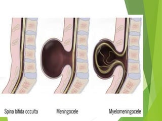
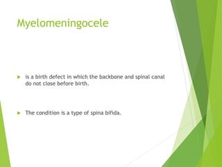
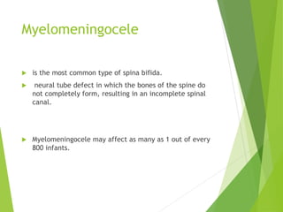
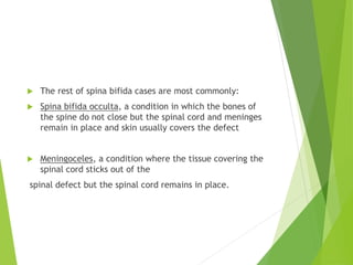
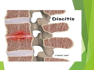
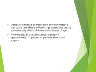
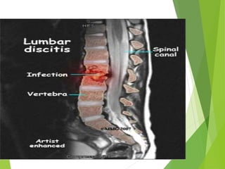
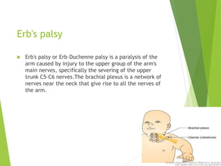
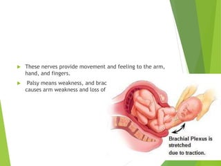
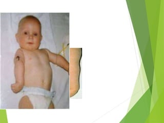
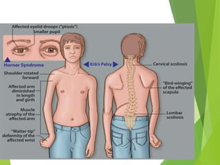
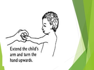
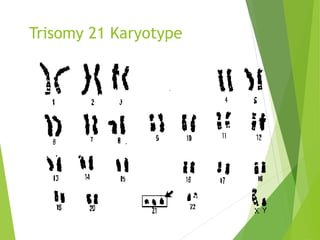
5. Myelomeningocele
is a birth defect in which the backbone and spinal canal
do not close before birth.
The condition is a type of spina bifida.
6. Myelomeningocele
is the most common type of spina bifida.
neural tube defect in which the bones of the spine do
not completely form, resulting in an incomplete spinal
canal.
Myelomeningocele may affect as many as 1 out of every
800 infants.
7. The rest of spina bifida cases are most commonly:
Spina bifida occulta, a condition in which the bones of
the spine do not close but the spinal cord and meninges
remain in place and skin usually covers the defect
Meningoceles, a condition where the tissue covering the
spinal cord sticks out of the
spinal defect but the spinal cord remains in place.
8. symptoms
A newborn may have a sac sticking out of the mid to
lower back.
The doctor cannot see through the sac when shining a
light behind it. Symptoms include:
Loss of bladder or bowel control
Partial or complete lack of sensation
Partial or complete paralysis of the legs
Weakness of the hips, legs, or feet of a newborn
9. Other symptoms :
Abnormal feet or legs, such as clubfoot
Build up of fluid inside the skull (hydrocephalus)
Hair at the back part of the pelvis called the sacral area
Dimpling of the sacral area
10. Serum AFP (alpha-fetoprotein) testing in conjunction
with a second trimester ultrasound detects over 90% of
infants affected with a neural tube defect.
23. Discitis or diskitis is an infection in the intervertebral
disc space that affects different age groups, but usually
spontaneously affects children under 8 years of age.
Nonetheless, discitis occurs post surgically in
approximately 1-2 percent of patients after spinal
surgery.
24. Symptoms
severe back pain, leading to lack of mobility.
Some very young children may refuse to walk and
arching of the back is possible.
In post-operative situations, the symptoms occur within
a week and result in severe low back pain or neck pain
(depending on the surgical location).
If untreated, the discitis may resolve on its own, cause
a chronic low grade infection, or progress
to osteomyelitis and possibly even an epidural abscess.
29. Risk factors:
Systemic:
Old age (>80 Y)
RA
DM
Immunosuppressive
Hemodyalisis
Malignancy
Local:
RA
OA
Prosthetic joint
30. Etiology (microbiology) Microbial agent:
Staphylococcus aureus: most common (75-80%)
Other organism in special patients:
Sexually active woman: Neisseria gonorrheae
Elderly, IV drug abuser, immunocompromised, UTI: Gram
negative (p. aeruginosa and E.coli)
SLE: Salmonella
HIV: Pneumococci, Salmonella, H. influenzae
Alcoholism, Humeral immunity abnormality,
Hemoglobinopathies: Pneumococcal infections
Primary immunoglobolin deficiency: Mycoplasma
31. Clinical manifestations
Fever (toxic)
Acute
Sever pain
Sever swelling of one joint
Sever tenderness
Warmth
Sever effusion
Sever limited ROM
32.
33.
34. Infections of prosthesis
implants
Many joint prostheses are used in orthopedic practice
and, whereas hip and knee joint replacement is
performed thousands of times per year throughout the
world.
shoulder, elbow, wrist, ankle, metacarpophalangeal or
interphalangeal joint replacement is much more recent
and experimental
. In 2004, hip and knee replacement procedures
accounted for 95% of the 1.07 million arthroplasty
procedures performed in the USA.
35. Although prosthetic joint implantations improve
patients' quality of life, these procedures are associated
with complications, including aseptic failure (i.e.,
aseptic loosening) and prosthetic joint infection (PJI).
More than 25% of all prostheses will eventually
demonstrate evidence of loosening, often necessitating
a revision arthroplasty.
Infection, although uncommon, is the most serious
complication following joint prosthesis implantation.
36. In patients with primary joint replacement, the
infection rate in the first 2 years is usually <1% in hip
and shoulder prostheses, <2% in knee prostheses, and
<9% in elbow prostheses.
The reported infection rates are probably
underestimated, since many cases of presumed aseptic
failure may be due to unrecognized infection.
In addition, infection rates after surgical revision are
usually considerably higher (up to 40%) than after
primary replacement.
38. remote infection,
prior native joint infection,
bacteremia (Staphylococcus aureus),
advanced HIV infection,
a revision surgery,
or preoperative use of low-molecular-weight heparin.
The mortality rate attributed to PJI may range between
0.4% in 65-year-old patients to 7% in 80-year-old
patients.
39. The most commonly isolated microorganisms are Gram-
positive cocci with coagulase-negative staphylococci, S.
aureus and enterococci accounting for 65% of cases.
Aerobic Gram-negative bacilli, including Escherichia
coli, Proteus mirabilis andPseudomonas aeruginosa, are
far less frequent causes of infection (6%).
Anaerobes, including Propionibacterium acnes, account
for 4% of infections.
40.
41. Erb's palsy
Erb's palsy or Erb–Duchenne palsy is a paralysis of the
arm caused by injury to the upper group of the arm's
main nerves, specifically the severing of the upper
trunk C5–C6 nerves.The brachial plexus is a network of
nerves near the neck that give rise to all the nerves of
the arm.
42. These nerves provide movement and feeling to the arm,
hand, and fingers.
Palsy means weakness, and brachial plexus birth palsy
causes arm weakness and loss of motion.
43. One or two of every 1,000 babies have this condition. It
is often caused when an infant's neck is stretched to the
side during a difficult delivery.
44. Causes:
difficult delivery
large baby,
a breech presentation,
prolonged labor.
Forceful delivery (If one side of the baby's neck is
stretched, the nerves may also be stretched, and injury
may result).
45. Symptoms
Weakness in one arm
Loss of feeling in the arm
Partial or total paralysis of the arm
The arm cannot be raised from the side; all power of
flexion of the elbow is lost, as is also supination of the
forearm".
The resulting biceps damage is the main cause of this
classic physical position commonly called "waiter's tip."
46.
47.
48.
49.
50. Down Syndrome
Genetics
All persons with Down syndrome have duplicated chromosome 21 material
The origin of the duplicated genetic material can vary
Nondisjunction (about 96% of cases)
The extra chromosome may arise by abnormal sorting to produce an extra, free-standing
chromosome (trisomy 21)
Translocation (3 % of cases)
a joining of chromosomes to produce extra chromosome 21 material that is attached to
another chromosome
Mosaicism (1-2%)
Patients with mixtures of normal and trisomic cells (mosaic Down syndrome) often have
milder phenotypes.
Percentages of normal cells within the blood sample used for chromosome studies may
differ from the percentages of normal cells in other tissues like brain or heart.
51. Genetics
Trisomy 21 (47, +21), - 94 %, The frequency of trisomy
increases with increasing maternal age.
Robertsonian translocation involving chromosome 21-
Approx. 3-4 %, not related to maternal age.
Trisomy 21 mosaicism – 2 to 3 % cases
54. Clinical Features
Head and neck
Brachycephaly
Up-slanting palpebral fissures
Epicanthal folds
Brushfield spots
Flat nasal bridge
Folded or dysplastic ears
Open mouth
Protruding tongue
Short neck
Excessive skin at the nape of
neck
Extremities
Short broad hands
Short fifth finger
Incurved fifth finger
Transverse palmer crease
Space between first and
second toe
Hyper flexibility of joints
56. Mental Retardation
Almost all DS babies have MR.
Mildly to moderately retarded .
Starts in the first year of life.
Average age of sitting(11 mon), and
walking (26 mon) is twice the typical
age.
First words at 18 months.
IQ declines through the first 10 years
of age, reaching a plateau in
adolescence that continues into
adulthood.
57. Growth
BW, length and HC are less in DS
Reduced growth rate
Prevalence of obesity is greater in DS
Weight is less than expected for length in infants with
DS, and then increases disproportion ally so that they
are obese by age 3-4 yrs
58. Diagnosis
Prenatal screening
If no screening – It is recognized from the characteristic
phenotypic features.
Confirmed by Karyotype.
59. Management
1. Growth – Measurements should be plotted on the
appropriate growth chart for children with DS.
This will help in prevention of obesity and early
diagnosis of celiac disease and hypothyroidism.
2. Cardiac disease – All newborns should be evaluated
by cardiac ECHO for CHD in consultation with
pediatric cardiologist.
3. Hearing – Screening to be done in the newborn
period, every 6 months until 3 yrs of age and then
annually.
60.
61. Arthrogryposis multiplex
congenita
rthrogryposis multiplex congenita (AMC) is a group of
nonprogressive conditions that cause multiple joint
contractures (stiff joints) and abnormal muscle
development.
62. signs and symptoms
AMC are present at birth but can vary greatly in
severity.
exact cause of AMC is not fully understood, but it is
thought to be associated with decreased movement or
limited space in utero, connective tissue disorders, or
maternal illness.
Sometimes AMC occurs as part of genetic syndrome.
63.
64. Treatment focuses on the specific symptoms
experienced by each individual and may include
physical therapy, removable splints, exercise, or
surgery.