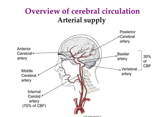
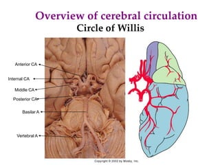
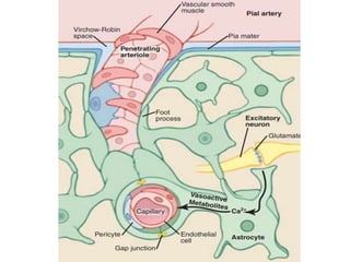
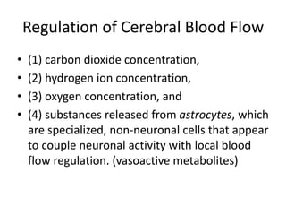
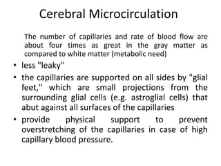
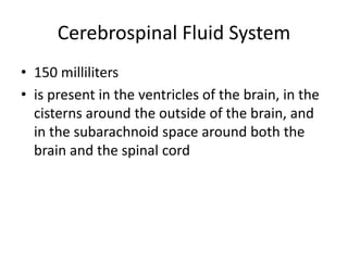
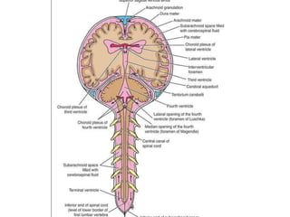
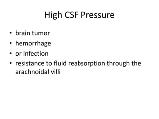
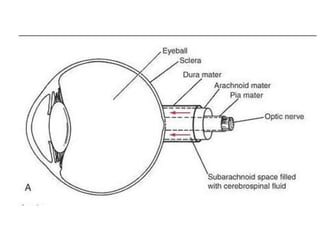
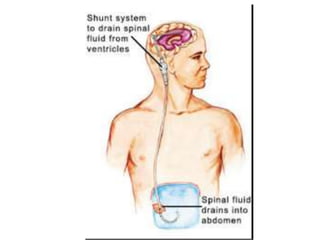
Cerebral blood flow& CSF
Overview of cerebral circulation
Anterior
Cerebral
artery
Middle
Cerebral
artery
Posterior
Cerebral
artery
Internal
Carotid
artery
(70% of CBF)
Vertebral
artery
Basilar
artery
30%
of
CBF
Arterial supply
Overview of cerebral circulation
Circle of Willis
Basilar A
Internal CA
Middle CA
Anterior CA
Vertebral A
Posterior CA
Normal Rate of Cerebral Blood Flow:
• Normal blood flow through the brain of the
adult person averages 50 to 65 milliliters per
100 grams of brain tissue per minute. (750 to
900 ml/min)
• Brain comprises only about 2 percent of the
body weight but receives 15 percent of the
resting cardiac output.
Regulation of Cerebral Blood Flow
• (1) carbon dioxide concentration,
• (2) hydrogen ion concentration,
• (3) oxygen concentration, and
• (4) substances released from astrocytes, which
are specialized, non-neuronal cells that appear
to couple neuronal activity with local blood
flow regulation. (vasoactive metabolites)
• Importance of Cerebral Blood Flow Control
by Carbon Dioxide and Hydrogen Ions
• Regulation by Oxygen deficiency (rate of
utilization of oxygen by the brain tissue
remains within narrow limits-almost exactly
3.5 (±0.2) milliliters of oxygen per 100 grams
of brain tissue per minute.
Cerebral Blood Flow Auto regulation
• Protects the Brain From Fluctuations in
Arterial Pressure Changes
cerebral blood flow is "auto regulated"
extremely well between arterial pressure
limits of 60 and 140 mm Hg.
• Role of Sympathetic Nervous System in
Controlling Cerebral Blood Flow
Cerebral Microcirculation
The number of capillaries and rate of blood flow are
about four times as great in the gray matter as
compared to white matter (metabolic need)
• less "leaky"
• the capillaries are supported on all sides by "glial
feet," which are small projections from the
surrounding glial cells (e.g. astroglial cells) that
abut against all surfaces of the capillaries
• provide physical support to prevent
overstretching of the capillaries in case of high
capillary blood pressure.
Cerebral "Stroke" Occurs When
Cerebral Blood Vessels Are Blocked
• arteriosclerotic plaques
• High BP
• Hemorrhage or infarction
Cerebrospinal Fluid System
• 150 milliliters
• is present in the ventricles of the brain, in the
cisterns around the outside of the brain, and
in the subarachnoid space around both the
brain and the spinal cord
CSF
• is formed at a rate of about 500 milliliters
each day
• secretion from the choroid plexuses in the
four ventricles, mainly in the two lateral
ventricles.
Secretion by the Choroid Plexus
• active transport of sodium ions through the
epithelial cells lining the outside of the plexus.
• The sodium ions in turn pull along large amounts
of chloride ions
• The two ions combined increase the quantity of
osmotically active sodium chloride and osmosis
of water through the membrane
• small amounts of glucose are added
• both potassium and bicarbonate ions are moved
out of the cerebrospinal fluid into the capillaries
Composition
• osmotic pressure, approximately equal to that
of plasma;
• sodium ion concentration, also approximately
equal to that of plasma;
• chloride ion, about 15 percent greater than in
plasma;
• potassium ion, approximately 40 percent less;
• and glucose, about 30 percent less.
CSF Pressure
The normal pressure in the cerebrospinal fluid
system when one is lying in a horizontal
position averages 130 mm of water (10 mm
Hg)
Cerebrospinal Fluid Pressure Very well
regulated:
1. The normal rate of cerebrospinal fluid
formation remains nearly constant
2. the arachnoidal villi function like "valves“.
Normally, this valve action of the villi allows
cerebrospinal fluid to begin to flow into the
blood when cerebrospinal fluid pressure is
about 1.5 mm Hg greater than the pressure of
the blood in the venous sinuses. Then, if the
cerebrospinal fluid pressure rises still higher,
the valves open more widely.
High CSF Pressure
• brain tumor
• hemorrhage
• or infection
• resistance to fluid reabsorption through the
arachnoidal villi
Edema of the Optic Disc-Papilledema
• (1) high cerebrospinal fluid pressure pushes fluid first
into the optic nerve sheath and then along the spaces
between the optic nerve fibers to the interior of the
eyeball;
• (2) the high pressure decreases outward fluid flow in
the optic nerves, causing accumulation of excess fluid
in the optic disc at the center of the retina; and
• (3) the pressure in the sheath also impedes flow of
blood in the retinal vein, thereby increasing the retinal
capillary pressure throughout the eye, which results in
still more retinal edema.
"Hydrocephalus”
• means excess water in the cranial vault
communicating hydrocephalus and
non communicating hydrocephalus
• In communicating hydrocephalus fluid flows
readily from the ventricular system into the
subarachnoid space, whereas in
noncommunicating hydrocephalus fluid flow out
of one or more of the ventricles is blocked.
• Usual cause of noncommunicating type is a
block in the aqueduct of Sylvius, resulting
from atresia (closure) before birth in many
babies or from blockage by a brain tumor at
any age.
• As fluid is formed by the choroid plexuses in
the two lateral and the third ventricles, the
volumes of these three ventricles increase
greatly.
• This flattens the brain into a thin shell against
the skull. In neonates, the increased pressure
also causes the whole head to swell because
the skull bones have not yet fused.
• The communicating type of hydrocephalus is
usually caused by blockage of fluid flow in the
subarachnoid spaces or by blockage of the
arachnoidal villi.
• Fluid therefore collects both on the outside of
the brain and to a lesser extent inside the
ventricles. This will also cause the head to
swell tremendously if it occurs in infancy when
the skull is still pliable and can be stretched,
and it can damage the brain at any age.
Treatment
• A therapy for many types of hydrocephalus is
surgical placement of a silicone tube shunt all
the way from one of the brain ventricles to the
peritoneal cavity where the excess fluid can be
absorbed into the blood.
Lumber puncture test
• Procedure
Significance:
• To measure CSF pressure
• Microscopy and chemical analysis is diagnostic
• Therapeutic
The Functions of the Cerebrospinal
Fluid
• Cushions and protects the central nervous system from
trauma
• Provides mechanical buoyancy and support for the
brain
• Serves as a reservoir and assists in the regulation of the
contents of the skull
• Nourishes the central nervous system
• Removes metabolites from the central nervous system
• Serves as a pathway for pineal secretions to reach the
pituitary gland
Cerebral blood flow

Cerebral blood flow

  • 1.
  • 2.
    Overview of cerebralcirculation Anterior Cerebral artery Middle Cerebral artery Posterior Cerebral artery Internal Carotid artery (70% of CBF) Vertebral artery Basilar artery 30% of CBF Arterial supply
  • 3.
    Overview of cerebralcirculation Circle of Willis Basilar A Internal CA Middle CA Anterior CA Vertebral A Posterior CA
  • 5.
    Normal Rate ofCerebral Blood Flow: • Normal blood flow through the brain of the adult person averages 50 to 65 milliliters per 100 grams of brain tissue per minute. (750 to 900 ml/min) • Brain comprises only about 2 percent of the body weight but receives 15 percent of the resting cardiac output.
  • 6.
    Regulation of CerebralBlood Flow • (1) carbon dioxide concentration, • (2) hydrogen ion concentration, • (3) oxygen concentration, and • (4) substances released from astrocytes, which are specialized, non-neuronal cells that appear to couple neuronal activity with local blood flow regulation. (vasoactive metabolites)
  • 8.
    • Importance ofCerebral Blood Flow Control by Carbon Dioxide and Hydrogen Ions • Regulation by Oxygen deficiency (rate of utilization of oxygen by the brain tissue remains within narrow limits-almost exactly 3.5 (±0.2) milliliters of oxygen per 100 grams of brain tissue per minute.
  • 9.
    Cerebral Blood FlowAuto regulation • Protects the Brain From Fluctuations in Arterial Pressure Changes cerebral blood flow is "auto regulated" extremely well between arterial pressure limits of 60 and 140 mm Hg. • Role of Sympathetic Nervous System in Controlling Cerebral Blood Flow
  • 10.
    Cerebral Microcirculation The numberof capillaries and rate of blood flow are about four times as great in the gray matter as compared to white matter (metabolic need) • less "leaky" • the capillaries are supported on all sides by "glial feet," which are small projections from the surrounding glial cells (e.g. astroglial cells) that abut against all surfaces of the capillaries • provide physical support to prevent overstretching of the capillaries in case of high capillary blood pressure.
  • 11.
    Cerebral "Stroke" OccursWhen Cerebral Blood Vessels Are Blocked • arteriosclerotic plaques • High BP • Hemorrhage or infarction
  • 12.
    Cerebrospinal Fluid System •150 milliliters • is present in the ventricles of the brain, in the cisterns around the outside of the brain, and in the subarachnoid space around both the brain and the spinal cord
  • 13.
    CSF • is formedat a rate of about 500 milliliters each day • secretion from the choroid plexuses in the four ventricles, mainly in the two lateral ventricles.
  • 15.
    Secretion by theChoroid Plexus • active transport of sodium ions through the epithelial cells lining the outside of the plexus. • The sodium ions in turn pull along large amounts of chloride ions • The two ions combined increase the quantity of osmotically active sodium chloride and osmosis of water through the membrane • small amounts of glucose are added • both potassium and bicarbonate ions are moved out of the cerebrospinal fluid into the capillaries
  • 16.
    Composition • osmotic pressure,approximately equal to that of plasma; • sodium ion concentration, also approximately equal to that of plasma; • chloride ion, about 15 percent greater than in plasma; • potassium ion, approximately 40 percent less; • and glucose, about 30 percent less.
  • 17.
    CSF Pressure The normalpressure in the cerebrospinal fluid system when one is lying in a horizontal position averages 130 mm of water (10 mm Hg)
  • 18.
    Cerebrospinal Fluid PressureVery well regulated: 1. The normal rate of cerebrospinal fluid formation remains nearly constant 2. the arachnoidal villi function like "valves“. Normally, this valve action of the villi allows cerebrospinal fluid to begin to flow into the blood when cerebrospinal fluid pressure is about 1.5 mm Hg greater than the pressure of the blood in the venous sinuses. Then, if the cerebrospinal fluid pressure rises still higher, the valves open more widely.
  • 19.
    High CSF Pressure •brain tumor • hemorrhage • or infection • resistance to fluid reabsorption through the arachnoidal villi
  • 20.
    Edema of theOptic Disc-Papilledema • (1) high cerebrospinal fluid pressure pushes fluid first into the optic nerve sheath and then along the spaces between the optic nerve fibers to the interior of the eyeball; • (2) the high pressure decreases outward fluid flow in the optic nerves, causing accumulation of excess fluid in the optic disc at the center of the retina; and • (3) the pressure in the sheath also impedes flow of blood in the retinal vein, thereby increasing the retinal capillary pressure throughout the eye, which results in still more retinal edema.
  • 22.
    "Hydrocephalus” • means excesswater in the cranial vault communicating hydrocephalus and non communicating hydrocephalus • In communicating hydrocephalus fluid flows readily from the ventricular system into the subarachnoid space, whereas in noncommunicating hydrocephalus fluid flow out of one or more of the ventricles is blocked.
  • 23.
    • Usual causeof noncommunicating type is a block in the aqueduct of Sylvius, resulting from atresia (closure) before birth in many babies or from blockage by a brain tumor at any age. • As fluid is formed by the choroid plexuses in the two lateral and the third ventricles, the volumes of these three ventricles increase greatly. • This flattens the brain into a thin shell against the skull. In neonates, the increased pressure also causes the whole head to swell because the skull bones have not yet fused.
  • 24.
    • The communicatingtype of hydrocephalus is usually caused by blockage of fluid flow in the subarachnoid spaces or by blockage of the arachnoidal villi. • Fluid therefore collects both on the outside of the brain and to a lesser extent inside the ventricles. This will also cause the head to swell tremendously if it occurs in infancy when the skull is still pliable and can be stretched, and it can damage the brain at any age.
  • 26.
    Treatment • A therapyfor many types of hydrocephalus is surgical placement of a silicone tube shunt all the way from one of the brain ventricles to the peritoneal cavity where the excess fluid can be absorbed into the blood.
  • 28.
    Lumber puncture test •Procedure Significance: • To measure CSF pressure • Microscopy and chemical analysis is diagnostic • Therapeutic
  • 29.
    The Functions ofthe Cerebrospinal Fluid • Cushions and protects the central nervous system from trauma • Provides mechanical buoyancy and support for the brain • Serves as a reservoir and assists in the regulation of the contents of the skull • Nourishes the central nervous system • Removes metabolites from the central nervous system • Serves as a pathway for pineal secretions to reach the pituitary gland