
Downloaded 140 times
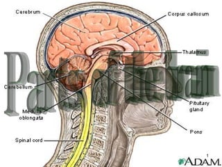
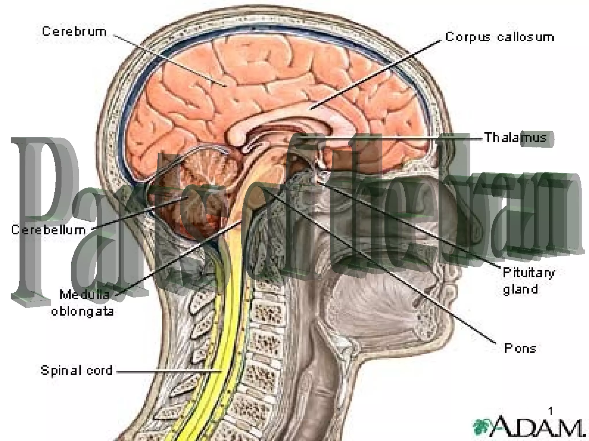
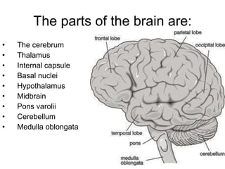
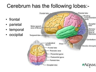

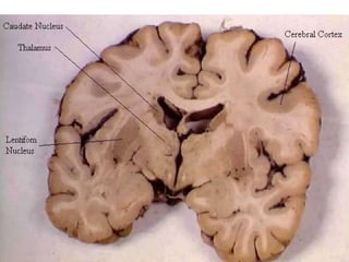
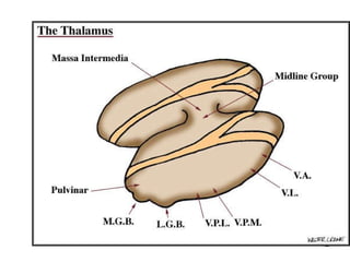
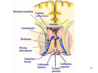
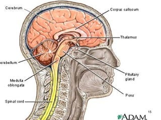
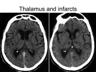
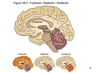
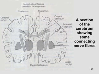

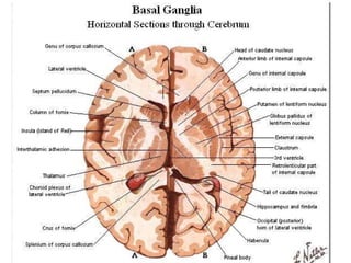

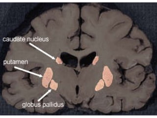
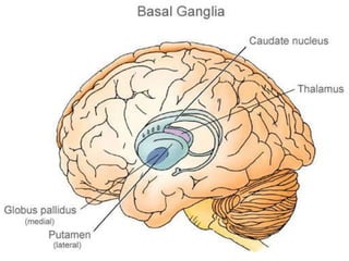
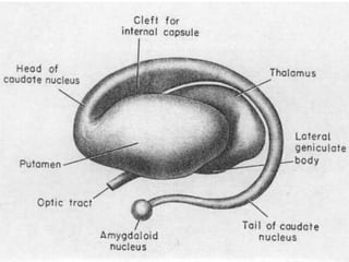
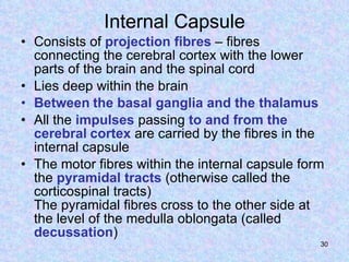
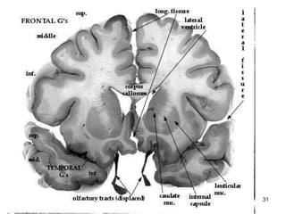
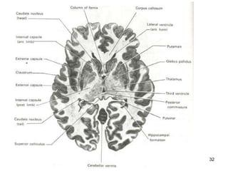
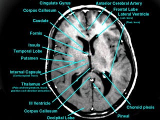

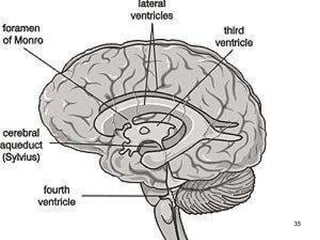
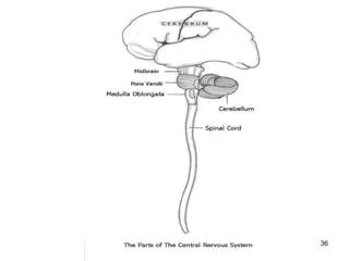
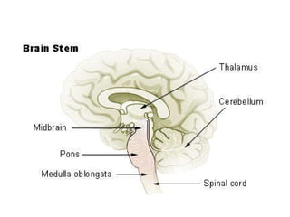
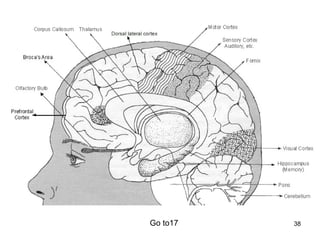
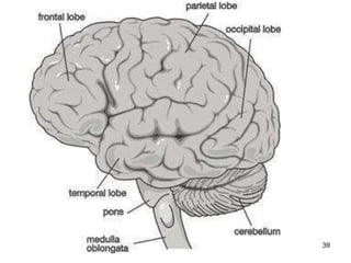
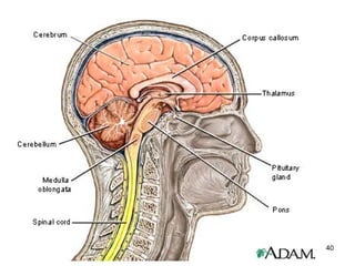
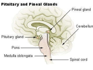

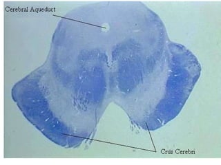
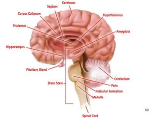


The parts of the brain include the cerebrum, thalamus, internal capsule, basal nuclei, hypothalamus, midbrain, pons varolii, cerebellum, and medulla oblongata. The cerebrum has lobes including the frontal, parietal, temporal, and occipital lobes. The thalamus relays sensory information to the cerebral cortex. The basal ganglia influence voluntary movements and posture. The internal capsule contains projection fibers connecting the cerebral cortex to lower parts of the brain. The cerebellum coordinates movement and balance.


