SlideShare a Scribd company logo
Sensory system
Dr. Dina Merzeban
Lecturer in physiology department
http://facebook.com/ merzeban physiology
www.youtube.com/physiology ‫تاني‬ ‫فكر‬
www.slideshare.net/merzeban
Sensory system
Consists of :
 Sensory receptors
 Sensory pathways
 Somatic sensory cortex
3
Receptors, Sensation,
and Perception
 Sensory receptors
 Specialized cells or multicellular structures
that collect information from the environment
 Stimulate neurons to send impulses along
sensory fibers to the brain
 Sensation
 A feeling that occurs when brain becomes
consciously aware of sensory impulse
 Perception
 A person’s understanding of the sensation; the
way the brain interprets the information
Sensory unit:
Single sensory afferent with all its receptor
ending.
Receptive field of a neuron:
Region cotaining all receptors which its
stimulation elicits stimulation in that neuron.
Receptive fields of adjacent neurons overlap
Classification of receptors
13
Anatomical classification
 Exteroceptors
 Senses associated with body surface such as
touch, pressure, temperature, and pain
 Interoceptors
 Senses associated with changes in the viscera such
as blood pressure stretching blood vessels and
ingestion of a meal
 Proprioceptors
 Senses associated with changes in muscles and
tendons such as at joints
Physiologically
 Mechanoreceptors
 Cutaneous (touch, pressure, vibration) eg. Pacinian,
Meissner’s corpuscle, free nerve endings
 Proprioceptors (joint position receptors) eg. Muscle
stretch receptors, tendon organs
 Baroreceptors
 Auditory/vestibular hair cells
•
•
 Chemoreceptors
 Taste buds and smell receptors
 Visceral chemoreceptors sensitive to
Pco2, pH, osmolality(osmoreceptors),
glucoreceptors .
•
 Thermoreceptors
 Cold and hot receptors
 Pain receptors(nociceptors)
 Photoreceptors: rods and cones
Properties of receptors
1-Specificity
Receptors are stimulated by a certain type of
energy (adequate stimulus).
Stimulation of a receptor usually produces only
one sensation
 modality specific
But some receptors are stimulated by more
than one sensory modality (polymodal)
 eg. free nerve endings
Receptors act as :
 detectors
 transducers
2-Excitability
• Receptor cells are specific cells that are
sensitive to different forms of energy from
the environment
• These cells when stimulated generate
receptor (or generator)potential
• They transform the stimulus into
electrical signals
What happens inside a receptor?
• TRANSDUCTION
Stimulus energy is converted to action
potentials
Inside the nervous system signals are always
action potentials
Language of the nervous system contains only
1 word: action potentials
• Receptors transform
an external signal into
a membrane potential
• Receptor Potential
- separate receptor
• Generator Potential
-a specialized ending
of an afferent neuron
7
Sensory Impulses
• Stimulation of receptor causes local change in its receptor potential
•A graded electrical current is generated that reflects intensityof
stimulation
•If receptor is part of a neuron, the membrane potential maygenerate
an action potential
•If receptor is not part of a neuron, the receptor potential mustbe
transferred to a neuron to trigger an action potential
• Peripheral nerves transmit impulses to CNS where they are analyzed
and interpreted in the brain
Receptor potential generation
in the Pacinian corpuscle
Resting
Physical Stimulus
Physical stimulus causing mechanical deformation on the capsule
Physical Stimulus
Mechanical deformation is transmitted to the inside
Opens up mechanosensitive Na+ channel
Causes depolarisation and thus receptor potential
Physical Stimulus
local current
Current flow through a local circuit
Physical Stimulus
Action Potentials
are generated
Opening of voltage gated Na+ channels causes generation of
action potentials
Characters of Receptor potentials
• Non propagated local excitatory state.
• This is a graded potential as It does not follow
“all-or-none law”
• Its amplitude depends on the strength of the
stimulus.
• Summated (no refractory period)
• Long duration
Stimulus
Receptor
potentials
Action
potentials
3-Relation between strength of stimulus and
magnitude of receptor potential.
Action Potentials
Resting
Membrane
Potential
-70
Threshold - 55
+30
Stimulus
Receptor potential
8
4-Sensory Adaptation
The ability to diminish the receptor potential
depolarization and number of action potentials in afferent
nerve despite sustained stimulus strength
 Types of receptors according to their speed of
adaptation
 Tonic :Do not adapt or slowly adapting receptors
e.g. pain receptors,muscle stretch receptors
and joint proprioceptors
 Moderately adapting receptors:
 temperature, smell and taste.
 Phasic: (Rapidly adapting receptors )
e.g. Tactile receptors
Pain
Time
Muscle
spindle
Temperature
Smell
taste
Touch and pressure
Pacinian corpuscles
• In the Pacinian corpuscle
Mechanical deformation is transmitted
throughout the capsule and pressure is
redistributed.
Na+ channels inactivates after some time
Impulse
Stimulus
Redistribution of pressure inside the capsule
Stimulus
No
Impulse
•Rapidly adapting receptors
phasic or rate or movement receptors
detect changes in stimulus strength
eg. Pacinian corpuscle, hair end-organ
•Slowly adapting receptors
tonic receptors
detect continuous stimulus strength
eg. muscle spindles, Golgi tendon organ, baroreceptors,
Ruffini endings and Merkel’s discs, pain receptors
Sensory coding
• A receptor must convey the
type of information it is
sending  the kind of
receptor activated
determined the signal
recognition by the brain
• It must convey the intensity
of the stimulus  the
stronger the signals, the
more frequent will be the
APs
• It must send information
about the location and
receptive field, characteristic
of the receptor
Sensory coding
Modality of sensation:
 Adequate stimulus
 Mullers law of specific nerve energy
 Labeled line principle
Locality of sensation
 Low of projection
phantom limb
Intensity of sensation
 Number of receptors
 Frequency of impulses :
 weber feshner principle
R=log S * K
 Stevens power principle
R=ASK
Tactile sensations
Classification of sensation
Special Sensations: Vision,
Hearing, Taste, Smell
General sensations:
Somatic sensation
Organic sensations: e.g. hunger
Emotional sensation: e.g. fear,
sadness
Somatic sensation
Mechanoreceptive sensations
Tactile sensations
Position sensations: static and
kinetic
Thermoreceptive sensations:
Cold, Warm
Pain sensation
Tactile sensations
 Touch :
Crude touch
Fine touch
Tactile localization
Tactile discrimination
Texture
 Stereognosis
 Pressure: crude & fine
 Vibration
 Itching & tickling
Transmission of somatic sensation
_ A fibers:Myelinated
• Subtypes: some overlap in ranges
• Fastest conducting and largest diameter – m/sec,
– B fibers: Slower myelinated
– C fibers: Un myelinated
• Slower conduction and smallest diameter (0.5 m/sec,
0.5 )
Nerve Fiber Classification
– I, II, III fibers: Myelinated
• Subtypes: Ia, Ib
• Fastest conducting and largest diameter – Ia
– IV fibers: Unmyelinated
• Slower conducting than IIIs and smallest diameter
Modality of sensation
Type of fiber
Proprioceptors
I
Aα
Fine touch, pressure, stereognosis, vibration
II
Aβ
Fast pain, temperature,crude touch
III
A
Slow pain, temp., tickle and itching
IV
C
Sensory pathway
• Once a receptor is stimulated
impulse travels through a particular pathway
known as sensory pathway or ascending
pathway up to the brain
Receptor
Sensory
modality
Sensory nerve
Ascending
Sensory pathway
Central Connections
Sensory area
in the brain
Touch stimulus
Sensory pathway
Two ascending pathways
• Dorsal column - medial lemniscus pathway
fast pathway
• Spinothalamic pathway
slow pathway
These two pathways come together at the level of thalamus
Posterior (dorsal)
Dorsal root ganglion
Dorsal root
Dorsal columns
Dorsal horn
Spinothalamic
tracts
Anterior (ventral)
Dorsal column pathway
Spinothalamic pathway
Lateral
Spinothalamic
tract
Anterior
Spinothalamic
tract
Dorsal column pathway
 Fine touch
 fine pressure
 stereognosis
 vibratory sense
 movement sense
 position sense
Spinothalamic pathway
• Pain
• Thermal sensations
• Crude touch & pressure
• tickle & itch
Dorsal column nuclei
(cuneate & gracile nucleus)
Dorsal column
Medial lemniscus
thalamus
thalamocortical tracts
internal capsule
1st
order
neuron
2nd
order
neuron
3rd
order
neuron
Dorsal column
medial lemniscus pathway
• after entering the spinal cord
•lateral branch: participates in spinal cord reflexes
•medial branch: turns upwards
• forms the dorsal columns
• spatial orientation:
•medial: lower parts of the body
•lateral: upper part of the body
dorsal column
medial lemniscus pathway
• synapse in the dorsal column nuclei
nucleus cuneatus & nucleus gracilus
• 2nd order neuron cross over to the opposite
side and ascends upwards as medial
lemniscus
• as this travels along the brain stem fibres
from head and neck are joined (trigeminal)
• ends in the thalamus (ventrobasal complex)
 ventral posterolateral nuclei
Spinothalamic pathway
• after entering the spinal cord
synapse in the dorsal horn
• cross over to the opposite side
• divide in to two tracts
lateral spinothalamic tract:
pain and temperature
anterior spinothalamic tract
crude touch
Thalamocortical tracts
from the thalamus 3rd order neuron
ascends up through the internal capsule
up to the sensory cortex
)thalamocortical radiation(
Sensory cortical areas
• parietal cortex
• a distinct spatial orientation exists
Sensory cortex
• Different areas of the body are represented
in different cortical areas in the sensory
cortex
• Sensory area
somatotopic representation
not proportionate
distorted map
upside down map
Representation
•upside down
•distorted
Map
Sensory homunculus
Sensory cortical areas
• Primary somatosensory cortex (SSI)
postcentral gyrus
(Brodmann areas 3, 1, 2)
• Secondary somatosensory cortex
and Somatosensory association
cortex (SSII)
Posterior parietal areas
(Brodmann areas 5, 7)
Somatosensory cortex
• Functions
To localise somatic sensations
To judge critical degree of pressure
To identify objects by their weight, shape,
form - stereognosis
To judge texture of materials
To localise pain & temperature
Somatosensory cortex
• Damage to the sensory cortex results in
decreased sensory thresholds
inability to discriminate the properties of
tactile stimuli
Inability to identify objects by touch
(astereognosis)
Secondary somatosensorycortex SS
II
 Occupies area 40
 Receives input from SSI
 Spatial orientation :face ant.
, arms centrally, legs post.
Somatosensory association cortex
• Located directly posterior to the
sensory cortex in the superior
parietal lobes
• Consists of areas 5 and 7
• Receives synthesized
connections from the primary
and secondary sensory cortices
• Neurons respond to several
types of inputs and are
involved in the understanding
of sensation
Secondary somatosensory cortex and
Somatosensory association cortex
• Damage can cause
Tactile agnosia
inability to recognize objects even though the objects can
be felt
Spatial neglect
This typically happens with non-dominant hemisphere
lesions
Neglect can be so severe that the individual even denies
that their left side belongs to them
Types of somatic sensations
 Mechanoreceptive sensations
 How to test
 Receptor
 Afferent
 pathway
Modality of sensation
Type of fiber
Proprioceptors
I
Aα
Fine touch, pressure, stereognosis, vibration
II
Aβ
Fast pain, temperature,crude touch
III
A delta
Slow pain, temp., tickle and itching
IV
C
Dorsal column pathway
 Fine touch
 fine pressure
 stereognosis
 vibratory sense
 movement sense
 position sense
Spinothalamic pathway
• Pain
• Thermal sensations
• Crude touch & pressure
• tickle & itch
pathway
afferent
receptor
Type of sensation
Dorsal column
Aβ
Meissner &merkel
Touch fine touch
Ant . spinothalamic
A delta
Free nerve ending
Crude
touch
Dorsal column
Aβ
A delta
Pacinian corpuscles&
spray type endings
pressure fine
crude
Dorsal column
Aβ
mixture
stereognosis
Dorsal column
Aβ
Pacinian 500hz
Meissners 80 hz
vibration
Dorsal column
Aα
Muscle spindles golgi
tendon organs
Ruffini endings
Prprioceptive
sensations
Ant . spinothalamic
C
Free nerve endings
Tickling and itching
Lat. spinothalamic
C
A delta& C
VR1 VRL1
CMR
Temp . Warm
cold
Lat. spinothalamic
A delta
C
Free nerve ending
Pain fast
slow
Receptive fields
• The receptor area which when stimulated
results in a response of a particular sensory
neuron
• Receptive fields of adjacent neurons overlap
Two-Point Discrimination
Whether a stimulus feels like one sensation or two
distinct sensations depends on:
• Number of receptors
• the size of the receptive fields of the sensory
receptors:Smaller receptive fields result in greater sensitivity
 Fingers are more sensitive than backs
• Degree of convergence within the pathway
• Area of representation
Sensory homunculus
Thermal sensations
Thermal receptors
a) Warm : free nerve ending ( C fibers )
b) Cold : free nerve endings ( C fibers and A
delta fibers )
c) Others :cold pain and warm pain
 Tested by using three test tubes ( 5 °C ,
25 °C & 40 °C )
 Touch any part of body by the test tube (
for 2 minutes )
 Ask him to feel hot or cold
Characters of thermal
receptors
 They respond to the
temperature of subcutaneous
tissue.
 Widely separated(wide area exposed for better
differentiation)
 Moderately adapting receptors
 distribution: lips > fingers >
trunk
 Cold sensation is more important than warm sensation
 Cold receptors:
- More numerous ( 4-10 times)
- More superficial (under skin )
- Adapt more slowly
- Carried on type C & A delta
Mechanism of stimulation of
thermal receptors
 Chemically by change in concentration of metabolites
 Respond markedly to changing temperature
Detection of thermal
sensation
 cold pain ( 5-15 °C) maximum
5 °C
 cold receptors ( 10-43°C) maximum
25°C
 warm receptors ( 30-50°C) maximum
45 °C
 warm pain above( 45°C )
 Paradoxical pain sensation
 At 0 °C : anaesthesia
Cold
pain
Warm
pain
Sensory
NERvous
system
Dr. Dina Merzeban
Lecturer in physiology department
http://facebook.com/ merzeban physiology
www.youtube.com/physiology ‫تاني‬ ‫فكر‬
www.slideshare.net/merzeban
Pain sensation
Pain is an, unpleasant emotional
experience associated with actual or
potential tissue damage - The International
Association for the Study of Pain (IASP)
Pain= tissue damage
Pain system:
 Pain receptors (nociceptors).
 Types of pain.
 Pain pathways.
 Pain control.
Nociceptors
 Free nerve endings attached to A delta & C,
slowly (non) adapting to prolonged stimulation
4 types:
Mechanical pain receptors.
Thermal pain receptors.
Chemical pain receptors.
Polymodal pain receptors
Distribution of pain receptors
Widely
distributed
Sup. Layers of skin
Pleura
Peritonium
periosteum
Arterial walls
Joint surface
Dura of cranial cavity
Less
distributed
Deep tissues
& Viscera
Absent
Liver
Parenchyma
Lung alveoli
Brain tissue
Stimulation of pain
sensation
 Damaged tissues----chemical mediators e.g. histamine,
serotonin, substance P, bradykinins, K+ -------
sensitizes painreceptors -------hyperalgesia
 1ry hyperalgesia
 2ry hyperalgesia
Pain pathways
 Two separate pathways:
I. Neospinothalamic pathway:
Conducting quick, localized pain
(Fast sharp pain)
II. Paleospinothalamic pathway:
For slow pain
Lateral
spino-
thalamic
tract
Ad fiber
glutamate
C Fibers
Subst. P
Substantia
Gelatinosa of
Rolandi (SGR)
glutamate
Neospinothalamic tract
Paleospinothalamic tract
 In the brain stem, 90% of fibers synpse on:
 Reticular formation
 Intralaminar (non specific ) nuclei
of thalamus
to activate the whole cortex
Reactions to pain
 Motor reflexes (AHCs)
 Arousal reaction (RAS)
 Autonomic reactions(hypothalamus)
 Emotional reactions(limbic system)
Perception of pain signals
Fast pain in thalamus & cortex
Slow pain in thalamus
Roles of the cortex in pain perception are
1.Localization of pain.
2.Discrimination of pain.
3.Modulation of pain.
Types of pain
According to oigin
 Cutaneous
 Deep
 Visceral
 Neuropathic
According to Quality :
1. Fast pain (sharp, acute, pricking,
immediate)
2. Slow pain (burning, chronic, dull
aching, throbbing
Slow pain
Fast pain
Felt within 1 sec or
more
Felt within 0.1 sec
May be prolonged
Short duration
Poorly localized
Well localized
All types of receptors
Mechanical or thermal
Skin, deep tissues &
viscera
Usually in skin, rare in
deep tissues
slow pain
Fast pain
Carried by C, blocked
by local anaesthetics
Carried by Ad, blocked
by pressure & O2 lack
C release Substance P
Ad release Glutamate
Transmitted by
Paleospinothalamic T
Transmitted by Neo-
spinothalamic T
End in RF Non-
specific thalamic nuclei
whole cortex
Its fibers end in
sensory cortex
Deep pain
 Felt in muscles , tendons , joints and bones
Causes:
 Trauma
 Inflammation
 Muscle spasm
 Ischemic pain
Ischemic pain
 Definitions : type of deep pain felt in
muscles when their blood supply is reduced
 Example : angina pectoris & intermittent
claudicating
 Causes
 Increase metabolites and proteolytic enzymes.
Intermittent claudication: recurrent pain in the calf
muscle during exertion, which stops on rest.
This is due to ischaemia of muscle which produces P-
factor – pain producing chemical agent which is
responsible for causation of pain. During rest the pain
stops because P factor is washed away by blood.
Eg:Thrombo Angitis Obliterans. (Buerger’s disease)
Coronary occlusion: In addition to P factor, there is
release of 5HT and plasma pain producing
polypeptide.
Inflammatory pain: - due to increased tension
causing pressure on nerve terminals as well as due
to release of a chemical pain producing factor.
Visceral pain
 Pain from viscera ( peritoneum , pleura
and pericardium )
 C – fiber pain (poorly localized)
 A delta fibers stimulated if from parietal
layers of viscera
 Most viscera contain only few pain
receptors
 Sharp cut doesn't cause pain ( injury of
few nerve endings )
 Diffuse stimulation cause severe pain
Visceral pain
Characters
a) Dull aching
b) Poorly localized
c) Autonomic responses : decrease
heart rate and ABP
d) Guarding (Rigidity)
e) Referred
Visceral pain
Causes
a) Over distension
b) Spasm
c) Ischemia
d) Inflammation and irritation
e) Infiltration by tumors
CAUSES OF VISCERAL PAIN
 Ischaemia: The substances released during ischaemic
reactions like bradykinin and proteolytic enzymes
stimulate the pain receptors of viscera.
 Chemical stimuli: The chemical substances like acidic
gastric juice leaks from ruptured ulcers into peritoneal
cavity and produce pain.
 Spasm of hollow organs:
spastic contraction of muscles in gastrointestinal tract
and other hollow organs of viscera cause pain by
stimulating the free nerve endings.
 Overdistension of hollow organs also cause pain.
Neuropathic pain
 Pain due to nerve fiber damage
 Examples :
a) Trigeminal neuralgia
b) Sciatica
c) Herpes zester
d) Diabetic neuropathy
Referred pain
 Definition: pain not felt in diseased
viscus and felt in corresponding
dermatome
 Examples:
 Cardiac : left shoulder
 Gall bladder : Right shoulder
 Appendix : at umbilicus
 Gastric : above umbilicus
 Kidney & pancreas : back
Referred pain
Heart pain is referred to the inner
aspect of left arm.
Diaphragmatic pain to the tip of the
shoulder
Ureteric pain to the testes in male and
the inner aspect of the thigh in female.
Gall bladder pain referred to epigastric
region.
Pain from the maxillary sinus referred to
the nearby tooth.
ReferredPain
1.Convergence Projection theory
DermatomalRule
 Referred pain is always felt over a structure
that developed from the same embryological
segment or dermatome from which the
organ which is the source of pain
developed.
 This phenomenon of referred pain is called
Dermatomal rule.
DermatomalRule
 During Embryonic development, the diaghragm
migrates from the neck region to its adult location
between the chest and abdomen and takes its nerve
supply, the phrenic nerve with it.
 Similarly the heart and the arm have the same
segmental origin.
 Testicles have migrated with its nerve supply from the
primitive urogenital ridge from which kidney and ureter
has developed.
CONVERGENCE THEORY
The number of peripheral pain exceed the number of
lateral spinothalamic tract. So, Both the somatic and
visceral afferents converge upon the same
spinothalamic neurons at the spinal cord level.
Hence when visceral pain impulses travel in the same
pathway along which impulses from the skin travels, the
individual gets the feeling that the pain originates in the
skin itself.
1.Convergence Projection theory
FACILITATION THEORY
Visceral and somatic pain afferents connect with separate
but adjoining spinothalamic neurons
and there may be some overlap of the neurons,
visceral afferents have collaterals connecting to the
spinopthalamic neurons receiving somatic pain afferents.
This causes impulses to travel
up the somatic spinothalamic
pathway and causes the sensation
of pain in the skin.
Facilitation theory
 Afferent from pain fibers of diseased viscera, give
subliminal fring to a nearby SGR (receiving afferents
from an area of skin), increasing its excitability.
 The result is pain felt in this skin area and lowering of
its pain threshold
2. Facilitation theory
Viscus
 Terminology
 Algesia =pain
 Analgesia = dec./loss of sensitivity to painful
stimuli
 Hyperalgesia = inc. sensitivity to painfulstimuli
 Allodynia =sensation of pain in response to
an innocuous stimulus
 Brown-Sequardsyndrome(hemisection of
the spinal cord.
 Syringomyelia(loss of pain & temp. with
sparing of touch & vibration.
Hyperalgesia
 Primary hyperalgesia
 Secondary hyperalgesia
Primary hyperalgesia
Secondary hyperalgesia
Ad fiber
glutamate
C Fibers
Subst. P
Substantia
Gelatinosa of
Rolandi (SGR)
glutamate
Somatosensory cortex
Other basal areas of brain
Thalamus – ventrobasal
complex
Spinothalamic tract Spinal
cord
(lamina I – lamina
marginalis)
Peripheral fibers Aδ fibers
Pain receptor(Free nerve
endings)
• Reticular nuclei
• Tectal area &
periaqueduvtal
grey region
• Thalamus
Other basal areas of
brain
Spinothalamic tract
Spinal cord
(lamina II & III –
substantia gelatinosa)
Peripheral fibers C
fibers
Pain receptor
(Free nerve endings)
Pain pathway
Pain Modulation
Pain Modulation
 Inhibitory neurotransmitters like
endogenous opioids work to hinder the
pain transmission.
 This inhibition of the pain impulse is known
as modulation
Physiological Basis of analgesia system
 The CNS has its own control system which inhibits the
impulse of pain sensation.
 This is also called Analagesia system.
 This control system is present in both brain and
spinal cord.
 Pain control complex in spinal cord:
 This is in dorsal grey horn.
 It is considered as the gateway for pain impulses to
reach the brain (via) spinothalamic tract.
PHYSIOLOGYOFPAIN PERCEPTION
Injury
Descending
Pathway
Peripheral
Nerve
Dorsal
Root
Ganglion
C-Fiber A-beta Fiber
A-delta Fiber
Ascendin
g
Pathways
Dorsal
Horn
Brain
Spinal Cord
GATE CONTROL THEORY
 Substantia Gelatinosa (SG) in dorsal horn of spinal cord
acts as a ‘gate’
 This gate can be closed by:
 Descending supraspinal inhibitory impulses
 A-beta neurons are stimulated which closes the gate
to A-delta & C neurons
Spinal pain inhibitory pathway
 Gate - located in the dorsal horn of the spinal cord
 Smaller, slower n. fibers carry pain impulses
 Larger, faster n. fibers carry other sensations
 Impulses from faster fibers arriving the gate 1st
inhibit pain impulses (acupuncture/pressure, cold,
heat, chem. skin irritation).
Brain
Pain
Heat, Cold,
Mechanical
Gate
(SG)
•Periaqueductal grey
Raphe magnus nucleus
(pain inhibitory complex in dorsal horn)
Hypothalamus
(periventricular nucleus )
Supraspinal inhibitory pathway
(Descending pain control mechanism)
Supraspinal inhibitory pathway
(Descending pain control mechanism)
Periaquaductal Gray Area (PGA)
release enkephalins
Nucleus Raphe Magnus (NRM)
release serotonin
Stimulation of presynaptic inhibitory intermediary
neuron
secreting enkephalin
Inhibit the release of substance P from SGR ascending
neurons
Hypothalamus
(periventricular nucleus )
Release endorphin
Spinal pain inhibitory pathway
 Gate - located in the dorsal horn of the spinal cord
 Smaller, slower n. fibers carry pain impulses
 Larger, faster n. fibers carry other sensations
 Impulses from faster fibers arriving the gate 1st
inhibit pain impulses (acupuncture/pressure, cold,
heat, chem. skin irritation).
Brain
Pain
Heat, Cold,
Mechanical
Gate
(SG)
Inhibition of pain transmission by tactile
sensory signals
 Rubbing the skin near painful areas and
applying liniments often relieves pain.
 This is due to the stimulation of Aβ sensory fibres
from peripheral tactile receptors depress
transmission of pain signals.
 This results from a type of local lateral
inhibition.
 Acupuncture is also used to relieve pain.
Acupuncture is also used to relieve pain based upon the pain
inhibitory mechanism
 Treatment for pain
 Remove the cause
 Drugs
 NSAIDs (inhibit COX)
 Opiates (inhibit NT release)
Surgical procedure that relieve pain
Different surgical procedures are done in the course of
pain pathway to relieve pain. They are
-Sympathectomy
-Cordotomy
-Thalamotomy
-Prefrontal lobotomy
Headache
 Def. Headache is a referred pain
 headache is pain referred to the surface of
the head from deep structures
 It may be
 frontal headache referred to areas supplied
by trigeminal nerve.
 occipital headache referred to areas of the
head supplied by the second cervical nerve
Headache
 the brain itself is insensitive to pain
 the intracranial pain sensitive areas
include
 dura and Tentorium
 nerves
 venous sinuses
 the dural arteries ( meningeal artery at the base of
the skull)

Distribution of pain receptors
Widely
distributed
Sup. Layers of skin
Pleura
Peritonium
periosteum
Arterial walls
Joint surface
Dura of cranial cavity
Less
distributed
Deep tissues
& Viscera
Absent
Liver
Parenchyma
Lung alveoli
Brain tissue
Headache
 it is caused by
 traction
 displacement
 inflammation
 vascular spasm
or distension of the pain sensitive structures in
the head or neck
 it may be from intracranial or extracranial origin
causes of intracranial headache
 meningitis or meningeal irritation ( constipation)
 intracranial mass ( brain tumours)
 drop of intracranial tension ( post lumbar puncture
headache )
 Rise of intracranial tension
 alcohol headache
causes of intracranial headache
 Trigeminal neuralgia is a facial pain confined
mainly to areas supplied by the second and third
divisions of the trigeminal nerve
 hypertension due to marked expansion of the
cerebral blood vs
 Migraine headache : unilateral headache
resulting from abnormal vascular Phenomenon
 First severe constriction of the cerebral blood vessels
followed by marked Dilatation of the cerebral blood
vessels
headache of of extracranial origin
 Tension headache due to spasm of the
neck and scalp muscles
 Eye disorders : errors of refraction &
glaucoma
 Nose : sinusitis
 Ear : otitis Media
 Mouth : Toothache
Headache
 Dura
 Cerebral blood vessels
 CSFpressure
 Nasal & accessory nasal structures
 Eye disorders
 Muscle spasm of head & neck muscles
 Alcohol
 constipation
Dr. Dina Merzeban
Lecturer in physiology department
http://facebook.com/ merzeban physiology
www.youtube.com/physiology ‫تاني‬ ‫فكر‬
www.slideshare.net/merzeban
Sensory abnormalities
• Various types of sensory abnormalities can
occur when the sensory pathways are
damaged
• Sensory loss, altered sensations or pain
could occur as a result
• In addition, motor pathways could also be
affected resulting in motor weakness
Types of sensory abnormalities
• Hypothesia
• Anaesthesia
absence of sensation
• Paraesthesia (numbness or pins-needles-
sensation) altered sensation
• Hemianaesthesia: Loss of sensation of one half of
the body
• Neuropathic pain
• Analgesia
• hyperalgesia
• Spatial neglect
Localisation of the abnormality
• Peripheral nerve
innervated area affected
• Roots
dermatomal pattern of
sensory loss
• Spinal cord
a sensorylevel
• Internal capsule
one half of the body
• Cortical areas
Other features
Examples of sensory lesions or sensory
disorders
•Polyneuropathy
All sensory nerves of both upper and
lower limbs are degenerated
Numbness of hands and feet
Glove and stocking type of sensory
loss
Diabetic or nutritional neuropathy
Examples of sensory lesions or
sensory disorders
• Sensory stroke
 Internal capsule lesion
 Numbness and sensory loss of one side of the body
Tabes dorsalis
• Dorsal column sensations
are affected
• Vibration, proprioception
affected early in disease
process
• Sensory ataxia
• Romberg sign
Syringomyelia
Spinal cord central canal lesion
Dissociated sensory loss
Temperature and pain sensations affected in
early in disease process and crude touch
affected later.
Touch and dorsal column functions not
affected
Dorsal column pathway
Spinothalamic pathway
Lateral
Spinothalamic
tract
Anterior
Spinothalamic
tract
Brown Sequard syndrome
 At the level of lesion
loss of all sensations supplied by
the affected dorsal roots
 Below level of lesion
On the same side :loss of dorsal column
sensations
On the opposite side: loss of
spinothalamic tract sensations
Dorsal column pathway
Spinothalamic pathway
Lateral
Spinothalamic
tract
Anterior
Spinothalamic
tract
Sensory abnormalities
• Various types of sensory abnormalities can
occur when the sensory pathways are
damaged
• Sensory loss, altered sensations or pain
could occur as a result
• In addition, motor pathways could also be
affected resulting in motor weakness
Types of sensory abnormalities
• Hypothesia
• Anaesthesia
absence of sensation
• Paraesthesia (numbness or pins-needles-
sensation) altered sensation
• Hemianaesthesia: Loss of sensation of one half of
the body
• Neuropathic pain
• Analgesia
• hyperalgesia
• Spatial neglect
Localisation of the abnormality
• Peripheral nerve
innervated area affected
• Roots
dermatomal pattern of sensory loss
• Spinal cord
a sensorylevel
• Internal capsule
one half of the body
• Cortical areas
Other features
•Polyneuropathy
All sensory nerves of both upper and
lower limbs are degenerated
Numbness of hands and feet
Glove and stocking type of sensory
loss
Diabetic or nutritional neuropathy
Internal capsule lesion
Numbness and sensory loss
of one side of the body
Sensory stroke
Tabes dorsalis
• Dorsal column sensations are affected
• Vibration, proprioception affected early in disease process
• Sensory ataxia
• Romberg sign
Syringomyelia
Spinal cord central canal lesion
Dissociated sensory loss
Temperature and pain sensations affected in
early in disease process and crude touch
affected later.
Touch and dorsal column functions not
affected
Posterior (dorsal)
Dorsal root ganglion
Dorsal root
Dorsal columns
Dorsal horn
Spinothalamic
tracts
Anterior (ventral)
Dorsal column pathway
Spinothalamic pathway
Lateral
Spinothalamic
tract
Anterior
Spinothalamic
tract
Brown Sequard
syndrome
 At the level of lesion
loss of all sensations
supplied by the
affected dorsal roots
 Below level of lesion
On the same side
:loss of dorsal column
sensations
On the opposite
side: loss of spinothalamic
tract sensations
Dorsal column pathway
Spinothalamic pathway
Lateral
Spinothalamic
tract
Anterior
Spinothalamic
tract
 Summation
• Spatial
• Temporal
 Relaying of signals through neuronal pools
• Threshold---Subthreshold stimuli
• Excitation or Facilitation
 Divergence and Convergence of signals passing
through neuronal pool
 After Discharge
• Synaptic after discharge
• Reverberatory circuit
• Continuous signal output
• Rhythmical signal output
Questions
 What are the characters of thermal
receptors?
 What is the mechanism of
thermoceptive stimulation?
 What are the types of pain receptors?
 Do you know the pain insensitive
structures?
 What are the types of pain
sensations?
 What is the neuropathic pain?
 Give example to neuropathic pain?
 What are causes of visceral pain?
 Mention the characters of visceral
pain
 Explain mechanisms of referred pain?
 Enumerate some examples of
referred pain?
physiology of Sensory nervous system,  updated 2021

More Related Content

What's hot

The special senses - Touch
The special senses - TouchThe special senses - Touch
Intd of sensory systemf
Intd of sensory systemfIntd of sensory systemf
Intd of sensory systemf
“Karishma R.Pandey”
 
Autonomic & Somatic Nervous Systems.
Autonomic &Somatic Nervous Systems.Autonomic &Somatic Nervous Systems.
Autonomic & Somatic Nervous Systems.
Syed Abdul Naveed
 
Cerebellum ppt
Cerebellum pptCerebellum ppt
Cerebellum ppt
Dr Nilesh Kate
 
Brainstem Physiology
Brainstem PhysiologyBrainstem Physiology
Brainstem Physiology
Dr. Rima Jani (PT)
 
Nervous system
Nervous systemNervous system
Nervous system
Ishfaq Ahmad
 
Gustaation
GustaationGustaation
Gustaation
vanajayarrlagadda
 
General Physiology - The nervous system, basic functions of synapses
General Physiology - The nervous system, basic functions of synapsesGeneral Physiology - The nervous system, basic functions of synapses
General Physiology - The nervous system, basic functions of synapses
Hamzeh AlBattikhi
 
Thalamus physiology
 Thalamus physiology Thalamus physiology
Thalamus physiology
Shivach549
 
Autonomic nervous system
Autonomic nervous systemAutonomic nervous system
Autonomic nervous system
Md Altamash Ahmad
 
The synapse
The synapseThe synapse
The synapse
presh_g
 
Sensory physiology
Sensory physiologySensory physiology
Sensory physiology
Chy Yong
 
General Physiology - Motor Functions of the Spinal Cord; the Cord Reflexes
General Physiology - Motor Functions of the Spinal Cord; the Cord ReflexesGeneral Physiology - Motor Functions of the Spinal Cord; the Cord Reflexes
General Physiology - Motor Functions of the Spinal Cord; the Cord Reflexes
Hamzeh AlBattikhi
 
Sensory Physiology
Sensory PhysiologySensory Physiology
Sensory Physiologyraj kumar
 
Organization of the nervous system
Organization of the nervous systemOrganization of the nervous system
Organization of the nervous system
Csilla Egri
 
Cerebellum
CerebellumCerebellum
Cerebrum
CerebrumCerebrum

What's hot (20)

The special senses - Touch
The special senses - TouchThe special senses - Touch
The special senses - Touch
 
Intd of sensory systemf
Intd of sensory systemfIntd of sensory systemf
Intd of sensory systemf
 
Autonomic & Somatic Nervous Systems.
Autonomic &Somatic Nervous Systems.Autonomic &Somatic Nervous Systems.
Autonomic & Somatic Nervous Systems.
 
Cerebellum ppt
Cerebellum pptCerebellum ppt
Cerebellum ppt
 
Brainstem Physiology
Brainstem PhysiologyBrainstem Physiology
Brainstem Physiology
 
Nervous system
Nervous systemNervous system
Nervous system
 
Gustaation
GustaationGustaation
Gustaation
 
General Physiology - The nervous system, basic functions of synapses
General Physiology - The nervous system, basic functions of synapsesGeneral Physiology - The nervous system, basic functions of synapses
General Physiology - The nervous system, basic functions of synapses
 
Thalamus physiology
 Thalamus physiology Thalamus physiology
Thalamus physiology
 
Neurons
NeuronsNeurons
Neurons
 
Autonomic nervous system
Autonomic nervous systemAutonomic nervous system
Autonomic nervous system
 
The synapse
The synapseThe synapse
The synapse
 
Cerebrum
CerebrumCerebrum
Cerebrum
 
Sonsory receptors
Sonsory receptorsSonsory receptors
Sonsory receptors
 
Sensory physiology
Sensory physiologySensory physiology
Sensory physiology
 
General Physiology - Motor Functions of the Spinal Cord; the Cord Reflexes
General Physiology - Motor Functions of the Spinal Cord; the Cord ReflexesGeneral Physiology - Motor Functions of the Spinal Cord; the Cord Reflexes
General Physiology - Motor Functions of the Spinal Cord; the Cord Reflexes
 
Sensory Physiology
Sensory PhysiologySensory Physiology
Sensory Physiology
 
Organization of the nervous system
Organization of the nervous systemOrganization of the nervous system
Organization of the nervous system
 
Cerebellum
CerebellumCerebellum
Cerebellum
 
Cerebrum
CerebrumCerebrum
Cerebrum
 

Similar to physiology of Sensory nervous system, updated 2021

Sensory system review
Sensory system reviewSensory system review
Sensory system review
dina merzeban
 
Tousif, role of receptors
Tousif, role of receptorsTousif, role of receptors
Tousif, role of receptorsfarhan_aq91
 
Sensory Receptors 2023 (2).pdf
Sensory Receptors 2023 (2).pdfSensory Receptors 2023 (2).pdf
Sensory Receptors 2023 (2).pdf
Anaaya Batool
 
Ch 13 lecture_outline_a
Ch 13 lecture_outline_aCh 13 lecture_outline_a
Ch 13 lecture_outline_aTheSlaps
 
Ch 13 lecture_outline_a
Ch 13 lecture_outline_aCh 13 lecture_outline_a
Ch 13 lecture_outline_aTheSlaps
 
Y2 s1 sensory system final
Y2 s1 sensory system finalY2 s1 sensory system final
Y2 s1 sensory system finalvajira54
 
Receptors 21.06.2023 physiology CNS-1.pptx
Receptors 21.06.2023 physiology CNS-1.pptxReceptors 21.06.2023 physiology CNS-1.pptx
Receptors 21.06.2023 physiology CNS-1.pptx
Drratnakumari
 
Psych b sensory system
Psych b sensory systemPsych b sensory system
Psych b sensory systemvajira54
 
L-2NS physiology.pptx
L-2NS physiology.pptxL-2NS physiology.pptx
L-2NS physiology.pptx
AmanuelDina
 
Sensory system and somatic sensation.pptx
Sensory system and somatic sensation.pptxSensory system and somatic sensation.pptx
Sensory system and somatic sensation.pptx
MuhammadMateen76
 
Ch 15 sensory pathways
Ch 15 sensory pathwaysCh 15 sensory pathways
Ch 15 sensory pathways
Ajaindu Shrivastava
 
Psych b sensory 2020 final
Psych b sensory 2020 finalPsych b sensory 2020 final
Psych b sensory 2020 final
vajira54
 
Sensory receptor.pptx
Sensory receptor.pptxSensory receptor.pptx
Sensory receptor.pptx
Nikhil Borode
 
The Nervous System:Part V
The Nervous System:Part VThe Nervous System:Part V
The Nervous System:Part V
Fawaz A.M.
 

Similar to physiology of Sensory nervous system, updated 2021 (20)

Sensory system review
Sensory system reviewSensory system review
Sensory system review
 
Sen recep bes
Sen recep besSen recep bes
Sen recep bes
 
Tousif, role of receptors
Tousif, role of receptorsTousif, role of receptors
Tousif, role of receptors
 
Sensory Receptors 2023 (2).pdf
Sensory Receptors 2023 (2).pdfSensory Receptors 2023 (2).pdf
Sensory Receptors 2023 (2).pdf
 
Ch 13 lecture_outline_a
Ch 13 lecture_outline_aCh 13 lecture_outline_a
Ch 13 lecture_outline_a
 
Ch 13 lecture_outline_a
Ch 13 lecture_outline_aCh 13 lecture_outline_a
Ch 13 lecture_outline_a
 
Y2 s1 sensory system final
Y2 s1 sensory system finalY2 s1 sensory system final
Y2 s1 sensory system final
 
Receptors 21.06.2023 physiology CNS-1.pptx
Receptors 21.06.2023 physiology CNS-1.pptxReceptors 21.06.2023 physiology CNS-1.pptx
Receptors 21.06.2023 physiology CNS-1.pptx
 
Psych b sensory system
Psych b sensory systemPsych b sensory system
Psych b sensory system
 
Human physiology part 5
Human physiology part 5Human physiology part 5
Human physiology part 5
 
L-2NS physiology.pptx
L-2NS physiology.pptxL-2NS physiology.pptx
L-2NS physiology.pptx
 
Sensory system and somatic sensation.pptx
Sensory system and somatic sensation.pptxSensory system and somatic sensation.pptx
Sensory system and somatic sensation.pptx
 
Cns 3
Cns 3Cns 3
Cns 3
 
Cns 3
Cns 3Cns 3
Cns 3
 
Cns 3
Cns 3Cns 3
Cns 3
 
Cns 3
Cns 3Cns 3
Cns 3
 
Ch 15 sensory pathways
Ch 15 sensory pathwaysCh 15 sensory pathways
Ch 15 sensory pathways
 
Psych b sensory 2020 final
Psych b sensory 2020 finalPsych b sensory 2020 final
Psych b sensory 2020 final
 
Sensory receptor.pptx
Sensory receptor.pptxSensory receptor.pptx
Sensory receptor.pptx
 
The Nervous System:Part V
The Nervous System:Part VThe Nervous System:Part V
The Nervous System:Part V
 

More from dina merzeban

Endocrine Physiology ca homeostasis.pptx
Endocrine Physiology  ca homeostasis.pptxEndocrine Physiology  ca homeostasis.pptx
Endocrine Physiology ca homeostasis.pptx
dina merzeban
 
Endocrine Physiology pituitary.
Endocrine Physiology  pituitary.Endocrine Physiology  pituitary.
Endocrine Physiology pituitary.
dina merzeban
 
Endocrine Physiology introduction.pptx
Endocrine Physiology  introduction.pptxEndocrine Physiology  introduction.pptx
Endocrine Physiology introduction.pptx
dina merzeban
 
Endocrine Physiology ADRENAL CORTEX.pptx
Endocrine Physiology  ADRENAL CORTEX.pptxEndocrine Physiology  ADRENAL CORTEX.pptx
Endocrine Physiology ADRENAL CORTEX.pptx
dina merzeban
 
Endocrine Physiology thyroid.
Endocrine Physiology  thyroid.Endocrine Physiology  thyroid.
Endocrine Physiology thyroid.
dina merzeban
 
physiology of trigeminal nerve, UMNL& LMNL facial paralysis, taste
physiology of  trigeminal nerve, UMNL& LMNL facial paralysis, tastephysiology of  trigeminal nerve, UMNL& LMNL facial paralysis, taste
physiology of trigeminal nerve, UMNL& LMNL facial paralysis, taste
dina merzeban
 
Muscle physiology
Muscle physiologyMuscle physiology
Muscle physiology
dina merzeban
 
Nerve physiology
Nerve physiologyNerve physiology
Nerve physiology
dina merzeban
 
Bodyfluids , measurement of compartmental volumes, and types of cell junctions
Bodyfluids , measurement of compartmental volumes, and types of cell junctionsBodyfluids , measurement of compartmental volumes, and types of cell junctions
Bodyfluids , measurement of compartmental volumes, and types of cell junctions
dina merzeban
 
Cerebellum
Cerebellum Cerebellum
Cerebellum
dina merzeban
 
Motor cortex areas and physiology of stroke
Motor cortex areas and physiology of strokeMotor cortex areas and physiology of stroke
Motor cortex areas and physiology of stroke
dina merzeban
 
Basalganglia
Basalganglia   Basalganglia
Basalganglia
dina merzeban
 
Renal physiology lecture 2 dina
Renal physiology lecture 2 dinaRenal physiology lecture 2 dina
Renal physiology lecture 2 dina
dina merzeban
 
Lecture 2 leucocytes
Lecture 2 leucocytesLecture 2 leucocytes
Lecture 2 leucocytes
dina merzeban
 
Lecture 1 platelet and hemostasis
Lecture 1 platelet and hemostasisLecture 1 platelet and hemostasis
Lecture 1 platelet and hemostasis
dina merzeban
 
Nervous system
Nervous system Nervous system
Nervous system
dina merzeban
 
Renal physiology lecture 1 dina
Renal physiology lecture 1 dinaRenal physiology lecture 1 dina
Renal physiology lecture 1 dina
dina merzeban
 
Peptic ulcer
Peptic ulcer Peptic ulcer
Peptic ulcer
dina merzeban
 
Digestive system
Digestive systemDigestive system
Digestive system
dina merzeban
 
Pathophysiology endocrine disorders nursery.
Pathophysiology endocrine disorders nursery.Pathophysiology endocrine disorders nursery.
Pathophysiology endocrine disorders nursery.
dina merzeban
 

More from dina merzeban (20)

Endocrine Physiology ca homeostasis.pptx
Endocrine Physiology  ca homeostasis.pptxEndocrine Physiology  ca homeostasis.pptx
Endocrine Physiology ca homeostasis.pptx
 
Endocrine Physiology pituitary.
Endocrine Physiology  pituitary.Endocrine Physiology  pituitary.
Endocrine Physiology pituitary.
 
Endocrine Physiology introduction.pptx
Endocrine Physiology  introduction.pptxEndocrine Physiology  introduction.pptx
Endocrine Physiology introduction.pptx
 
Endocrine Physiology ADRENAL CORTEX.pptx
Endocrine Physiology  ADRENAL CORTEX.pptxEndocrine Physiology  ADRENAL CORTEX.pptx
Endocrine Physiology ADRENAL CORTEX.pptx
 
Endocrine Physiology thyroid.
Endocrine Physiology  thyroid.Endocrine Physiology  thyroid.
Endocrine Physiology thyroid.
 
physiology of trigeminal nerve, UMNL& LMNL facial paralysis, taste
physiology of  trigeminal nerve, UMNL& LMNL facial paralysis, tastephysiology of  trigeminal nerve, UMNL& LMNL facial paralysis, taste
physiology of trigeminal nerve, UMNL& LMNL facial paralysis, taste
 
Muscle physiology
Muscle physiologyMuscle physiology
Muscle physiology
 
Nerve physiology
Nerve physiologyNerve physiology
Nerve physiology
 
Bodyfluids , measurement of compartmental volumes, and types of cell junctions
Bodyfluids , measurement of compartmental volumes, and types of cell junctionsBodyfluids , measurement of compartmental volumes, and types of cell junctions
Bodyfluids , measurement of compartmental volumes, and types of cell junctions
 
Cerebellum
Cerebellum Cerebellum
Cerebellum
 
Motor cortex areas and physiology of stroke
Motor cortex areas and physiology of strokeMotor cortex areas and physiology of stroke
Motor cortex areas and physiology of stroke
 
Basalganglia
Basalganglia   Basalganglia
Basalganglia
 
Renal physiology lecture 2 dina
Renal physiology lecture 2 dinaRenal physiology lecture 2 dina
Renal physiology lecture 2 dina
 
Lecture 2 leucocytes
Lecture 2 leucocytesLecture 2 leucocytes
Lecture 2 leucocytes
 
Lecture 1 platelet and hemostasis
Lecture 1 platelet and hemostasisLecture 1 platelet and hemostasis
Lecture 1 platelet and hemostasis
 
Nervous system
Nervous system Nervous system
Nervous system
 
Renal physiology lecture 1 dina
Renal physiology lecture 1 dinaRenal physiology lecture 1 dina
Renal physiology lecture 1 dina
 
Peptic ulcer
Peptic ulcer Peptic ulcer
Peptic ulcer
 
Digestive system
Digestive systemDigestive system
Digestive system
 
Pathophysiology endocrine disorders nursery.
Pathophysiology endocrine disorders nursery.Pathophysiology endocrine disorders nursery.
Pathophysiology endocrine disorders nursery.
 

Recently uploaded

For Better Surat #ℂall #Girl Service ❤85270-49040❤ Surat #ℂall #Girls
For Better Surat #ℂall #Girl Service ❤85270-49040❤ Surat #ℂall #GirlsFor Better Surat #ℂall #Girl Service ❤85270-49040❤ Surat #ℂall #Girls
For Better Surat #ℂall #Girl Service ❤85270-49040❤ Surat #ℂall #Girls
Savita Shen $i11
 
KDIGO 2024 guidelines for diabetologists
KDIGO 2024 guidelines for diabetologistsKDIGO 2024 guidelines for diabetologists
KDIGO 2024 guidelines for diabetologists
د.محمود نجيب
 
263778731218 Abortion Clinic /Pills In Harare ,
263778731218 Abortion Clinic /Pills In Harare ,263778731218 Abortion Clinic /Pills In Harare ,
263778731218 Abortion Clinic /Pills In Harare ,
sisternakatoto
 
TEST BANK for Operations Management, 14th Edition by William J. Stevenson, Ve...
TEST BANK for Operations Management, 14th Edition by William J. Stevenson, Ve...TEST BANK for Operations Management, 14th Edition by William J. Stevenson, Ve...
TEST BANK for Operations Management, 14th Edition by William J. Stevenson, Ve...
kevinkariuki227
 
Couples presenting to the infertility clinic- Do they really have infertility...
Couples presenting to the infertility clinic- Do they really have infertility...Couples presenting to the infertility clinic- Do they really have infertility...
Couples presenting to the infertility clinic- Do they really have infertility...
Sujoy Dasgupta
 
BRACHYTHERAPY OVERVIEW AND APPLICATORS
BRACHYTHERAPY OVERVIEW  AND  APPLICATORSBRACHYTHERAPY OVERVIEW  AND  APPLICATORS
BRACHYTHERAPY OVERVIEW AND APPLICATORS
Krishan Murari
 
Non-respiratory Functions of the Lungs.pdf
Non-respiratory Functions of the Lungs.pdfNon-respiratory Functions of the Lungs.pdf
Non-respiratory Functions of the Lungs.pdf
MedicoseAcademics
 
BENIGN PROSTATIC HYPERPLASIA.BPH. BPHpdf
BENIGN PROSTATIC HYPERPLASIA.BPH. BPHpdfBENIGN PROSTATIC HYPERPLASIA.BPH. BPHpdf
BENIGN PROSTATIC HYPERPLASIA.BPH. BPHpdf
DR SETH JOTHAM
 
The Normal Electrocardiogram - Part I of II
The Normal Electrocardiogram - Part I of IIThe Normal Electrocardiogram - Part I of II
The Normal Electrocardiogram - Part I of II
MedicoseAcademics
 
Lung Cancer: Artificial Intelligence, Synergetics, Complex System Analysis, S...
Lung Cancer: Artificial Intelligence, Synergetics, Complex System Analysis, S...Lung Cancer: Artificial Intelligence, Synergetics, Complex System Analysis, S...
Lung Cancer: Artificial Intelligence, Synergetics, Complex System Analysis, S...
Oleg Kshivets
 
Pulmonary Thromboembolism - etilogy, types, medical- Surgical and nursing man...
Pulmonary Thromboembolism - etilogy, types, medical- Surgical and nursing man...Pulmonary Thromboembolism - etilogy, types, medical- Surgical and nursing man...
Pulmonary Thromboembolism - etilogy, types, medical- Surgical and nursing man...
VarunMahajani
 
Superficial & Deep Fascia of the NECK.pptx
Superficial & Deep Fascia of the NECK.pptxSuperficial & Deep Fascia of the NECK.pptx
Superficial & Deep Fascia of the NECK.pptx
Dr. Rabia Inam Gandapore
 
Hemodialysis: Chapter 3, Dialysis Water Unit - Dr.Gawad
Hemodialysis: Chapter 3, Dialysis Water Unit - Dr.GawadHemodialysis: Chapter 3, Dialysis Water Unit - Dr.Gawad
Hemodialysis: Chapter 3, Dialysis Water Unit - Dr.Gawad
NephroTube - Dr.Gawad
 
Physiology of Special Chemical Sensation of Taste
Physiology of Special Chemical Sensation of TastePhysiology of Special Chemical Sensation of Taste
Physiology of Special Chemical Sensation of Taste
MedicoseAcademics
 
Knee anatomy and clinical tests 2024.pdf
Knee anatomy and clinical tests 2024.pdfKnee anatomy and clinical tests 2024.pdf
Knee anatomy and clinical tests 2024.pdf
vimalpl1234
 
Are There Any Natural Remedies To Treat Syphilis.pdf
Are There Any Natural Remedies To Treat Syphilis.pdfAre There Any Natural Remedies To Treat Syphilis.pdf
Are There Any Natural Remedies To Treat Syphilis.pdf
Little Cross Family Clinic
 
Evaluation of antidepressant activity of clitoris ternatea in animals
Evaluation of antidepressant activity of clitoris ternatea in animalsEvaluation of antidepressant activity of clitoris ternatea in animals
Evaluation of antidepressant activity of clitoris ternatea in animals
Shweta
 
ACUTE SCROTUM.....pdf. ACUTE SCROTAL CONDITIOND
ACUTE SCROTUM.....pdf. ACUTE SCROTAL CONDITIONDACUTE SCROTUM.....pdf. ACUTE SCROTAL CONDITIOND
ACUTE SCROTUM.....pdf. ACUTE SCROTAL CONDITIOND
DR SETH JOTHAM
 
Report Back from SGO 2024: What’s the Latest in Cervical Cancer?
Report Back from SGO 2024: What’s the Latest in Cervical Cancer?Report Back from SGO 2024: What’s the Latest in Cervical Cancer?
Report Back from SGO 2024: What’s the Latest in Cervical Cancer?
bkling
 
Ophthalmology Clinical Tests for OSCE exam
Ophthalmology Clinical Tests for OSCE examOphthalmology Clinical Tests for OSCE exam
Ophthalmology Clinical Tests for OSCE exam
KafrELShiekh University
 

Recently uploaded (20)

For Better Surat #ℂall #Girl Service ❤85270-49040❤ Surat #ℂall #Girls
For Better Surat #ℂall #Girl Service ❤85270-49040❤ Surat #ℂall #GirlsFor Better Surat #ℂall #Girl Service ❤85270-49040❤ Surat #ℂall #Girls
For Better Surat #ℂall #Girl Service ❤85270-49040❤ Surat #ℂall #Girls
 
KDIGO 2024 guidelines for diabetologists
KDIGO 2024 guidelines for diabetologistsKDIGO 2024 guidelines for diabetologists
KDIGO 2024 guidelines for diabetologists
 
263778731218 Abortion Clinic /Pills In Harare ,
263778731218 Abortion Clinic /Pills In Harare ,263778731218 Abortion Clinic /Pills In Harare ,
263778731218 Abortion Clinic /Pills In Harare ,
 
TEST BANK for Operations Management, 14th Edition by William J. Stevenson, Ve...
TEST BANK for Operations Management, 14th Edition by William J. Stevenson, Ve...TEST BANK for Operations Management, 14th Edition by William J. Stevenson, Ve...
TEST BANK for Operations Management, 14th Edition by William J. Stevenson, Ve...
 
Couples presenting to the infertility clinic- Do they really have infertility...
Couples presenting to the infertility clinic- Do they really have infertility...Couples presenting to the infertility clinic- Do they really have infertility...
Couples presenting to the infertility clinic- Do they really have infertility...
 
BRACHYTHERAPY OVERVIEW AND APPLICATORS
BRACHYTHERAPY OVERVIEW  AND  APPLICATORSBRACHYTHERAPY OVERVIEW  AND  APPLICATORS
BRACHYTHERAPY OVERVIEW AND APPLICATORS
 
Non-respiratory Functions of the Lungs.pdf
Non-respiratory Functions of the Lungs.pdfNon-respiratory Functions of the Lungs.pdf
Non-respiratory Functions of the Lungs.pdf
 
BENIGN PROSTATIC HYPERPLASIA.BPH. BPHpdf
BENIGN PROSTATIC HYPERPLASIA.BPH. BPHpdfBENIGN PROSTATIC HYPERPLASIA.BPH. BPHpdf
BENIGN PROSTATIC HYPERPLASIA.BPH. BPHpdf
 
The Normal Electrocardiogram - Part I of II
The Normal Electrocardiogram - Part I of IIThe Normal Electrocardiogram - Part I of II
The Normal Electrocardiogram - Part I of II
 
Lung Cancer: Artificial Intelligence, Synergetics, Complex System Analysis, S...
Lung Cancer: Artificial Intelligence, Synergetics, Complex System Analysis, S...Lung Cancer: Artificial Intelligence, Synergetics, Complex System Analysis, S...
Lung Cancer: Artificial Intelligence, Synergetics, Complex System Analysis, S...
 
Pulmonary Thromboembolism - etilogy, types, medical- Surgical and nursing man...
Pulmonary Thromboembolism - etilogy, types, medical- Surgical and nursing man...Pulmonary Thromboembolism - etilogy, types, medical- Surgical and nursing man...
Pulmonary Thromboembolism - etilogy, types, medical- Surgical and nursing man...
 
Superficial & Deep Fascia of the NECK.pptx
Superficial & Deep Fascia of the NECK.pptxSuperficial & Deep Fascia of the NECK.pptx
Superficial & Deep Fascia of the NECK.pptx
 
Hemodialysis: Chapter 3, Dialysis Water Unit - Dr.Gawad
Hemodialysis: Chapter 3, Dialysis Water Unit - Dr.GawadHemodialysis: Chapter 3, Dialysis Water Unit - Dr.Gawad
Hemodialysis: Chapter 3, Dialysis Water Unit - Dr.Gawad
 
Physiology of Special Chemical Sensation of Taste
Physiology of Special Chemical Sensation of TastePhysiology of Special Chemical Sensation of Taste
Physiology of Special Chemical Sensation of Taste
 
Knee anatomy and clinical tests 2024.pdf
Knee anatomy and clinical tests 2024.pdfKnee anatomy and clinical tests 2024.pdf
Knee anatomy and clinical tests 2024.pdf
 
Are There Any Natural Remedies To Treat Syphilis.pdf
Are There Any Natural Remedies To Treat Syphilis.pdfAre There Any Natural Remedies To Treat Syphilis.pdf
Are There Any Natural Remedies To Treat Syphilis.pdf
 
Evaluation of antidepressant activity of clitoris ternatea in animals
Evaluation of antidepressant activity of clitoris ternatea in animalsEvaluation of antidepressant activity of clitoris ternatea in animals
Evaluation of antidepressant activity of clitoris ternatea in animals
 
ACUTE SCROTUM.....pdf. ACUTE SCROTAL CONDITIOND
ACUTE SCROTUM.....pdf. ACUTE SCROTAL CONDITIONDACUTE SCROTUM.....pdf. ACUTE SCROTAL CONDITIOND
ACUTE SCROTUM.....pdf. ACUTE SCROTAL CONDITIOND
 
Report Back from SGO 2024: What’s the Latest in Cervical Cancer?
Report Back from SGO 2024: What’s the Latest in Cervical Cancer?Report Back from SGO 2024: What’s the Latest in Cervical Cancer?
Report Back from SGO 2024: What’s the Latest in Cervical Cancer?
 
Ophthalmology Clinical Tests for OSCE exam
Ophthalmology Clinical Tests for OSCE examOphthalmology Clinical Tests for OSCE exam
Ophthalmology Clinical Tests for OSCE exam
 

physiology of Sensory nervous system, updated 2021

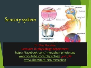
  • 1. Sensory system Dr. Dina Merzeban Lecturer in physiology department http://facebook.com/ merzeban physiology www.youtube.com/physiology ‫تاني‬ ‫فكر‬ www.slideshare.net/merzeban
  • 2. Sensory system Consists of :  Sensory receptors  Sensory pathways  Somatic sensory cortex
  • 3. 3 Receptors, Sensation, and Perception  Sensory receptors  Specialized cells or multicellular structures that collect information from the environment  Stimulate neurons to send impulses along sensory fibers to the brain  Sensation  A feeling that occurs when brain becomes consciously aware of sensory impulse  Perception  A person’s understanding of the sensation; the way the brain interprets the information
  • 4. Sensory unit: Single sensory afferent with all its receptor ending. Receptive field of a neuron: Region cotaining all receptors which its stimulation elicits stimulation in that neuron. Receptive fields of adjacent neurons overlap
  • 5.
  • 7. 13 Anatomical classification  Exteroceptors  Senses associated with body surface such as touch, pressure, temperature, and pain  Interoceptors  Senses associated with changes in the viscera such as blood pressure stretching blood vessels and ingestion of a meal  Proprioceptors  Senses associated with changes in muscles and tendons such as at joints
  • 8. Physiologically  Mechanoreceptors  Cutaneous (touch, pressure, vibration) eg. Pacinian, Meissner’s corpuscle, free nerve endings  Proprioceptors (joint position receptors) eg. Muscle stretch receptors, tendon organs  Baroreceptors  Auditory/vestibular hair cells • •
  • 9.  Chemoreceptors  Taste buds and smell receptors  Visceral chemoreceptors sensitive to Pco2, pH, osmolality(osmoreceptors), glucoreceptors . •  Thermoreceptors  Cold and hot receptors
  • 10.  Pain receptors(nociceptors)  Photoreceptors: rods and cones
  • 11.
  • 13. 1-Specificity Receptors are stimulated by a certain type of energy (adequate stimulus). Stimulation of a receptor usually produces only one sensation  modality specific But some receptors are stimulated by more than one sensory modality (polymodal)  eg. free nerve endings
  • 14. Receptors act as :  detectors  transducers
  • 15. 2-Excitability • Receptor cells are specific cells that are sensitive to different forms of energy from the environment • These cells when stimulated generate receptor (or generator)potential • They transform the stimulus into electrical signals
  • 16. What happens inside a receptor? • TRANSDUCTION Stimulus energy is converted to action potentials Inside the nervous system signals are always action potentials Language of the nervous system contains only 1 word: action potentials
  • 17. • Receptors transform an external signal into a membrane potential • Receptor Potential - separate receptor • Generator Potential -a specialized ending of an afferent neuron
  • 18. 7 Sensory Impulses • Stimulation of receptor causes local change in its receptor potential •A graded electrical current is generated that reflects intensityof stimulation •If receptor is part of a neuron, the membrane potential maygenerate an action potential •If receptor is not part of a neuron, the receptor potential mustbe transferred to a neuron to trigger an action potential • Peripheral nerves transmit impulses to CNS where they are analyzed and interpreted in the brain
  • 19. Receptor potential generation in the Pacinian corpuscle
  • 20.
  • 22. Physical Stimulus Physical stimulus causing mechanical deformation on the capsule
  • 23. Physical Stimulus Mechanical deformation is transmitted to the inside Opens up mechanosensitive Na+ channel Causes depolarisation and thus receptor potential
  • 24. Physical Stimulus local current Current flow through a local circuit
  • 25. Physical Stimulus Action Potentials are generated Opening of voltage gated Na+ channels causes generation of action potentials
  • 26. Characters of Receptor potentials • Non propagated local excitatory state. • This is a graded potential as It does not follow “all-or-none law” • Its amplitude depends on the strength of the stimulus. • Summated (no refractory period) • Long duration
  • 27. Stimulus Receptor potentials Action potentials 3-Relation between strength of stimulus and magnitude of receptor potential.
  • 29. 8 4-Sensory Adaptation The ability to diminish the receptor potential depolarization and number of action potentials in afferent nerve despite sustained stimulus strength
  • 30.  Types of receptors according to their speed of adaptation  Tonic :Do not adapt or slowly adapting receptors e.g. pain receptors,muscle stretch receptors and joint proprioceptors  Moderately adapting receptors:  temperature, smell and taste.  Phasic: (Rapidly adapting receptors ) e.g. Tactile receptors
  • 32.
  • 33. • In the Pacinian corpuscle Mechanical deformation is transmitted throughout the capsule and pressure is redistributed. Na+ channels inactivates after some time
  • 34. Impulse Stimulus Redistribution of pressure inside the capsule Stimulus No Impulse
  • 35. •Rapidly adapting receptors phasic or rate or movement receptors detect changes in stimulus strength eg. Pacinian corpuscle, hair end-organ •Slowly adapting receptors tonic receptors detect continuous stimulus strength eg. muscle spindles, Golgi tendon organ, baroreceptors, Ruffini endings and Merkel’s discs, pain receptors
  • 36. Sensory coding • A receptor must convey the type of information it is sending  the kind of receptor activated determined the signal recognition by the brain • It must convey the intensity of the stimulus  the stronger the signals, the more frequent will be the APs • It must send information about the location and receptive field, characteristic of the receptor
  • 37. Sensory coding Modality of sensation:  Adequate stimulus  Mullers law of specific nerve energy  Labeled line principle
  • 38. Locality of sensation  Low of projection phantom limb
  • 39. Intensity of sensation  Number of receptors  Frequency of impulses :  weber feshner principle R=log S * K  Stevens power principle R=ASK Tactile sensations
  • 40. Classification of sensation Special Sensations: Vision, Hearing, Taste, Smell General sensations: Somatic sensation Organic sensations: e.g. hunger Emotional sensation: e.g. fear, sadness
  • 41. Somatic sensation Mechanoreceptive sensations Tactile sensations Position sensations: static and kinetic Thermoreceptive sensations: Cold, Warm Pain sensation
  • 42. Tactile sensations  Touch : Crude touch Fine touch Tactile localization Tactile discrimination Texture  Stereognosis  Pressure: crude & fine  Vibration  Itching & tickling
  • 43. Transmission of somatic sensation _ A fibers:Myelinated • Subtypes: some overlap in ranges • Fastest conducting and largest diameter – m/sec, – B fibers: Slower myelinated – C fibers: Un myelinated • Slower conduction and smallest diameter (0.5 m/sec, 0.5 ) Nerve Fiber Classification
  • 44. Nerve Fiber Classification – I, II, III fibers: Myelinated • Subtypes: Ia, Ib • Fastest conducting and largest diameter – Ia – IV fibers: Unmyelinated • Slower conducting than IIIs and smallest diameter
  • 45. Modality of sensation Type of fiber Proprioceptors I Aα Fine touch, pressure, stereognosis, vibration II Aβ Fast pain, temperature,crude touch III A Slow pain, temp., tickle and itching IV C
  • 46. Sensory pathway • Once a receptor is stimulated impulse travels through a particular pathway known as sensory pathway or ascending pathway up to the brain
  • 47. Receptor Sensory modality Sensory nerve Ascending Sensory pathway Central Connections Sensory area in the brain Touch stimulus Sensory pathway
  • 48. Two ascending pathways • Dorsal column - medial lemniscus pathway fast pathway • Spinothalamic pathway slow pathway These two pathways come together at the level of thalamus
  • 49. Posterior (dorsal) Dorsal root ganglion Dorsal root Dorsal columns Dorsal horn Spinothalamic tracts Anterior (ventral)
  • 50. Dorsal column pathway Spinothalamic pathway Lateral Spinothalamic tract Anterior Spinothalamic tract
  • 51. Dorsal column pathway  Fine touch  fine pressure  stereognosis  vibratory sense  movement sense  position sense Spinothalamic pathway • Pain • Thermal sensations • Crude touch & pressure • tickle & itch
  • 52. Dorsal column nuclei (cuneate & gracile nucleus) Dorsal column Medial lemniscus thalamus thalamocortical tracts internal capsule 1st order neuron 2nd order neuron 3rd order neuron
  • 53. Dorsal column medial lemniscus pathway • after entering the spinal cord •lateral branch: participates in spinal cord reflexes •medial branch: turns upwards • forms the dorsal columns • spatial orientation: •medial: lower parts of the body •lateral: upper part of the body
  • 54. dorsal column medial lemniscus pathway • synapse in the dorsal column nuclei nucleus cuneatus & nucleus gracilus • 2nd order neuron cross over to the opposite side and ascends upwards as medial lemniscus • as this travels along the brain stem fibres from head and neck are joined (trigeminal) • ends in the thalamus (ventrobasal complex)  ventral posterolateral nuclei
  • 55. Spinothalamic pathway • after entering the spinal cord synapse in the dorsal horn • cross over to the opposite side • divide in to two tracts lateral spinothalamic tract: pain and temperature anterior spinothalamic tract crude touch
  • 56.
  • 57. Thalamocortical tracts from the thalamus 3rd order neuron ascends up through the internal capsule up to the sensory cortex )thalamocortical radiation(
  • 58. Sensory cortical areas • parietal cortex • a distinct spatial orientation exists
  • 59.
  • 60.
  • 61.
  • 62. Sensory cortex • Different areas of the body are represented in different cortical areas in the sensory cortex • Sensory area somatotopic representation not proportionate distorted map upside down map
  • 65. Sensory cortical areas • Primary somatosensory cortex (SSI) postcentral gyrus (Brodmann areas 3, 1, 2) • Secondary somatosensory cortex and Somatosensory association cortex (SSII) Posterior parietal areas (Brodmann areas 5, 7)
  • 66. Somatosensory cortex • Functions To localise somatic sensations To judge critical degree of pressure To identify objects by their weight, shape, form - stereognosis To judge texture of materials To localise pain & temperature
  • 67. Somatosensory cortex • Damage to the sensory cortex results in decreased sensory thresholds inability to discriminate the properties of tactile stimuli Inability to identify objects by touch (astereognosis)
  • 68. Secondary somatosensorycortex SS II  Occupies area 40  Receives input from SSI  Spatial orientation :face ant. , arms centrally, legs post.
  • 69. Somatosensory association cortex • Located directly posterior to the sensory cortex in the superior parietal lobes • Consists of areas 5 and 7 • Receives synthesized connections from the primary and secondary sensory cortices • Neurons respond to several types of inputs and are involved in the understanding of sensation
  • 70. Secondary somatosensory cortex and Somatosensory association cortex • Damage can cause Tactile agnosia inability to recognize objects even though the objects can be felt Spatial neglect This typically happens with non-dominant hemisphere lesions Neglect can be so severe that the individual even denies that their left side belongs to them
  • 71. Types of somatic sensations  Mechanoreceptive sensations  How to test  Receptor  Afferent  pathway
  • 72. Modality of sensation Type of fiber Proprioceptors I Aα Fine touch, pressure, stereognosis, vibration II Aβ Fast pain, temperature,crude touch III A delta Slow pain, temp., tickle and itching IV C
  • 73. Dorsal column pathway  Fine touch  fine pressure  stereognosis  vibratory sense  movement sense  position sense Spinothalamic pathway • Pain • Thermal sensations • Crude touch & pressure • tickle & itch
  • 74. pathway afferent receptor Type of sensation Dorsal column Aβ Meissner &merkel Touch fine touch Ant . spinothalamic A delta Free nerve ending Crude touch Dorsal column Aβ A delta Pacinian corpuscles& spray type endings pressure fine crude Dorsal column Aβ mixture stereognosis Dorsal column Aβ Pacinian 500hz Meissners 80 hz vibration Dorsal column Aα Muscle spindles golgi tendon organs Ruffini endings Prprioceptive sensations Ant . spinothalamic C Free nerve endings Tickling and itching Lat. spinothalamic C A delta& C VR1 VRL1 CMR Temp . Warm cold Lat. spinothalamic A delta C Free nerve ending Pain fast slow
  • 75. Receptive fields • The receptor area which when stimulated results in a response of a particular sensory neuron • Receptive fields of adjacent neurons overlap
  • 76.
  • 77. Two-Point Discrimination Whether a stimulus feels like one sensation or two distinct sensations depends on: • Number of receptors • the size of the receptive fields of the sensory receptors:Smaller receptive fields result in greater sensitivity  Fingers are more sensitive than backs • Degree of convergence within the pathway • Area of representation
  • 78.
  • 79.
  • 82. Thermal receptors a) Warm : free nerve ending ( C fibers ) b) Cold : free nerve endings ( C fibers and A delta fibers ) c) Others :cold pain and warm pain
  • 83.  Tested by using three test tubes ( 5 °C , 25 °C & 40 °C )  Touch any part of body by the test tube ( for 2 minutes )  Ask him to feel hot or cold
  • 84. Characters of thermal receptors  They respond to the temperature of subcutaneous tissue.  Widely separated(wide area exposed for better differentiation)  Moderately adapting receptors  distribution: lips > fingers > trunk
  • 85.  Cold sensation is more important than warm sensation  Cold receptors: - More numerous ( 4-10 times) - More superficial (under skin ) - Adapt more slowly - Carried on type C & A delta
  • 86. Mechanism of stimulation of thermal receptors  Chemically by change in concentration of metabolites  Respond markedly to changing temperature
  • 87. Detection of thermal sensation  cold pain ( 5-15 °C) maximum 5 °C  cold receptors ( 10-43°C) maximum 25°C  warm receptors ( 30-50°C) maximum 45 °C  warm pain above( 45°C )  Paradoxical pain sensation  At 0 °C : anaesthesia
  • 89. Sensory NERvous system Dr. Dina Merzeban Lecturer in physiology department http://facebook.com/ merzeban physiology www.youtube.com/physiology ‫تاني‬ ‫فكر‬ www.slideshare.net/merzeban
  • 91. Pain is an, unpleasant emotional experience associated with actual or potential tissue damage - The International Association for the Study of Pain (IASP) Pain= tissue damage
  • 92. Pain system:  Pain receptors (nociceptors).  Types of pain.  Pain pathways.  Pain control.
  • 93. Nociceptors  Free nerve endings attached to A delta & C, slowly (non) adapting to prolonged stimulation 4 types: Mechanical pain receptors. Thermal pain receptors. Chemical pain receptors. Polymodal pain receptors
  • 94. Distribution of pain receptors Widely distributed Sup. Layers of skin Pleura Peritonium periosteum Arterial walls Joint surface Dura of cranial cavity Less distributed Deep tissues & Viscera Absent Liver Parenchyma Lung alveoli Brain tissue
  • 95. Stimulation of pain sensation  Damaged tissues----chemical mediators e.g. histamine, serotonin, substance P, bradykinins, K+ ------- sensitizes painreceptors -------hyperalgesia  1ry hyperalgesia  2ry hyperalgesia
  • 96.
  • 97. Pain pathways  Two separate pathways: I. Neospinothalamic pathway: Conducting quick, localized pain (Fast sharp pain) II. Paleospinothalamic pathway: For slow pain Lateral spino- thalamic tract
  • 98. Ad fiber glutamate C Fibers Subst. P Substantia Gelatinosa of Rolandi (SGR) glutamate
  • 100. Paleospinothalamic tract  In the brain stem, 90% of fibers synpse on:  Reticular formation  Intralaminar (non specific ) nuclei of thalamus to activate the whole cortex
  • 101. Reactions to pain  Motor reflexes (AHCs)  Arousal reaction (RAS)  Autonomic reactions(hypothalamus)  Emotional reactions(limbic system)
  • 102. Perception of pain signals Fast pain in thalamus & cortex Slow pain in thalamus Roles of the cortex in pain perception are 1.Localization of pain. 2.Discrimination of pain. 3.Modulation of pain.
  • 103. Types of pain According to oigin  Cutaneous  Deep  Visceral  Neuropathic
  • 104. According to Quality : 1. Fast pain (sharp, acute, pricking, immediate) 2. Slow pain (burning, chronic, dull aching, throbbing
  • 105. Slow pain Fast pain Felt within 1 sec or more Felt within 0.1 sec May be prolonged Short duration Poorly localized Well localized All types of receptors Mechanical or thermal Skin, deep tissues & viscera Usually in skin, rare in deep tissues
  • 106. slow pain Fast pain Carried by C, blocked by local anaesthetics Carried by Ad, blocked by pressure & O2 lack C release Substance P Ad release Glutamate Transmitted by Paleospinothalamic T Transmitted by Neo- spinothalamic T End in RF Non- specific thalamic nuclei whole cortex Its fibers end in sensory cortex
  • 107. Deep pain  Felt in muscles , tendons , joints and bones Causes:  Trauma  Inflammation  Muscle spasm  Ischemic pain
  • 108. Ischemic pain  Definitions : type of deep pain felt in muscles when their blood supply is reduced  Example : angina pectoris & intermittent claudicating  Causes  Increase metabolites and proteolytic enzymes.
  • 109. Intermittent claudication: recurrent pain in the calf muscle during exertion, which stops on rest. This is due to ischaemia of muscle which produces P- factor – pain producing chemical agent which is responsible for causation of pain. During rest the pain stops because P factor is washed away by blood. Eg:Thrombo Angitis Obliterans. (Buerger’s disease)
  • 110. Coronary occlusion: In addition to P factor, there is release of 5HT and plasma pain producing polypeptide. Inflammatory pain: - due to increased tension causing pressure on nerve terminals as well as due to release of a chemical pain producing factor.
  • 111. Visceral pain  Pain from viscera ( peritoneum , pleura and pericardium )  C – fiber pain (poorly localized)  A delta fibers stimulated if from parietal layers of viscera  Most viscera contain only few pain receptors  Sharp cut doesn't cause pain ( injury of few nerve endings )  Diffuse stimulation cause severe pain
  • 112. Visceral pain Characters a) Dull aching b) Poorly localized c) Autonomic responses : decrease heart rate and ABP d) Guarding (Rigidity) e) Referred
  • 113. Visceral pain Causes a) Over distension b) Spasm c) Ischemia d) Inflammation and irritation e) Infiltration by tumors
  • 114. CAUSES OF VISCERAL PAIN  Ischaemia: The substances released during ischaemic reactions like bradykinin and proteolytic enzymes stimulate the pain receptors of viscera.  Chemical stimuli: The chemical substances like acidic gastric juice leaks from ruptured ulcers into peritoneal cavity and produce pain.
  • 115.  Spasm of hollow organs: spastic contraction of muscles in gastrointestinal tract and other hollow organs of viscera cause pain by stimulating the free nerve endings.  Overdistension of hollow organs also cause pain.
  • 116. Neuropathic pain  Pain due to nerve fiber damage  Examples : a) Trigeminal neuralgia b) Sciatica c) Herpes zester d) Diabetic neuropathy
  • 117. Referred pain  Definition: pain not felt in diseased viscus and felt in corresponding dermatome  Examples:  Cardiac : left shoulder  Gall bladder : Right shoulder  Appendix : at umbilicus  Gastric : above umbilicus  Kidney & pancreas : back
  • 118. Referred pain Heart pain is referred to the inner aspect of left arm. Diaphragmatic pain to the tip of the shoulder Ureteric pain to the testes in male and the inner aspect of the thigh in female. Gall bladder pain referred to epigastric region. Pain from the maxillary sinus referred to the nearby tooth.
  • 121. DermatomalRule  Referred pain is always felt over a structure that developed from the same embryological segment or dermatome from which the organ which is the source of pain developed.  This phenomenon of referred pain is called Dermatomal rule.
  • 122. DermatomalRule  During Embryonic development, the diaghragm migrates from the neck region to its adult location between the chest and abdomen and takes its nerve supply, the phrenic nerve with it.  Similarly the heart and the arm have the same segmental origin.  Testicles have migrated with its nerve supply from the primitive urogenital ridge from which kidney and ureter has developed.
  • 123. CONVERGENCE THEORY The number of peripheral pain exceed the number of lateral spinothalamic tract. So, Both the somatic and visceral afferents converge upon the same spinothalamic neurons at the spinal cord level. Hence when visceral pain impulses travel in the same pathway along which impulses from the skin travels, the individual gets the feeling that the pain originates in the skin itself.
  • 125. FACILITATION THEORY Visceral and somatic pain afferents connect with separate but adjoining spinothalamic neurons and there may be some overlap of the neurons, visceral afferents have collaterals connecting to the spinopthalamic neurons receiving somatic pain afferents. This causes impulses to travel up the somatic spinothalamic pathway and causes the sensation of pain in the skin.
  • 126. Facilitation theory  Afferent from pain fibers of diseased viscera, give subliminal fring to a nearby SGR (receiving afferents from an area of skin), increasing its excitability.  The result is pain felt in this skin area and lowering of its pain threshold
  • 128.  Terminology  Algesia =pain  Analgesia = dec./loss of sensitivity to painful stimuli  Hyperalgesia = inc. sensitivity to painfulstimuli  Allodynia =sensation of pain in response to an innocuous stimulus  Brown-Sequardsyndrome(hemisection of the spinal cord.  Syringomyelia(loss of pain & temp. with sparing of touch & vibration.
  • 129. Hyperalgesia  Primary hyperalgesia  Secondary hyperalgesia
  • 132. Ad fiber glutamate C Fibers Subst. P Substantia Gelatinosa of Rolandi (SGR) glutamate
  • 133. Somatosensory cortex Other basal areas of brain Thalamus – ventrobasal complex Spinothalamic tract Spinal cord (lamina I – lamina marginalis) Peripheral fibers Aδ fibers Pain receptor(Free nerve endings)
  • 134. • Reticular nuclei • Tectal area & periaqueduvtal grey region • Thalamus Other basal areas of brain Spinothalamic tract Spinal cord (lamina II & III – substantia gelatinosa) Peripheral fibers C fibers Pain receptor (Free nerve endings)
  • 137.
  • 138. Pain Modulation  Inhibitory neurotransmitters like endogenous opioids work to hinder the pain transmission.  This inhibition of the pain impulse is known as modulation
  • 139. Physiological Basis of analgesia system  The CNS has its own control system which inhibits the impulse of pain sensation.  This is also called Analagesia system.  This control system is present in both brain and spinal cord.  Pain control complex in spinal cord:  This is in dorsal grey horn.  It is considered as the gateway for pain impulses to reach the brain (via) spinothalamic tract.
  • 140. PHYSIOLOGYOFPAIN PERCEPTION Injury Descending Pathway Peripheral Nerve Dorsal Root Ganglion C-Fiber A-beta Fiber A-delta Fiber Ascendin g Pathways Dorsal Horn Brain Spinal Cord
  • 141. GATE CONTROL THEORY  Substantia Gelatinosa (SG) in dorsal horn of spinal cord acts as a ‘gate’  This gate can be closed by:  Descending supraspinal inhibitory impulses  A-beta neurons are stimulated which closes the gate to A-delta & C neurons
  • 142. Spinal pain inhibitory pathway  Gate - located in the dorsal horn of the spinal cord  Smaller, slower n. fibers carry pain impulses  Larger, faster n. fibers carry other sensations  Impulses from faster fibers arriving the gate 1st inhibit pain impulses (acupuncture/pressure, cold, heat, chem. skin irritation). Brain Pain Heat, Cold, Mechanical Gate (SG)
  • 143.
  • 144.
  • 145.
  • 146.
  • 147. •Periaqueductal grey Raphe magnus nucleus (pain inhibitory complex in dorsal horn) Hypothalamus (periventricular nucleus ) Supraspinal inhibitory pathway (Descending pain control mechanism)
  • 148. Supraspinal inhibitory pathway (Descending pain control mechanism) Periaquaductal Gray Area (PGA) release enkephalins Nucleus Raphe Magnus (NRM) release serotonin Stimulation of presynaptic inhibitory intermediary neuron secreting enkephalin Inhibit the release of substance P from SGR ascending neurons Hypothalamus (periventricular nucleus ) Release endorphin
  • 149. Spinal pain inhibitory pathway  Gate - located in the dorsal horn of the spinal cord  Smaller, slower n. fibers carry pain impulses  Larger, faster n. fibers carry other sensations  Impulses from faster fibers arriving the gate 1st inhibit pain impulses (acupuncture/pressure, cold, heat, chem. skin irritation). Brain Pain Heat, Cold, Mechanical Gate (SG)
  • 150.
  • 151. Inhibition of pain transmission by tactile sensory signals  Rubbing the skin near painful areas and applying liniments often relieves pain.  This is due to the stimulation of Aβ sensory fibres from peripheral tactile receptors depress transmission of pain signals.  This results from a type of local lateral inhibition.  Acupuncture is also used to relieve pain.
  • 152. Acupuncture is also used to relieve pain based upon the pain inhibitory mechanism  Treatment for pain  Remove the cause  Drugs  NSAIDs (inhibit COX)  Opiates (inhibit NT release)
  • 153. Surgical procedure that relieve pain Different surgical procedures are done in the course of pain pathway to relieve pain. They are -Sympathectomy -Cordotomy -Thalamotomy -Prefrontal lobotomy
  • 154. Headache  Def. Headache is a referred pain  headache is pain referred to the surface of the head from deep structures  It may be  frontal headache referred to areas supplied by trigeminal nerve.  occipital headache referred to areas of the head supplied by the second cervical nerve
  • 155. Headache  the brain itself is insensitive to pain  the intracranial pain sensitive areas include  dura and Tentorium  nerves  venous sinuses  the dural arteries ( meningeal artery at the base of the skull) 
  • 156. Distribution of pain receptors Widely distributed Sup. Layers of skin Pleura Peritonium periosteum Arterial walls Joint surface Dura of cranial cavity Less distributed Deep tissues & Viscera Absent Liver Parenchyma Lung alveoli Brain tissue
  • 157. Headache  it is caused by  traction  displacement  inflammation  vascular spasm or distension of the pain sensitive structures in the head or neck
  • 158.  it may be from intracranial or extracranial origin causes of intracranial headache  meningitis or meningeal irritation ( constipation)  intracranial mass ( brain tumours)  drop of intracranial tension ( post lumbar puncture headache )  Rise of intracranial tension  alcohol headache
  • 159. causes of intracranial headache  Trigeminal neuralgia is a facial pain confined mainly to areas supplied by the second and third divisions of the trigeminal nerve  hypertension due to marked expansion of the cerebral blood vs  Migraine headache : unilateral headache resulting from abnormal vascular Phenomenon  First severe constriction of the cerebral blood vessels followed by marked Dilatation of the cerebral blood vessels
  • 160. headache of of extracranial origin  Tension headache due to spasm of the neck and scalp muscles  Eye disorders : errors of refraction & glaucoma  Nose : sinusitis  Ear : otitis Media  Mouth : Toothache
  • 161. Headache  Dura  Cerebral blood vessels  CSFpressure  Nasal & accessory nasal structures  Eye disorders  Muscle spasm of head & neck muscles  Alcohol  constipation
  • 162. Dr. Dina Merzeban Lecturer in physiology department http://facebook.com/ merzeban physiology www.youtube.com/physiology ‫تاني‬ ‫فكر‬ www.slideshare.net/merzeban
  • 163. Sensory abnormalities • Various types of sensory abnormalities can occur when the sensory pathways are damaged • Sensory loss, altered sensations or pain could occur as a result • In addition, motor pathways could also be affected resulting in motor weakness
  • 164. Types of sensory abnormalities • Hypothesia • Anaesthesia absence of sensation • Paraesthesia (numbness or pins-needles- sensation) altered sensation • Hemianaesthesia: Loss of sensation of one half of the body • Neuropathic pain • Analgesia • hyperalgesia • Spatial neglect
  • 165. Localisation of the abnormality • Peripheral nerve innervated area affected • Roots dermatomal pattern of sensory loss • Spinal cord a sensorylevel • Internal capsule one half of the body • Cortical areas Other features
  • 166. Examples of sensory lesions or sensory disorders •Polyneuropathy All sensory nerves of both upper and lower limbs are degenerated Numbness of hands and feet Glove and stocking type of sensory loss Diabetic or nutritional neuropathy
  • 167. Examples of sensory lesions or sensory disorders • Sensory stroke  Internal capsule lesion  Numbness and sensory loss of one side of the body
  • 168. Tabes dorsalis • Dorsal column sensations are affected • Vibration, proprioception affected early in disease process • Sensory ataxia • Romberg sign
  • 169. Syringomyelia Spinal cord central canal lesion Dissociated sensory loss Temperature and pain sensations affected in early in disease process and crude touch affected later. Touch and dorsal column functions not affected
  • 170. Dorsal column pathway Spinothalamic pathway Lateral Spinothalamic tract Anterior Spinothalamic tract
  • 171.
  • 172. Brown Sequard syndrome  At the level of lesion loss of all sensations supplied by the affected dorsal roots  Below level of lesion On the same side :loss of dorsal column sensations On the opposite side: loss of spinothalamic tract sensations
  • 173. Dorsal column pathway Spinothalamic pathway Lateral Spinothalamic tract Anterior Spinothalamic tract
  • 174.
  • 175.
  • 177. Sensory abnormalities • Various types of sensory abnormalities can occur when the sensory pathways are damaged • Sensory loss, altered sensations or pain could occur as a result • In addition, motor pathways could also be affected resulting in motor weakness
  • 178. Types of sensory abnormalities • Hypothesia • Anaesthesia absence of sensation • Paraesthesia (numbness or pins-needles- sensation) altered sensation • Hemianaesthesia: Loss of sensation of one half of the body • Neuropathic pain • Analgesia • hyperalgesia • Spatial neglect
  • 179. Localisation of the abnormality • Peripheral nerve innervated area affected • Roots dermatomal pattern of sensory loss • Spinal cord a sensorylevel • Internal capsule one half of the body • Cortical areas Other features
  • 180. •Polyneuropathy All sensory nerves of both upper and lower limbs are degenerated Numbness of hands and feet Glove and stocking type of sensory loss Diabetic or nutritional neuropathy
  • 181. Internal capsule lesion Numbness and sensory loss of one side of the body Sensory stroke
  • 182. Tabes dorsalis • Dorsal column sensations are affected • Vibration, proprioception affected early in disease process • Sensory ataxia • Romberg sign
  • 183. Syringomyelia Spinal cord central canal lesion Dissociated sensory loss Temperature and pain sensations affected in early in disease process and crude touch affected later. Touch and dorsal column functions not affected
  • 184. Posterior (dorsal) Dorsal root ganglion Dorsal root Dorsal columns Dorsal horn Spinothalamic tracts Anterior (ventral)
  • 185. Dorsal column pathway Spinothalamic pathway Lateral Spinothalamic tract Anterior Spinothalamic tract
  • 186.
  • 187. Brown Sequard syndrome  At the level of lesion loss of all sensations supplied by the affected dorsal roots  Below level of lesion On the same side :loss of dorsal column sensations On the opposite side: loss of spinothalamic tract sensations
  • 188. Dorsal column pathway Spinothalamic pathway Lateral Spinothalamic tract Anterior Spinothalamic tract
  • 189.  Summation • Spatial • Temporal  Relaying of signals through neuronal pools • Threshold---Subthreshold stimuli • Excitation or Facilitation  Divergence and Convergence of signals passing through neuronal pool  After Discharge • Synaptic after discharge • Reverberatory circuit • Continuous signal output • Rhythmical signal output
  • 190. Questions  What are the characters of thermal receptors?  What is the mechanism of thermoceptive stimulation?  What are the types of pain receptors?  Do you know the pain insensitive structures?  What are the types of pain sensations?
  • 191.  What is the neuropathic pain?  Give example to neuropathic pain?  What are causes of visceral pain?  Mention the characters of visceral pain  Explain mechanisms of referred pain?  Enumerate some examples of referred pain?