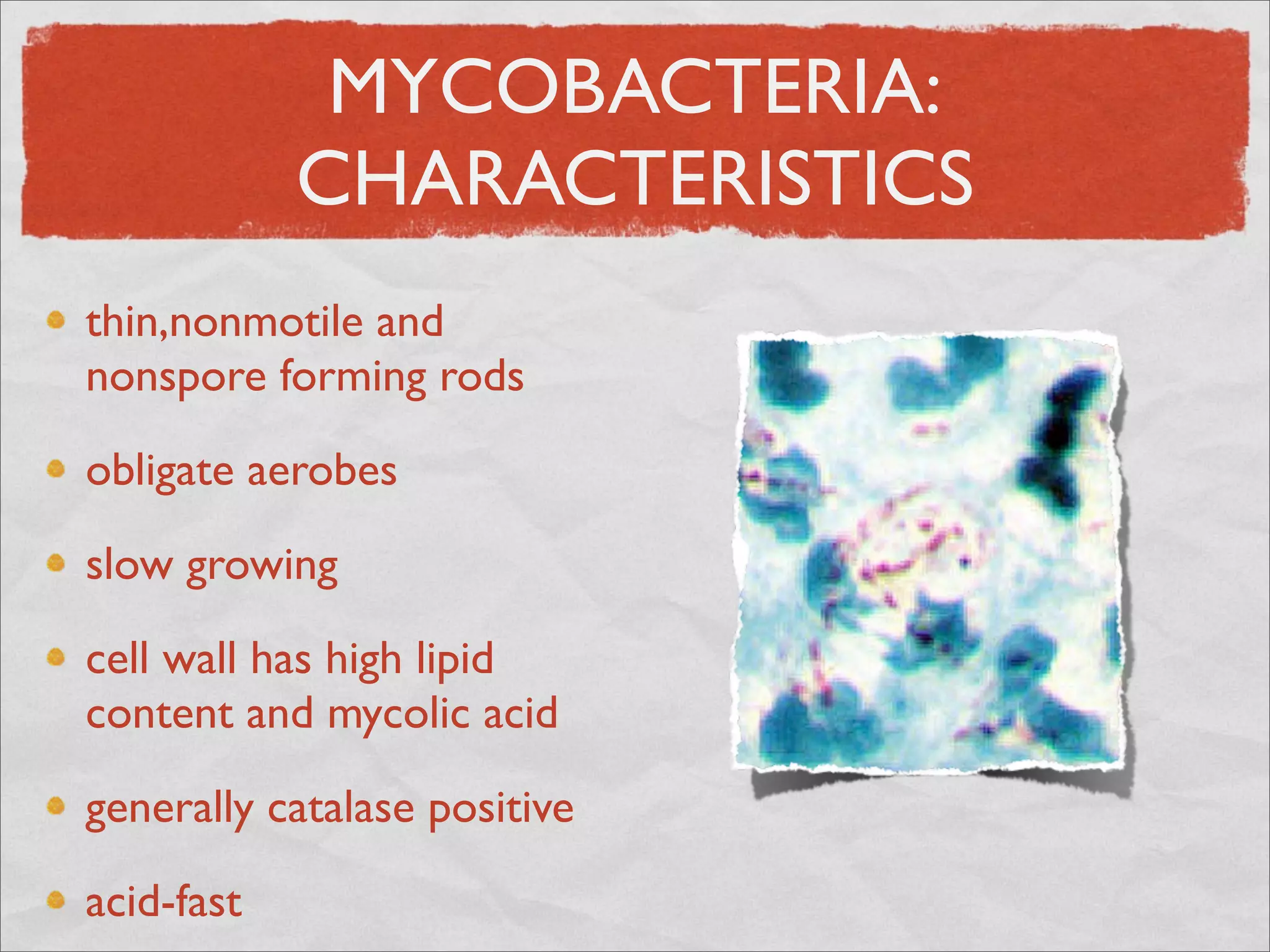
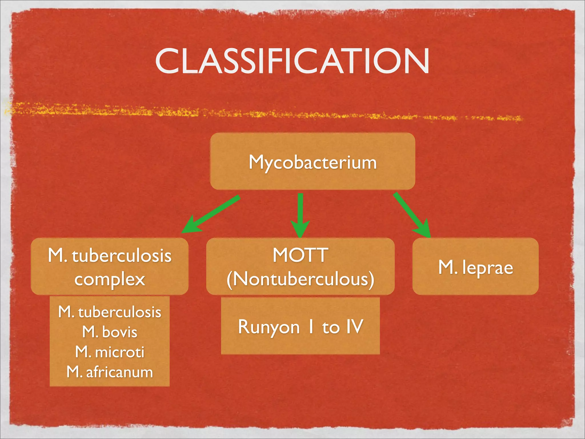
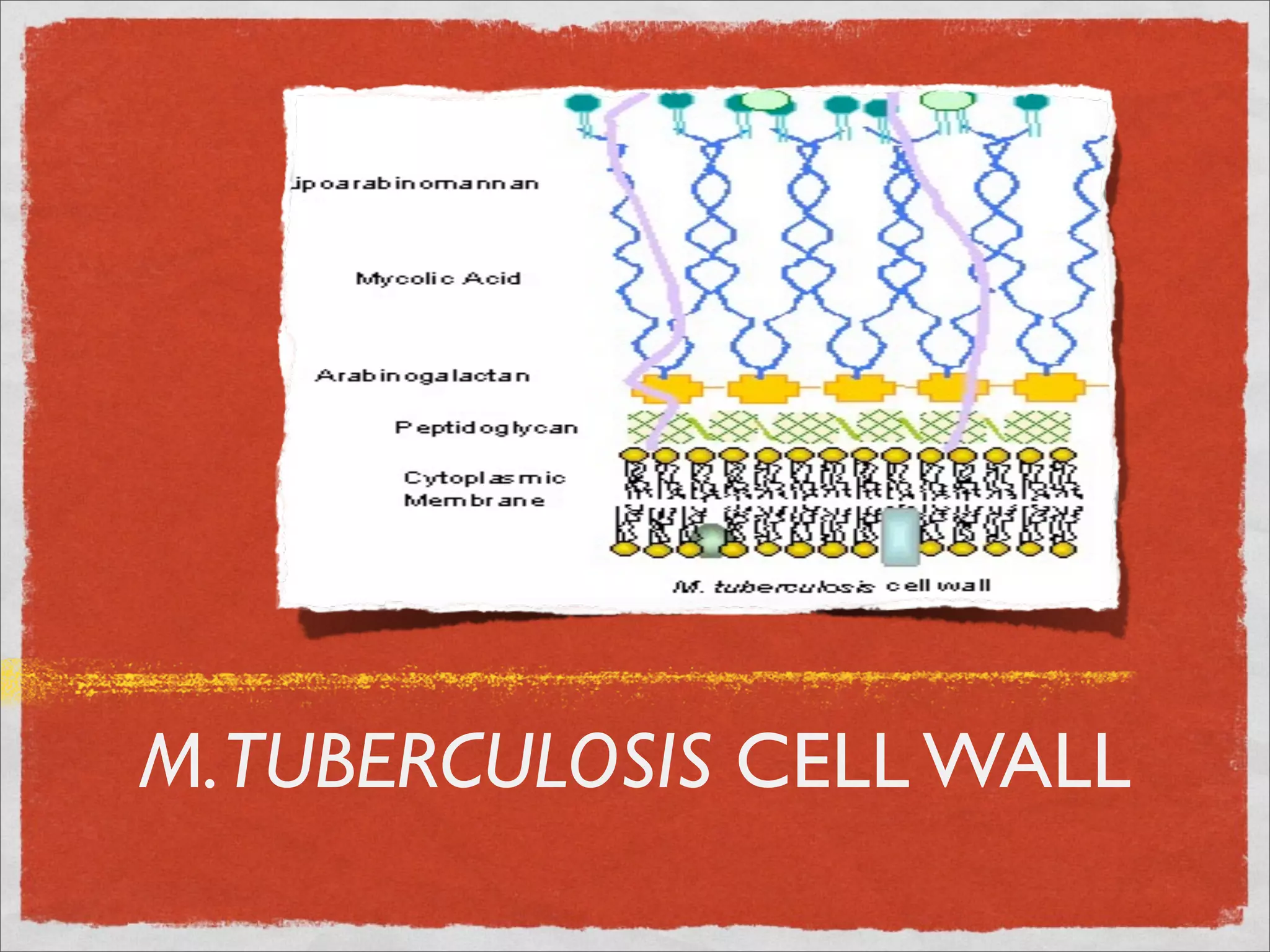
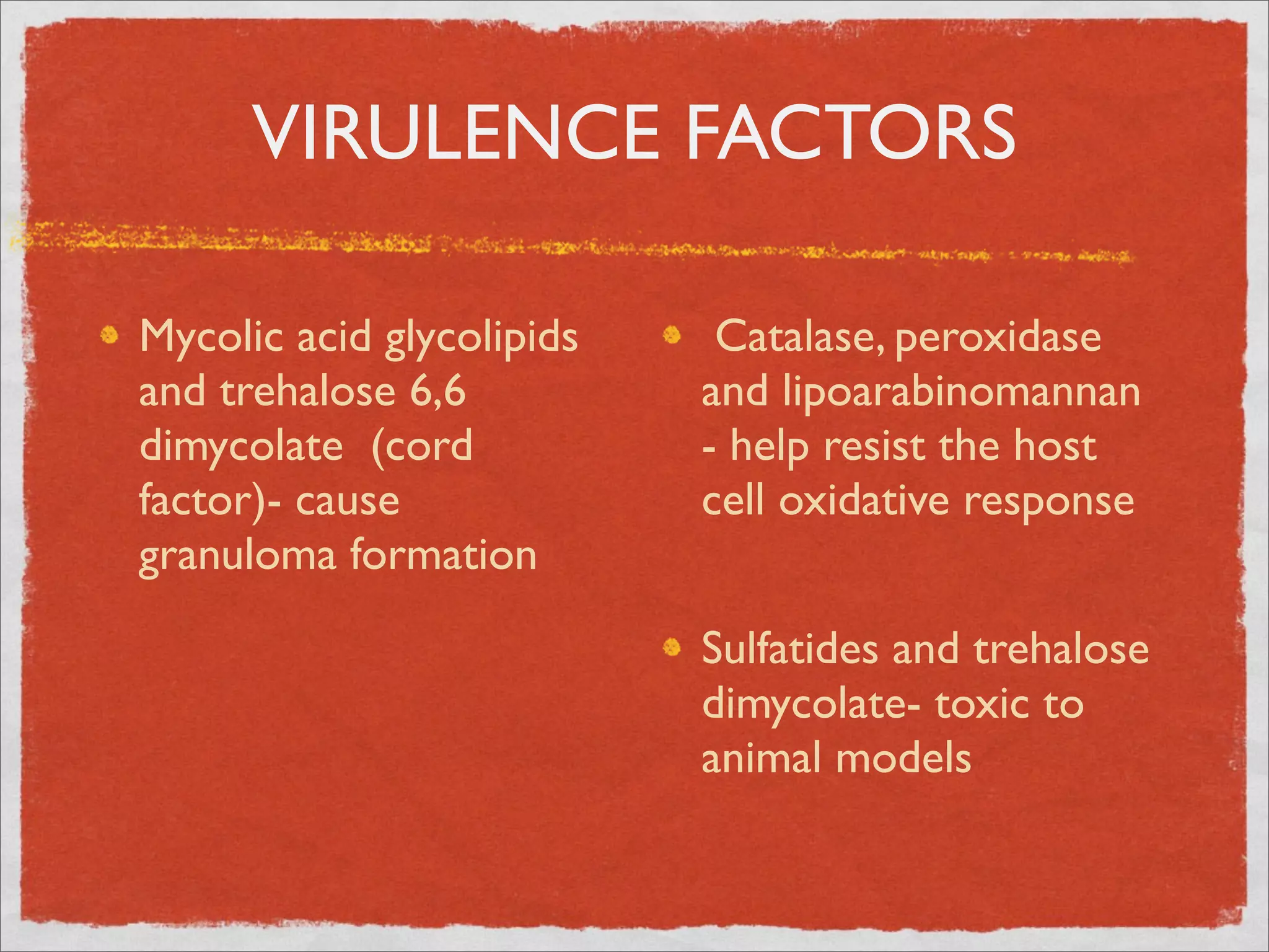
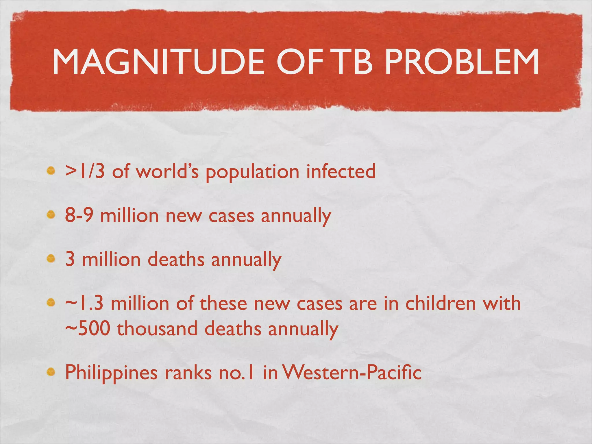
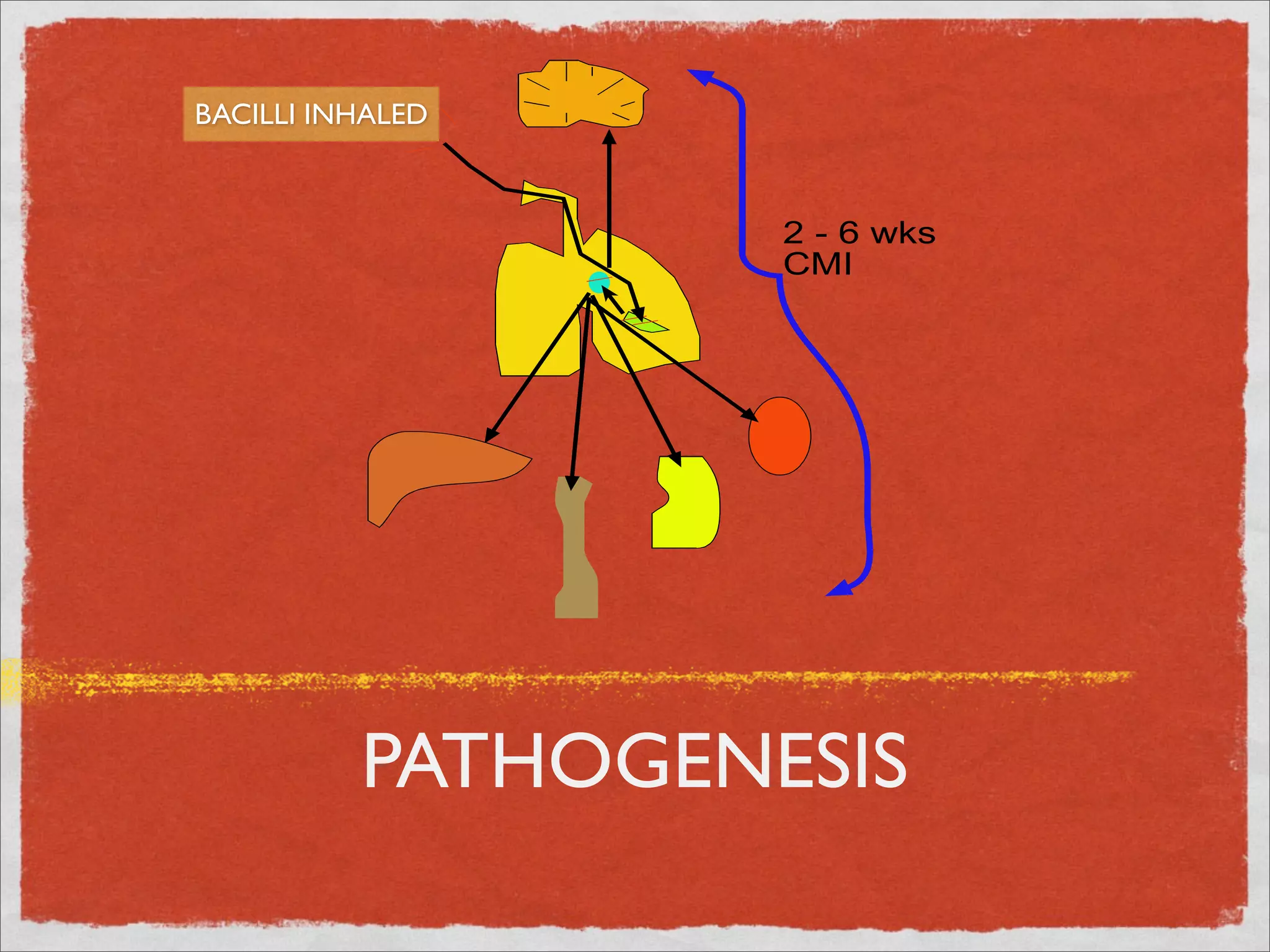
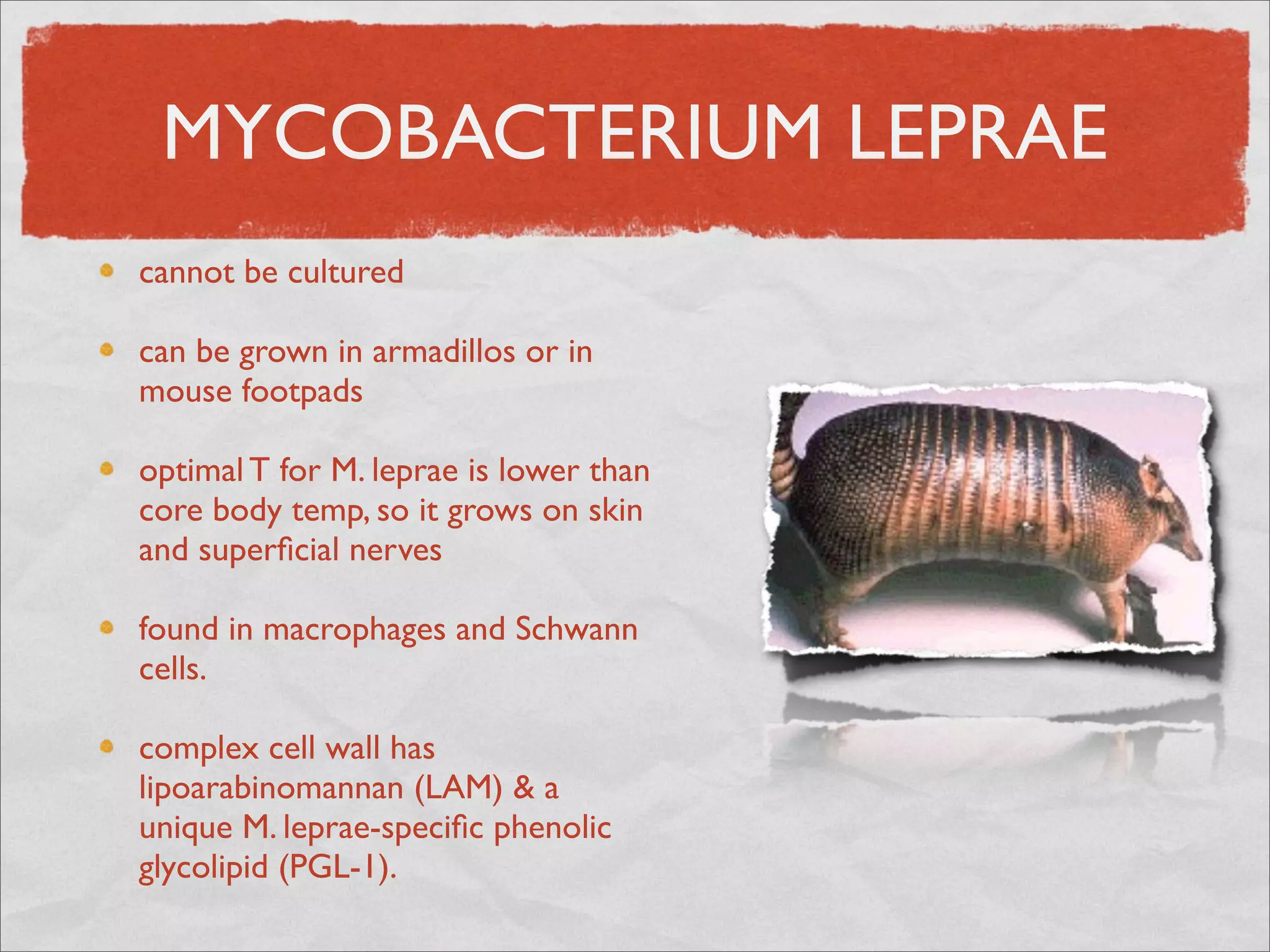
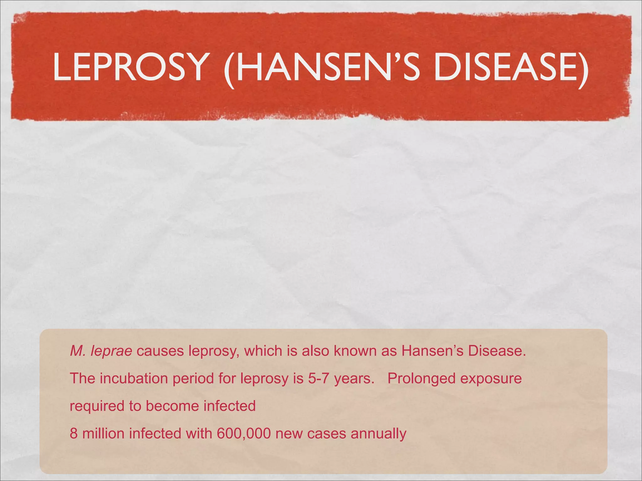
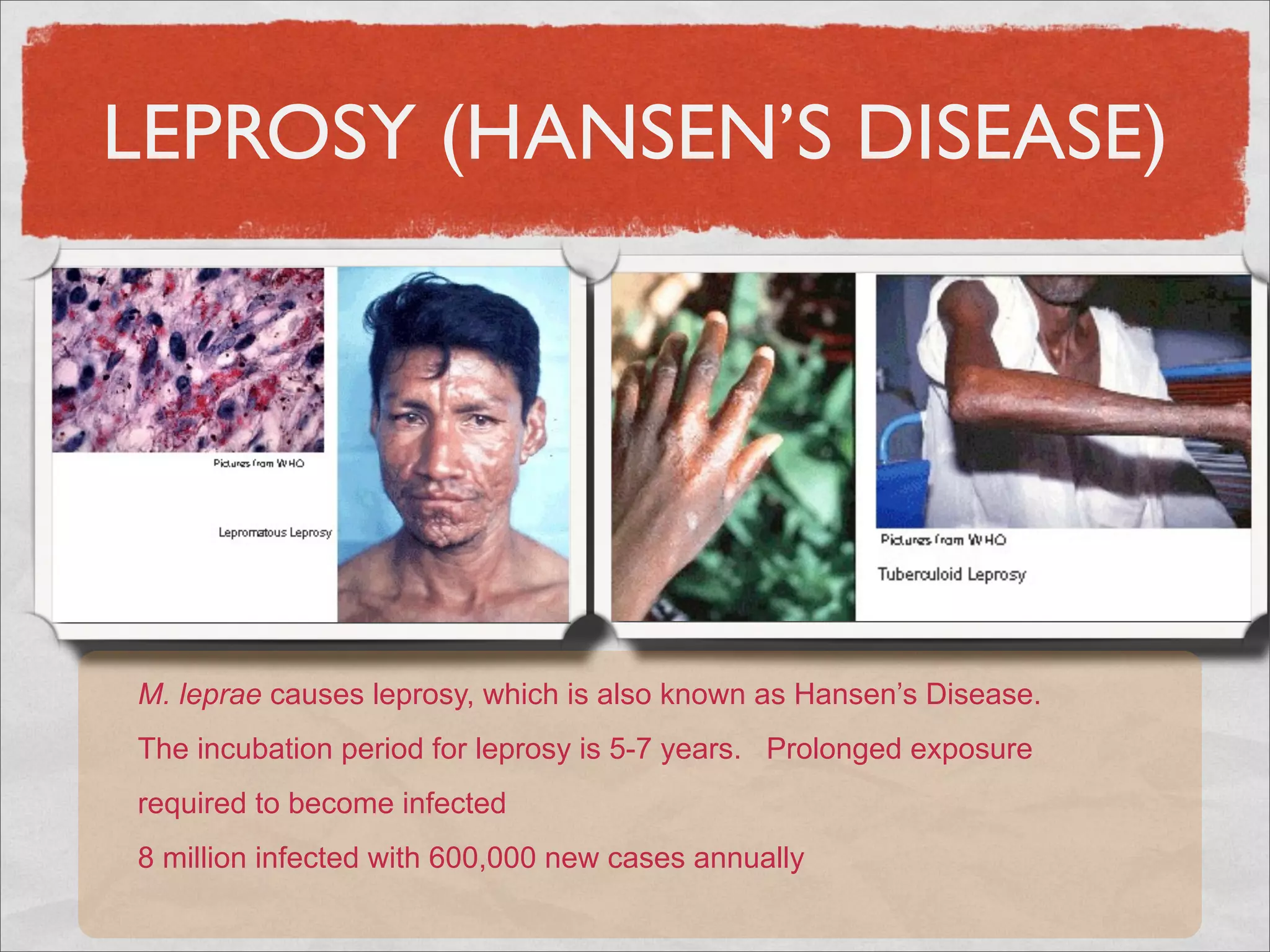
MYCOBACTERIA
   Jaime A. Santos
MYCOBACTERIA:
            CHARACTERISTICS
thin,nonmotile and
nonspore forming rods

obligate aerobes

slow growing

cell wall has high lipid
content and mycolic acid

generally catalase positive

acid-fast
MYCOBACTERIA:
            CHARACTERISTICS
thin,nonmotile and
nonspore forming rods

obligate aerobes

slow growing

cell wall has high lipid
content and mycolic acid

generally catalase positive

acid-fast
CLASSIFICATION

    Mycobacterium
CLASSIFICATION

                     Mycobacterium


M. tuberculosis        MOTT
                                      M. leprae
   complex         (Nontuberculous)
 M. tuberculosis
     M. bovis       Runyon 1 to IV
   M. microti
  M. africanum
M. TUBERCULOSIS

optimal growth:  37 C/ 5-10% CO2/ pH 6.0-7.6.

in vivo, it can use a variety of enzymes for anaerobic
metabolism

requires complex media such as Löwenstein Jensen
doubling time of ~18 hours.  Colonies visible in 3-6
weeks

multiplies intracellularly in phagosome and prevents
phagolysosome fusion
M. TUBERCULOSIS CELL WALL
VIRULENCE FACTORS

Mycolic acid glycolipids      Catalase, peroxidase
and trehalose 6,6          and lipoarabinomannan
dimycolate  (cord          - help resist the host
factor)- cause             cell oxidative response 
granuloma formation        	


                           Sulfatides and trehalose
                           dimycolate- toxic to
                           animal models
MAGNITUDE OF TB PROBLEM

>1/3 of world’s population infected

8-9 million new cases annually

3 million deaths annually

~1.3 million of these new cases are in children with
~500 thousand deaths annually

Philippines ranks no.1 in Western-Pacific
BACILLI INHALED


                   2 - 6 wks
                   CMI


          PATHOGENESIS
IMMUNE RESPONSE
IMMUNE RESPONSE
IMMUNE RESPONSE
IMMUNE RESPONSE
CLINICAL MANIFESTATIONS

asymptomatic     other symtoms and
                 signs depending on the
fever            organ involved e.g.
                 CNS, bone, renal
cough >3 weeks

chest pain

hemoptysis

lymphadenpathy
CHILDHOOD TB

asymtomatic ~50%           failure to make a quick
                           return to health after an
cough/wheezing > 2         infection e.g.
weeks                      measles,tonsillitis or
                           pertussis
fever > 2 weeks
                           failure to respond to
painless cervical and/or
                           appropriate antibiotics
other lymphadenopathy
                           as in AOM or
poor weight gain           pneumonia
DIAGNOSIS

SIGNS AND SYMPTOMS

HISTORY OF EXPOSURE

CHEST X-RAY

TUBERCULIN TEST

BACTERIOLOGIC DIAGNOSIS: SMEAR, CULTURE,
PCR

HISTOLOGIC
CHEST X-RAY
CHEST X-RAY
CHEST X-RAY
TUBERCULIN TEST
Mantoux test

0.1 ml of solution
containing or equivalent
to1g ( 5 TU PPD-S)

read at 48-72 hours using
ballpoint pen method

results recorded in mm

delayed-type
hypersensitivity
TUBERCULIN TEST
Mantoux test

0.1 ml of solution
containing or equivalent
to1g ( 5 TU PPD-S)

read at 48-72 hours using
ballpoint pen method

results recorded in mm

delayed-type
hypersensitivity
CULTURE

incubated at 35° to 37° C in an atmosphere of 5 to
10% CO2

cultures should be examined weekly for 8 weeks.

solid media e.g. Lowenstein-Jensen allows visualization
of colony morphology but requires 3-4 weeks

broth systems detecting 14C labelled CO2
(BACTEC) require only 5-12 days
ANTI-TB DRUGS

isoniazid (H) - 5 to 10 mg/kg ( max 300 mg)

rifampicin (R) - 10 to 15 mg/kg (max 600 mg)

pyrazinamide (Z) - 15 to 30 mg/kg (max 2 gm)

ethambutol (E) - 15 to 25 mg/kg (max 2.5 gm)

streptomycin (S) - 20 to 30 mg/kg (max 1 gm)

second-line drugs

PROBLEM OF DRUG RESISTANCE
MOTT

Nontuberculous mycobacteria (NTM) are soil and
water organisms

noncommunicable

Disease develops in setting of trauma/surgery or
immunosuppression.

INH resistant

Diagnosis is by acid fast staining of the specimen;
followed by culture and/or 16s rRNA probes culture
RUNYON CLASSIFICATION

Group     Growth               Pigment                Examples                   Disease

                                                                             1. similar to TB
                         yellow-orange on light      1. M. kansasii
  I         slow            (photochromogen)         2. M. marinum
                                                                            2. swimming pool
                                                                                granuloma
                         yellow-orange in light
  II        slow                or dark            M. scrofulaceum          cervical adenitis
                          (scotochromogen)

                                                  M. avium intracellulare    similar to TB,
 III        slow             no pigment              complex (MAC)            esp. in AIDS

                                                     M. fortuitum           soft tissue, lung, bone,
 IV     rapid (5 days)       no pigment              M. cheilonae            CNS, eye infections
MYCOBACTERIUM LEPRAE
cannot be cultured

can be grown in armadillos or in
mouse footpads

optimal T for M. leprae is lower than
core body temp, so it grows on skin
and superficial nerves

found in macrophages and Schwann
cells.

complex cell wall has
lipoarabinomannan (LAM) & a
unique M. leprae-specific phenolic
glycolipid (PGL-1).
LEPROSY


anesthetic plaques,                 symmetric skin
  and asymmetric                   nodules, plaques,
 peripheral nerve               leonine (i.e., lion-like)
trunk involvement,                   facies, loss of
   paucibacillary                eyelashes and body
                                  hair, multibacillary
LEPROSY


tuberculoid                         lepromatous


anesthetic plaques,                 symmetric skin
  and asymmetric                   nodules, plaques,
 peripheral nerve               leonine (i.e., lion-like)
trunk involvement,                   facies, loss of
   paucibacillary                eyelashes and body
                                  hair, multibacillary
LEPROSY (HANSEN’S DISEASE)


M. leprae causes leprosy, which is also known as Hansen’s Disease.
The incubation period for leprosy is 5-7 years. Prolonged exposure
required to become infected
8 million infected with 600,000 new cases annually
LEPROSY (HANSEN’S DISEASE)


M. leprae causes leprosy, which is also known as Hansen’s Disease.
The incubation period for leprosy is 5-7 years. Prolonged exposure
required to become infected
8 million infected with 600,000 new cases annually
LEPROSY AND THE IMMUNE
         SYSTEM
DIAGNOSIS


clinical signs

lepromin skin test (mainly of immune status)

biopsy and histology
TREATMENT
dapsone, rifampin, clofazimine, and either ethionamide
or prothionamide

Paucibacillary cases (tuberculoid and borderline
tuberculoid) x 6 months, dapsone alone is usually given
for up to 3 years after disease inactivity

lepromatous or borderline lepromatous leprosy may
require primary treatment for 3 years, with dapsone
alone continued for the rest of the patient's life

antiinflammatory drugs; wound care
Thank you!

Tb

  • 1.
    MYCOBACTERIA Jaime A. Santos
  • 2.
    MYCOBACTERIA: CHARACTERISTICS thin,nonmotile and nonspore forming rods obligate aerobes slow growing cell wall has high lipid content and mycolic acid generally catalase positive acid-fast
  • 3.
    MYCOBACTERIA: CHARACTERISTICS thin,nonmotile and nonspore forming rods obligate aerobes slow growing cell wall has high lipid content and mycolic acid generally catalase positive acid-fast
  • 4.
    CLASSIFICATION Mycobacterium
  • 5.
    CLASSIFICATION Mycobacterium M. tuberculosis MOTT M. leprae complex (Nontuberculous) M. tuberculosis M. bovis Runyon 1 to IV M. microti M. africanum
  • 6.
    M. TUBERCULOSIS optimal growth: 37 C/ 5-10% CO2/ pH 6.0-7.6. in vivo, it can use a variety of enzymes for anaerobic metabolism requires complex media such as Löwenstein Jensen doubling time of ~18 hours.  Colonies visible in 3-6 weeks multiplies intracellularly in phagosome and prevents phagolysosome fusion
  • 7.
  • 8.
  • 9.
    VIRULENCE FACTORS Mycolic acidglycolipids Catalase, peroxidase and trehalose 6,6 and lipoarabinomannan dimycolate  (cord - help resist the host factor)- cause cell oxidative response  granuloma formation Sulfatides and trehalose dimycolate- toxic to animal models
  • 10.
    MAGNITUDE OF TBPROBLEM >1/3 of world’s population infected 8-9 million new cases annually 3 million deaths annually ~1.3 million of these new cases are in children with ~500 thousand deaths annually Philippines ranks no.1 in Western-Pacific
  • 11.
    BACILLI INHALED 2 - 6 wks CMI PATHOGENESIS
  • 12.
  • 13.
  • 14.
  • 15.
  • 16.
    CLINICAL MANIFESTATIONS asymptomatic other symtoms and signs depending on the fever organ involved e.g. CNS, bone, renal cough >3 weeks chest pain hemoptysis lymphadenpathy
  • 17.
    CHILDHOOD TB asymtomatic ~50% failure to make a quick return to health after an cough/wheezing > 2 infection e.g. weeks measles,tonsillitis or pertussis fever > 2 weeks failure to respond to painless cervical and/or appropriate antibiotics other lymphadenopathy as in AOM or poor weight gain pneumonia
  • 18.
    DIAGNOSIS SIGNS AND SYMPTOMS HISTORYOF EXPOSURE CHEST X-RAY TUBERCULIN TEST BACTERIOLOGIC DIAGNOSIS: SMEAR, CULTURE, PCR HISTOLOGIC
  • 19.
  • 20.
  • 21.
  • 22.
    TUBERCULIN TEST Mantoux test 0.1ml of solution containing or equivalent to1g ( 5 TU PPD-S) read at 48-72 hours using ballpoint pen method results recorded in mm delayed-type hypersensitivity
  • 23.
    TUBERCULIN TEST Mantoux test 0.1ml of solution containing or equivalent to1g ( 5 TU PPD-S) read at 48-72 hours using ballpoint pen method results recorded in mm delayed-type hypersensitivity
  • 24.
    CULTURE incubated at 35°to 37° C in an atmosphere of 5 to 10% CO2 cultures should be examined weekly for 8 weeks. solid media e.g. Lowenstein-Jensen allows visualization of colony morphology but requires 3-4 weeks broth systems detecting 14C labelled CO2 (BACTEC) require only 5-12 days
  • 25.
    ANTI-TB DRUGS isoniazid (H)- 5 to 10 mg/kg ( max 300 mg) rifampicin (R) - 10 to 15 mg/kg (max 600 mg) pyrazinamide (Z) - 15 to 30 mg/kg (max 2 gm) ethambutol (E) - 15 to 25 mg/kg (max 2.5 gm) streptomycin (S) - 20 to 30 mg/kg (max 1 gm) second-line drugs PROBLEM OF DRUG RESISTANCE
  • 26.
    MOTT Nontuberculous mycobacteria (NTM)are soil and water organisms noncommunicable Disease develops in setting of trauma/surgery or immunosuppression. INH resistant Diagnosis is by acid fast staining of the specimen; followed by culture and/or 16s rRNA probes culture
  • 27.
    RUNYON CLASSIFICATION Group Growth Pigment Examples Disease 1. similar to TB yellow-orange on light 1. M. kansasii I slow (photochromogen) 2. M. marinum 2. swimming pool granuloma yellow-orange in light II slow or dark M. scrofulaceum cervical adenitis (scotochromogen) M. avium intracellulare similar to TB, III slow no pigment complex (MAC) esp. in AIDS M. fortuitum soft tissue, lung, bone, IV rapid (5 days) no pigment M. cheilonae CNS, eye infections
  • 28.
    MYCOBACTERIUM LEPRAE cannot becultured can be grown in armadillos or in mouse footpads optimal T for M. leprae is lower than core body temp, so it grows on skin and superficial nerves found in macrophages and Schwann cells. complex cell wall has lipoarabinomannan (LAM) & a unique M. leprae-specific phenolic glycolipid (PGL-1).
  • 29.
    MYCOBACTERIUM LEPRAE cannot becultured can be grown in armadillos or in mouse footpads optimal T for M. leprae is lower than core body temp, so it grows on skin and superficial nerves found in macrophages and Schwann cells. complex cell wall has lipoarabinomannan (LAM) & a unique M. leprae-specific phenolic glycolipid (PGL-1).
  • 30.
    LEPROSY anesthetic plaques, symmetric skin and asymmetric nodules, plaques, peripheral nerve leonine (i.e., lion-like) trunk involvement, facies, loss of paucibacillary eyelashes and body hair, multibacillary
  • 31.
    LEPROSY tuberculoid lepromatous anesthetic plaques, symmetric skin and asymmetric nodules, plaques, peripheral nerve leonine (i.e., lion-like) trunk involvement, facies, loss of paucibacillary eyelashes and body hair, multibacillary
  • 32.
    LEPROSY (HANSEN’S DISEASE) M.leprae causes leprosy, which is also known as Hansen’s Disease. The incubation period for leprosy is 5-7 years. Prolonged exposure required to become infected 8 million infected with 600,000 new cases annually
  • 33.
    LEPROSY (HANSEN’S DISEASE) M.leprae causes leprosy, which is also known as Hansen’s Disease. The incubation period for leprosy is 5-7 years. Prolonged exposure required to become infected 8 million infected with 600,000 new cases annually
  • 34.
    LEPROSY AND THEIMMUNE SYSTEM
  • 35.
    DIAGNOSIS clinical signs lepromin skintest (mainly of immune status) biopsy and histology
  • 36.
    TREATMENT dapsone, rifampin, clofazimine,and either ethionamide or prothionamide Paucibacillary cases (tuberculoid and borderline tuberculoid) x 6 months, dapsone alone is usually given for up to 3 years after disease inactivity lepromatous or borderline lepromatous leprosy may require primary treatment for 3 years, with dapsone alone continued for the rest of the patient's life antiinflammatory drugs; wound care
  • 37.