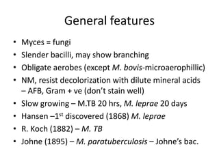
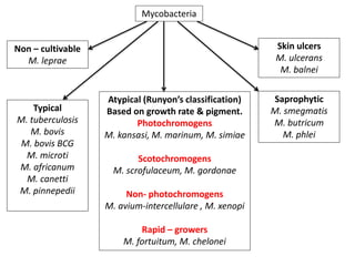
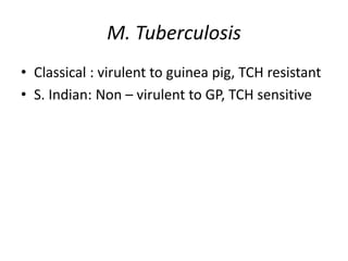
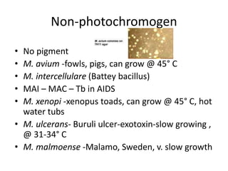
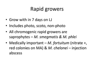
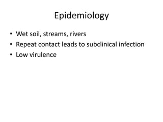
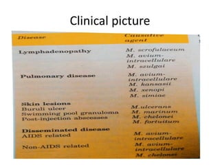
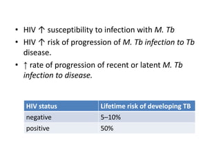
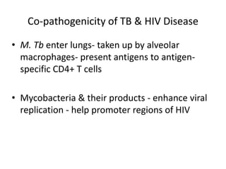
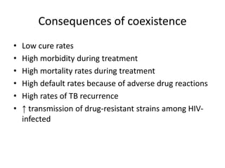
This document discusses Mycobacteria, which are slender bacilli that are slow growing and acid-fast. It covers the classification of mycobacteria including typical pathogens like M. tuberculosis and M. leprae, as well as atypical or environmental mycobacteria. Key details about morphology, culture techniques, biochemical reactions, pathogenesis, diagnosis and treatment of tuberculosis are provided. The relationship between tuberculosis and HIV is also summarized.
2. General features
• Myces = fungi
• Slender bacilli, may show branching
• Obligate aerobes (except M. bovis-microaerophillic)
• NM, resist decolorization with dilute mineral acids
– AFB, Gram + ve (don’t stain well)
• Slow growing – M.TB 20 hrs, M. leprae 20 days
• Hansen –1st discovered (1868) M. leprae
• R. Koch (1882) – M. TB
• Johne (1895) – M. paratuberculosis – Johne’s bac.
3. Mycobacteria
Non – cultivable
M. leprae
Typical
M. tuberculosis
M. bovis
M. bovis BCG
M. microti
M. africanum
M. canetti
M. pinnepedii
Skin ulcers
M. ulcerans
M. balnei
Saprophytic
M. smegmatis
M. butricum
M. phlei
Atypical (Runyon’s classification)
Based on growth rate & pigment.
Photochromogens
M. kansasi, M. marinum, M. simiae
Scotochromogens
M. scrofulaceum, M. gordonae
Non- photochromogens
M. avium-intercellulare , M. xenopi
Rapid – growers
M. fortuitum, M. chelonei
4. M. Tuberculosis
• Classical : virulent to guinea pig, TCH resistant
• S. Indian: Non – virulent to GP, TCH sensitive
9. Culture
• Upto 6-8 wks @ 37° C
• Lowenstein – Jensen Medium: Eggs,
asparagine, mineral salts, malachite green,
glycerol – Inspisation (80 ° C x 30 m x 3 days)
• Ruff, tuff, buff, wrinkled colonies – not easily
emulsified
10. • Liquid media: hydrophobic walls – surface pellicle
• Tween 80 for diffuse growth
• Petergananim Dorset’s – egg based medias
• Middlebrook 7H10 & 7H11
• Dubo’s
• Virulent strains: serpentine cords in liquid media
– mycolic acids linked together with trehalose
11.
12. Biochemical reactions
• Niacin test: niacin produced – free niacin to
ribonucleotide, M. tb does not reduce it –
hence it accumulates – gives positive reaction
– Canary red color
• Arylsulphatase test: negative
13. • Neutral red test : Virulent strains bind neutral
red (M. tb, M. bovis, M. avium, ulcerans)
• Catalase – peroxidase test : atypical – catalase
+++, peroxidase – ve , typical – catalase +,
peroxidase – ve (INH resistant – both negative)
• Amidase: Nicotinamide & PYZ reduced
• Nitrate reduction: M.tb, kansasi, fortuitum,
chelonae are +ve
• TCH : classical – resistant , S. Indian - Sensitive
14. Mycobacteriophages
• Temperate phages
• Not true lysogeny – as phage genome acts as
independent plasmid – pseudolysogeny
• Phage types – A, B, C & I
• I is m/c in India
• Phage 33d (environmental) that lyses all
strains of M. tb except BCG
15. Koch’s phenomenon
• Non-immune guinea pigs – virulent strains –
local lesion heals quickly – after 10 -14 days
nodule – ulceration – progresses to Tb & death
• Immune GP – 1 – 2 days indurated lesion –
necrosis / ulcer - healing without LN
involvement
• Three components – induration & necrosis,
local acute congestion or h’age, fever
16. Tuberculin test
• Old tuberculin – filtrate of 6-8 weeks old culture
• PPD – Purified Protein derivative – refined by
Seibert
• Mantoux test, Heaf test – Dx of latent Tb
• Positive = sensitization
• false negative – Milliary Tb (Low immunity)
• False positive – technique or atypical Tb
• Uses: Prevalence, Dx of active infection in
children & indicator of successful BCG vaccination
17.
18. Lab diagnosis
Sputum (S & M) in clean, leak proof container, CSF, 3 whole EMU, Pus
ZN / Auramine – Rhodamine staining (≈50,000 bacilli/ml),
For Urine – acid-alcohol (M. smegmatis – acid fast only )
Homogenization, decontamination & Concentration
(Petroff’s or NALC-NaOH method)
Culture –LJ 2 slopes @ 37 C x 8-12 wks, Automation (BACTEC,
BacT ALERT) - ( 10-100 bacilli/ml. Of sputum)
Biochemical reactions, Mycobacteriophages, Molecular techniques
AST – Resistance ratio, absolute conc. Proportion method, Dilution
methods, phage luciferase gene, MTBDRplus
19. Treatment
• First line drugs: Isoniazid (H), Rifampicin (R),
Ethambutol (E), Streptomycin (S), Pyrazinamide
(Z)
• Second Line drugs: Thioacetazone (Thz), PAS ,
Ethionamide (Eto), Cycloserine (Cs), Kanamycin
(Km), Amikacin (Am), Capreomycin (Cm)
• Newer agents: Ciprofloxacin ,Ofloxacin,
Clarithromycin, Azithromycin, Rifabutin
20. BCG
• Bacillus Calmette & Guerin
• M. bovis – grown in glycerol potato medium – s/c
every week x 239 times (≈13 years) 1908 – 1921 –
until attenuated
• Widely used strains – Pasteur117 3P2, Tokyo 172,
Copenhagen 1331 & Glaxo 1077
• Intradermal 0.1 @ birth or 1st year of life
• Nodule in 2-3wks - ↑ size - ≈ 4-8mm x 5 wks –
ulcerates – scar
• Immunity ≈ 10 years
21. Non-tubercular mycobacteria
• a/k/a MOTT, atypical/ opportunistic/ anonymous
• mostly saprophytes grow in +nce of PNB
• Runyon’s classification- Pigment & Growth
• Photochromogens – pigment only when exposed
to light & air x 1 hr
• Scotochromogens-Pigment even in dark
• Non-photochromogens – no pigment
• Rapid growers – growth in <7 days
22. Photochromogens
• Slow growers , but grow faster than typical,
yellow – orange pigment (β-carotein)
• M. kansasi: disseminated disease in IC
resembling Tb
• M. marinum: swimming pool or fish tank
granuloma, grows @ 33° C
• M. simiae: monkeys, very rare human
infections
23.
24. Scotochromogens
• Yellow, orange, red pigment – even in dark
• M. scrofulaceum – scrofulae in kids – cervical
LAP, found in water
• M. gordonae – water, rare pathogen,
contaminates clinical samples
• M. szulgai - scotochromogen @37° C &
photochromogen @ 25° C
25. Non-photochromogen
• No pigment
• M. avium -fowls, pigs, can grow @ 45° C
• M. intercellulare (Battey bacillus)
• MAI – MAC – Tb in AIDS
• M. xenopi -xenopus toads, can grow @ 45° C, hot
water tubs
• M. ulcerans- Buruli ulcer-exotoxin-slow growing ,
@ 31-34° C
• M. malmoense -Malamo, Sweden, v. slow growth
26. Rapid growers
• Grow with in 7 days on LJ
• Includes photo, scoto, non-photo
• All chromogenic rapid growers are
saprophytes – M. smegmatis & M. phlei
• Medically important – M. fortuitum (nitrate +,
red colonies on MA) & M. chelonei – injection
abscess
29. Lab diagnosis
• ZN stain
• Culture in dark & light
• Biochemical reactions
• Treatment – species dependent
30. TB/HIV
• TB and HIV are closely interlinked.
• TB is leading cause of HIV-related morbidity &
mortality.
• HIV is most important factor fuelling TB epidemic
• HIV is most powerful factor known to ↑ risk of TB.
• TB can occur at any point in the course of
progression of HIV infection
• A person infected with HIV has a 10 times ↑ risk of
developing TB.
31. • HIV ↑ susceptibility to infection with M. Tb
• HIV ↑ risk of progression of M. Tb infection to Tb
disease.
• ↑ rate of progression of recent or latent M. Tb
infection to disease.
HIV status Lifetime risk of developing TB
negative 5–10%
positive 50%
32. Co-pathogenicity of TB & HIV Disease
• M. Tb enter lungs- taken up by alveolar
macrophages- present antigens to antigen-
specific CD4+ T cells
• Mycobacteria & their products - enhance viral
replication - help promoter regions of HIV
33. Consequences of coexistence
• Low cure rates
• High morbidity during treatment
• High mortality rates during treatment
• High default rates because of adverse drug reactions
• High rates of TB recurrence
• ↑ transmission of drug-resistant strains among HIV-
infected