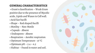
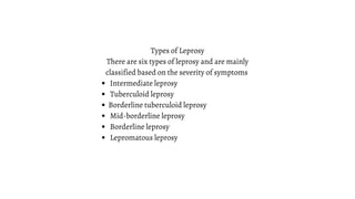
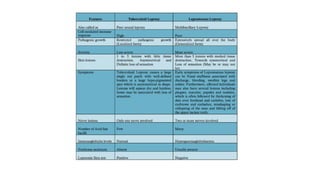
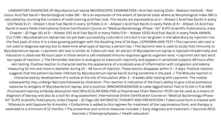
MYCOBACTERIUM
MARIAM NOORU
Mycobacterium is a genus of Mycobacteriaceae family
They are nonmotile, non–spore-forming, aerobic bacilli.
They are slightly curved rods with filamentous and branching forms on occasion.
The cell wall is thick, complex, and lipid-rich, resulting in a hydrophobic surface.
They do not stain readily, but once stained, resist decolourisation with dilute
mineral acids. Hence they are called ‘Acid fast bacilli’.
Bacteria are classified in the genus Mycobacterium on the basis of
(1) their acid-fastness,
(2) the presence of cell wall mycolic acids
(3) a high guanine plus cytosine (G+C) content in their deoxyribonucleic acid
(DNA).
Growth properties and colonial morphology ( preliminary classification of
mycobacteria).
INTRODUCTION
Pathogenic Mycobacterium
M. tuberculosis Complex
◦M. tuberculosis - Common
◦M. leprae - Uncommon
◦M. africanum
◦M. bovis
◦M. ulcerans
All are Strictly Pathogenic
Runyon Group I (Slow growing
photochromogens)
◦M. kanasii - Common
◦M. marinum
◦M. simae Uncommon
All are usually pathogenic (not strictly)
Runyon Group II (Slow growing
scotochromogens)
◦M. szulgai
◦M. scrofulaceum Uncommon
◦M. xenopi
Usually pathogenic Sometimes pathogenic
Runyon Group III (Slow growing
nonchromogens)
◦M. avium complex – common
◦M. genavense
◦M. hemophilum uncommon
◦M. malmoense
Strictly pathogenic Usually pathogenic
Runyon Group IV (Rapid growers)
◦M. fortuitum
◦M. chelonae Common
◦M. abscessus
◦M. mucogenicum Uncommon
Sometimes pathogenic
Mycobacterium tuberculosis
GENERAL CHARACTERISTICS
• Gram’s classification – Weak Gram
positive due to the presence of Mycolic
acids, Lipids and Waxes in Cell wall.
• Acid fast bacilli
• Shape – Rod shaped Bacilli
• Motility – Non-Motile
• Capsule - Absent
• Endospores - Absent
• Respiration – Aerobic respiration
• Optimum Temperature - 37 °C
• Optimum pH – 5.4 – 6.5
• Habitat – Found in water and soil.
Trehalose Di mycolate (Cord Factor)
Sulfatides
Catalase peroxidase
PATHOGENICITY OF Mycobacterium tuberculosis
DISEASE TRANSMISSION - Person-to-person spread by infectious Air borne
aerosols.
INCUBATION PERIOD - 2 to 6 Weeks
VIRULENCE FACTORS OF Mycobacterium tuberculosis
Mycobacteria
⬇
pulmonary alveoli
⬇
replicate within macrophages
⬇
picked up by dendritic cells
⬇
transport to local LN
⬇
spread through bloodstream to other tissues/organs
⬇
secondary TB lesions
primary site of infection : upper part of the lower lobe, or lower part of the upper lobe of lung
secondary TB lesions: apex of the upper lobes , peripheral lymph nodes, kidneys, brain, and bone
Pathogenesis
Primary pulmonary tuberculosis occurs in non sensitized hosts.
This may occur in any organ such as lungs, tonsils, intestine or skin
Among children the common site is lungs.
The inhaled bacilli are engulfed by alveolar macrophages in which they multiply to form
initial lesions called GHON FOCI.
Most frequently it occurs in the lower lobe or lower part of the upper lobe.
From here some bacilli are transported to hilar lymph node and causing
lymphadenopathy.
The ghon foci together with the enlarged hilar lymph node forms primary infecton.
In most cases the primary infection is asymptomatic
Occasionally the primary infection may spread through lymph nodes and causes bone &
join tuberculosis, renal tuberculosis, meningeal tuberculosis, endometrial tuberculosis
and testicular tuberculosis.
PRIMARY TUBERCULOSIS
This type of infection is mainly caused by reactivation of primary lesions or by bacilli
that are inhaled or ingested from the environment.
It is otherwise known as post primary tuberculosis or adult tuberculosis.
It mostly involves lungs and lesions are produced in the apical region ( apex) of the
lungs and can be transmitted to kidney, meninges, bones and other organs.
Formation of granuloma occurs and the necrotic elements of the reaction cause
destruction of the tissues and large areas of caseation, termed as tuberculomas
The activated macrophages secrete the enzyme protease that causes softening and
liquefaction of necrosis.
the necrosis is entered to the bronchus from the lungs and leaving a cavity (cavity
TBC)  The necrosis then enters to the blood vessels and spreading the bacilli through
out the body.
SECONDARY TUBERCULOSIS
Coughing that lasts three or more weeks
Coughing up blood
Chest pain, or pain with breathing or
coughing
Unintentional weight loss
Fatigue
Fever
Night sweats
Chills
Loss of appetite
Signs and symptoms of active TB
include:
Laboratory diagnosis of M. tuberculosis:
Specimens:
PTB:- sputum,
Tubercular meningitis:- CSF
Genitourinary TB:- urine.
Others- largyngeal swabs, gastric lavage, pleural fluid, pus sample,
nasopharyngeal aspirates.
MICROSCOPIC EXAMINATION
• Acid fast stating (Ziehl - Neelson method) – Red colour Acid fast bacilli
COLONY MORPHOLOGY ON CULTURE MEDIUM
• Lowenstein Jensen (LJ) Medium – Mycobacterium tuberculosis appears as brown,
granular colonies (sometimes called "buff, rough and tough")
BIOCHEMICAL TESTS
Catalase test - Negative
Oxidase test – Negative
Urease test – Positive
Indole test - Negative
Methyl Red (MR) test - Negative
Voges Proskauer (VP) test - Negative
Citrate utilization test – Negative
Nitrate reduction – Positive
Niacin test - Positive
Neutral Red test - Positive
Mantoux test is a Tuberculin skin test used for the diagnosis of
TB.
In this test, 0.1 ml or 5 tuberculin units of PPD (purified protein
derivative) is injected intradermally into the volar aspect of the
forearm using a 27-G needle.
PPD should be injected between the layers of the skin and not
subcutaneously. The results is read after 48-72 hours.
IMMUNODIAGNOSIS
(i) Tuberculin Skin Test:
Anti-tuberculosis drugs are divided as-
First line drugs:
Isoniazid, Rifampicin, Ethambutol, Streptomycin (injection) and Pyrazinamide
Second line drugs:
Used for the cases of TB where first line drugs are ineffective.
Includes ciprofloxacin, cycloserine, ethionmide, kanamycin, ofloxacin, levofloxacin, capreomycin and
others.
Treatment of tuberculosis:
Mycobacterium leprae
GENERAL CHARACTERISTICS
• Gram’s classification – Weak Gram positive due to the presence
of Mycolic acids, Lipids and Waxes in Cell wall. Also called as
Hansen’s Bacillus Spirilly
• Acid fast bacilli
• Shape – Rod shaped Bacilli. Sometimes Pleomorphic in nature.
• Intracellular parasite. Unable to be cultured on artificial media.
• Motility – Non-Motile
• Capsule - Absent
• Endospores - Absent
• Respiration – Microaerophilic respiration
• Optimum Temperature – 27 °C to 30 °C
• Optimum pH – 7.0
• Habitat – Found in air, water and soil.
• Mycobacterium leprae has a long generation time of about 12
days.
Fibronectin
Secreted Proteins
Phenolic Glycolipid (PGL-1)
Lipoarabinomannan (LAM)
PATHOGENICITY OF Mycobacterium leprae
DISEASE TRANSMISSION: Person to person spread by infectious Air borne Nasal secretions
or Droplets or Aerosols.
INCUBATION PERIOD: Mycobacterium leprae multiplies slowly and the incubation period
of the disease on average is 5 years.
VIRULENCE FACTORS OF Mycobacterium leprae
Mycobacterium leprae enter the body usually through Respiratory system.
Mycobacterium leprae migrate towards the Neural tissue (present in CNS) and enter the
Schwann cells.
After entering the Schwann cells or Macrophage, Mycobacterium leprae start
multiplying slowly within the cells, get liberated from the destroyed cells and enter other
unaffected cells. Person remains free from signs and symptoms of Leprosy at this stage.
As the Mycobacterium leprae multiply, bacterial load increases in the body and infection
is recognized by the immunological system.
Lymphocytes and Histiocytes invade the infected tissue. At this stage clinical
manifestation may appear as involvement of nerves with impairment of sensation or
skin patch.
If it is not diagnosed and treated in the early stages, further progress of the diseases is
determined by the strength of the patient’s Cell mediated immune response.
PATHOGENESIS
Granuloma formation occurs in Cutaneous nerve. Cutaneous nerve swell and gets destroyed.
Severe inflammation may result in Caseous necrosis (a unique form of cell death in which the
tissue maintains a cheese-like appearance) within the nerve.
Mycobacterium leprae may escape from nerve to adjacent skin at any time and cause classical
skin lesions.
Good Cell Mediated Immunity successfully limits the disease to the nerve Schwann cell resulting
in occurrence of Tuberculoid Leprosy.
Mycobacterium leprae entering the Schwann cells multiply unchecked and destroy the nerve.
Mycobacterium leprae liberated by infected and destroyed cells are engulfed by Histiocytes
(Tissue Macrophage).
Mycobacterium leprae multiply inside these macrophages and travel to other tissues, through
blood, lymph or tissue fluid.
a)InPersonswithstrongCellMedicatedImmunity(PureneuralleprosyorTuberculoidLeprosy)
b)InpersonswithdepressedCellMedicatedImmunity(MultibacillaryLeprosyorLepromatousLeprosy)
Intermediate leprosy
Tuberculoid leprosy
Borderline tuberculoid leprosy
Mid-borderline leprosy
Borderline leprosy
Lepromatous leprosy
Types of Leprosy
There are six types of leprosy and are mainly
classified based on the severity of symptoms
LABORATORY DIAGNOSIS OF Mycobacterium leprae MICROSCOPIC EXAMINATION • Acid fast stating (Ziehl - Neelson method) – Red
colour Acid fast bacilli • Bacteriological index (BI) – BI is an expression of the extent of bacterial loads where as Morphological index (MI) is
calculated by counting the numbers of solid-staining acid-fast rods. The results are expressed as a) 1+ - Atleast 1 Acid Fast Bacilli in every
100 fields b) 2+ - Atleast 1 Acid Fast Bacilli in every 10 fields c) 3+ - Atleast 1 Acid Fast Bacilli in every fields d) 4+ - Atleast 10 Acid Fast
Bacilli in every fields International Online Certification Course on “Medical Bacteriology (Phase - IV)” ©JPS Scientific Publications, India
Chapter - 32 Page 161 e) 5+ - Atleast 100 Acid Fast Bacilli in every fields f) 6+ - Atleast 1000 Acid Fast Bacilli in every fields ANIMAL
CULTURE • Mycobacterium leprae has not yet been successfully cultured in vitro but it can be grown in the laboratory by injection into
the foot pads of mice. It is a slow growing pathogen with the doubling time of 14 days. LEPROMIN SKIN TEST • The Lepromin skin test is
not used to diagnose leprosy but to determine what type of leprosy a person has. • The lepromin test is used to study host immunity to
Mycobacterium leprae. • Lepromin skin test is similar to Tuberculin test. An extract of Mycobacterium leprae is injected intradermally and
induration is observed 48 hours later in those whom a cell-mediated immune response against organism exist. • Lepromin skin test elicit
two types of reaction: ✓The Fernandez reaction is analogous to tuberculin reactivity and appears in sensitized subjects 48 hours after
skin testing. Positive reaction is characterized by the appearance of a localized area of inflammation with congestion and edema
measuring 10 mm and more in diameter during 24 – 48 hours of injection. These lesions disappear within 3 – 4 days. Positive reaction
suggests that the patient has been infected by Mycobacterium leprae bacilli during sometime in the past. ✓The Mitsuda reaction is
characterized by development of a nodule at the site of inoculation after 3 – 4 weeks after testing with Lepromin. The nodule
subsequently may undergo Necrosis followed by Ulceration. This reaction is indicative of the host’s ability to give a Granulomatous
response to antigens of Mycobacterium leprae, and is positive. IMMUNODIAGNOSIS a) Latex Agglutination Test b) ELISA c) FLA-ABS
(Fluorescent leprosy antibody absorption test) MOLECULAR ANALYSIS a) Polymerase Chain Reaction (PCR) can be used as a means of
diagnosis of leprosy and also as a tool for drug assessment. International Online Certification Course on “Medical Bacteriology (Phase -
IV)” ©JPS Scientific Publications, India Chapter - 32 Page 162 ANTIBIOTIC THERAPY AND PREVENTION • Tuberculoid form is treated with
Rifampicin and Dapsone for 6 months. • Clofazimine is added to this regimen for treatment of the Lepromatous form, and therapy is
extended to a minimum of 12 months. • The preventive and control measures includes ✓Early diagnosis and treatment ✓Vaccines (BCG
Vaccine) ✓Chemoprophylaxis ✓Health education

Mycobacterium

  • 1.
  • 2.
    Mycobacterium is agenus of Mycobacteriaceae family They are nonmotile, non–spore-forming, aerobic bacilli. They are slightly curved rods with filamentous and branching forms on occasion. The cell wall is thick, complex, and lipid-rich, resulting in a hydrophobic surface. They do not stain readily, but once stained, resist decolourisation with dilute mineral acids. Hence they are called ‘Acid fast bacilli’. Bacteria are classified in the genus Mycobacterium on the basis of (1) their acid-fastness, (2) the presence of cell wall mycolic acids (3) a high guanine plus cytosine (G+C) content in their deoxyribonucleic acid (DNA). Growth properties and colonial morphology ( preliminary classification of mycobacteria). INTRODUCTION
  • 3.
    Pathogenic Mycobacterium M. tuberculosisComplex ◦M. tuberculosis - Common ◦M. leprae - Uncommon ◦M. africanum ◦M. bovis ◦M. ulcerans All are Strictly Pathogenic
  • 4.
    Runyon Group I(Slow growing photochromogens) ◦M. kanasii - Common ◦M. marinum ◦M. simae Uncommon All are usually pathogenic (not strictly) Runyon Group II (Slow growing scotochromogens) ◦M. szulgai ◦M. scrofulaceum Uncommon ◦M. xenopi Usually pathogenic Sometimes pathogenic Runyon Group III (Slow growing nonchromogens) ◦M. avium complex – common ◦M. genavense ◦M. hemophilum uncommon ◦M. malmoense Strictly pathogenic Usually pathogenic Runyon Group IV (Rapid growers) ◦M. fortuitum ◦M. chelonae Common ◦M. abscessus ◦M. mucogenicum Uncommon Sometimes pathogenic
  • 6.
  • 7.
    GENERAL CHARACTERISTICS • Gram’sclassification – Weak Gram positive due to the presence of Mycolic acids, Lipids and Waxes in Cell wall. • Acid fast bacilli • Shape – Rod shaped Bacilli • Motility – Non-Motile • Capsule - Absent • Endospores - Absent • Respiration – Aerobic respiration • Optimum Temperature - 37 °C • Optimum pH – 5.4 – 6.5 • Habitat – Found in water and soil.
  • 8.
    Trehalose Di mycolate(Cord Factor) Sulfatides Catalase peroxidase PATHOGENICITY OF Mycobacterium tuberculosis DISEASE TRANSMISSION - Person-to-person spread by infectious Air borne aerosols. INCUBATION PERIOD - 2 to 6 Weeks VIRULENCE FACTORS OF Mycobacterium tuberculosis
  • 9.
    Mycobacteria ⬇ pulmonary alveoli ⬇ replicate withinmacrophages ⬇ picked up by dendritic cells ⬇ transport to local LN ⬇ spread through bloodstream to other tissues/organs ⬇ secondary TB lesions primary site of infection : upper part of the lower lobe, or lower part of the upper lobe of lung secondary TB lesions: apex of the upper lobes , peripheral lymph nodes, kidneys, brain, and bone Pathogenesis
  • 11.
    Primary pulmonary tuberculosisoccurs in non sensitized hosts. This may occur in any organ such as lungs, tonsils, intestine or skin Among children the common site is lungs. The inhaled bacilli are engulfed by alveolar macrophages in which they multiply to form initial lesions called GHON FOCI. Most frequently it occurs in the lower lobe or lower part of the upper lobe. From here some bacilli are transported to hilar lymph node and causing lymphadenopathy. The ghon foci together with the enlarged hilar lymph node forms primary infecton. In most cases the primary infection is asymptomatic Occasionally the primary infection may spread through lymph nodes and causes bone & join tuberculosis, renal tuberculosis, meningeal tuberculosis, endometrial tuberculosis and testicular tuberculosis. PRIMARY TUBERCULOSIS
  • 12.
    This type ofinfection is mainly caused by reactivation of primary lesions or by bacilli that are inhaled or ingested from the environment. It is otherwise known as post primary tuberculosis or adult tuberculosis. It mostly involves lungs and lesions are produced in the apical region ( apex) of the lungs and can be transmitted to kidney, meninges, bones and other organs. Formation of granuloma occurs and the necrotic elements of the reaction cause destruction of the tissues and large areas of caseation, termed as tuberculomas The activated macrophages secrete the enzyme protease that causes softening and liquefaction of necrosis. the necrosis is entered to the bronchus from the lungs and leaving a cavity (cavity TBC)  The necrosis then enters to the blood vessels and spreading the bacilli through out the body. SECONDARY TUBERCULOSIS
  • 13.
    Coughing that laststhree or more weeks Coughing up blood Chest pain, or pain with breathing or coughing Unintentional weight loss Fatigue Fever Night sweats Chills Loss of appetite Signs and symptoms of active TB include:
  • 14.
    Laboratory diagnosis ofM. tuberculosis: Specimens: PTB:- sputum, Tubercular meningitis:- CSF Genitourinary TB:- urine. Others- largyngeal swabs, gastric lavage, pleural fluid, pus sample, nasopharyngeal aspirates. MICROSCOPIC EXAMINATION • Acid fast stating (Ziehl - Neelson method) – Red colour Acid fast bacilli COLONY MORPHOLOGY ON CULTURE MEDIUM • Lowenstein Jensen (LJ) Medium – Mycobacterium tuberculosis appears as brown, granular colonies (sometimes called "buff, rough and tough")
  • 15.
    BIOCHEMICAL TESTS Catalase test- Negative Oxidase test – Negative Urease test – Positive Indole test - Negative Methyl Red (MR) test - Negative Voges Proskauer (VP) test - Negative Citrate utilization test – Negative Nitrate reduction – Positive Niacin test - Positive Neutral Red test - Positive Mantoux test is a Tuberculin skin test used for the diagnosis of TB. In this test, 0.1 ml or 5 tuberculin units of PPD (purified protein derivative) is injected intradermally into the volar aspect of the forearm using a 27-G needle. PPD should be injected between the layers of the skin and not subcutaneously. The results is read after 48-72 hours. IMMUNODIAGNOSIS (i) Tuberculin Skin Test: Anti-tuberculosis drugs are divided as- First line drugs: Isoniazid, Rifampicin, Ethambutol, Streptomycin (injection) and Pyrazinamide Second line drugs: Used for the cases of TB where first line drugs are ineffective. Includes ciprofloxacin, cycloserine, ethionmide, kanamycin, ofloxacin, levofloxacin, capreomycin and others. Treatment of tuberculosis:
  • 16.
  • 17.
    GENERAL CHARACTERISTICS • Gram’sclassification – Weak Gram positive due to the presence of Mycolic acids, Lipids and Waxes in Cell wall. Also called as Hansen’s Bacillus Spirilly • Acid fast bacilli • Shape – Rod shaped Bacilli. Sometimes Pleomorphic in nature. • Intracellular parasite. Unable to be cultured on artificial media. • Motility – Non-Motile • Capsule - Absent • Endospores - Absent • Respiration – Microaerophilic respiration • Optimum Temperature – 27 °C to 30 °C • Optimum pH – 7.0 • Habitat – Found in air, water and soil. • Mycobacterium leprae has a long generation time of about 12 days.
  • 18.
    Fibronectin Secreted Proteins Phenolic Glycolipid(PGL-1) Lipoarabinomannan (LAM) PATHOGENICITY OF Mycobacterium leprae DISEASE TRANSMISSION: Person to person spread by infectious Air borne Nasal secretions or Droplets or Aerosols. INCUBATION PERIOD: Mycobacterium leprae multiplies slowly and the incubation period of the disease on average is 5 years. VIRULENCE FACTORS OF Mycobacterium leprae
  • 19.
    Mycobacterium leprae enterthe body usually through Respiratory system. Mycobacterium leprae migrate towards the Neural tissue (present in CNS) and enter the Schwann cells. After entering the Schwann cells or Macrophage, Mycobacterium leprae start multiplying slowly within the cells, get liberated from the destroyed cells and enter other unaffected cells. Person remains free from signs and symptoms of Leprosy at this stage. As the Mycobacterium leprae multiply, bacterial load increases in the body and infection is recognized by the immunological system. Lymphocytes and Histiocytes invade the infected tissue. At this stage clinical manifestation may appear as involvement of nerves with impairment of sensation or skin patch. If it is not diagnosed and treated in the early stages, further progress of the diseases is determined by the strength of the patient’s Cell mediated immune response. PATHOGENESIS
  • 20.
    Granuloma formation occursin Cutaneous nerve. Cutaneous nerve swell and gets destroyed. Severe inflammation may result in Caseous necrosis (a unique form of cell death in which the tissue maintains a cheese-like appearance) within the nerve. Mycobacterium leprae may escape from nerve to adjacent skin at any time and cause classical skin lesions. Good Cell Mediated Immunity successfully limits the disease to the nerve Schwann cell resulting in occurrence of Tuberculoid Leprosy. Mycobacterium leprae entering the Schwann cells multiply unchecked and destroy the nerve. Mycobacterium leprae liberated by infected and destroyed cells are engulfed by Histiocytes (Tissue Macrophage). Mycobacterium leprae multiply inside these macrophages and travel to other tissues, through blood, lymph or tissue fluid. a)InPersonswithstrongCellMedicatedImmunity(PureneuralleprosyorTuberculoidLeprosy) b)InpersonswithdepressedCellMedicatedImmunity(MultibacillaryLeprosyorLepromatousLeprosy)
  • 21.
    Intermediate leprosy Tuberculoid leprosy Borderlinetuberculoid leprosy Mid-borderline leprosy Borderline leprosy Lepromatous leprosy Types of Leprosy There are six types of leprosy and are mainly classified based on the severity of symptoms
  • 23.
    LABORATORY DIAGNOSIS OFMycobacterium leprae MICROSCOPIC EXAMINATION • Acid fast stating (Ziehl - Neelson method) – Red colour Acid fast bacilli • Bacteriological index (BI) – BI is an expression of the extent of bacterial loads where as Morphological index (MI) is calculated by counting the numbers of solid-staining acid-fast rods. The results are expressed as a) 1+ - Atleast 1 Acid Fast Bacilli in every 100 fields b) 2+ - Atleast 1 Acid Fast Bacilli in every 10 fields c) 3+ - Atleast 1 Acid Fast Bacilli in every fields d) 4+ - Atleast 10 Acid Fast Bacilli in every fields International Online Certification Course on “Medical Bacteriology (Phase - IV)” ©JPS Scientific Publications, India Chapter - 32 Page 161 e) 5+ - Atleast 100 Acid Fast Bacilli in every fields f) 6+ - Atleast 1000 Acid Fast Bacilli in every fields ANIMAL CULTURE • Mycobacterium leprae has not yet been successfully cultured in vitro but it can be grown in the laboratory by injection into the foot pads of mice. It is a slow growing pathogen with the doubling time of 14 days. LEPROMIN SKIN TEST • The Lepromin skin test is not used to diagnose leprosy but to determine what type of leprosy a person has. • The lepromin test is used to study host immunity to Mycobacterium leprae. • Lepromin skin test is similar to Tuberculin test. An extract of Mycobacterium leprae is injected intradermally and induration is observed 48 hours later in those whom a cell-mediated immune response against organism exist. • Lepromin skin test elicit two types of reaction: ✓The Fernandez reaction is analogous to tuberculin reactivity and appears in sensitized subjects 48 hours after skin testing. Positive reaction is characterized by the appearance of a localized area of inflammation with congestion and edema measuring 10 mm and more in diameter during 24 – 48 hours of injection. These lesions disappear within 3 – 4 days. Positive reaction suggests that the patient has been infected by Mycobacterium leprae bacilli during sometime in the past. ✓The Mitsuda reaction is characterized by development of a nodule at the site of inoculation after 3 – 4 weeks after testing with Lepromin. The nodule subsequently may undergo Necrosis followed by Ulceration. This reaction is indicative of the host’s ability to give a Granulomatous response to antigens of Mycobacterium leprae, and is positive. IMMUNODIAGNOSIS a) Latex Agglutination Test b) ELISA c) FLA-ABS (Fluorescent leprosy antibody absorption test) MOLECULAR ANALYSIS a) Polymerase Chain Reaction (PCR) can be used as a means of diagnosis of leprosy and also as a tool for drug assessment. International Online Certification Course on “Medical Bacteriology (Phase - IV)” ©JPS Scientific Publications, India Chapter - 32 Page 162 ANTIBIOTIC THERAPY AND PREVENTION • Tuberculoid form is treated with Rifampicin and Dapsone for 6 months. • Clofazimine is added to this regimen for treatment of the Lepromatous form, and therapy is extended to a minimum of 12 months. • The preventive and control measures includes ✓Early diagnosis and treatment ✓Vaccines (BCG Vaccine) ✓Chemoprophylaxis ✓Health education