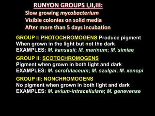
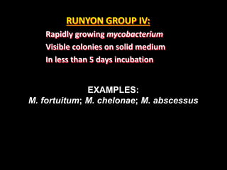
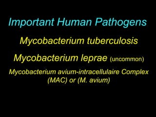
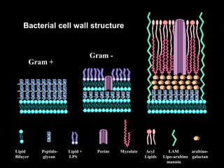
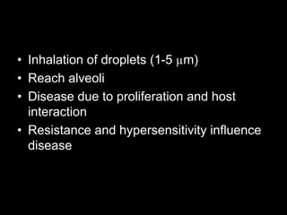
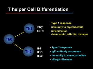
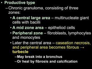
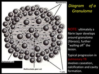
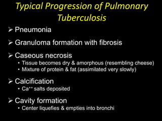
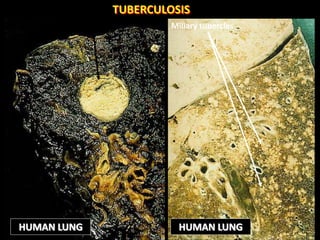
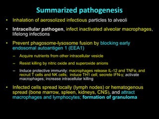
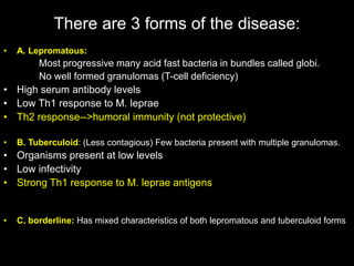
MYCOBACTERIA
OF
MEDICAL IMPORTANCE
Prof M.I.N. Matee
Department of Microbiology and
Immunology,
MUHAS
Venue: CPL
Date: 5th December 2017
MYCOBACTERIUM
THIS GENUS IS COMPOSED OF:
Strictly aerobic, acid-fast rods, does not
stain well (gram stain indeterminant),
DNA has high g+c content, unique cell wall,
Mycolic acid carbon chain length > c60
Relatively slow growth (two groups)
A. RAPID GROWERS (Visible colonies in <5 days)
B. SLOW GROWERS (Visible colonies in > 5 days)
TYPE SPECIES: Mycobacterium tuberculosis
THE GENUS MYCOBACTERIUM CAN BE DIVIDED
INTO FOUR BROAD GROUPS
1. THE TUBERCULOSIS COMPLEX
2. SLOW GROWING MYCOBACTERIA
OTHER THAN TUBERCULOSIS (MOTT)
3. RAPIDLY GROWING MYCOBACTERIA
4. MYCOBACTERIUM LEPRAE
THE TUBERCULOSIS COMPLEX
(Organisms that resemble M. tuberculosis;
Causing a similar type of disease in humans)
1. M. tuberculosis
2. M. bovis
3. M. africanum
THE RUNYON GROUPING:
An older idea that
Remains useful for differentiating mott
RUNYON GROUPS I,II,III:
Slow growing mycobacterium
Visible colonies on solid media
After more than 5 days incubation
GROUP I: PHOTOCHROMOGENS Produce pigment
When grown in the light but not the dark
EXAMPLES: M. kansasii; M. marinum; M. simiae
GROUP II: SCOTOCHROMOGENS
Pigment when grown in both light and dark
EXAMPLES: M. scrofulaceum; M. szulgai; M. xenopi
GROUP III: NONCHROMOGENS
No pigment when grown in both light and dark
EXAMPLES: M. avium-intracellulare; M. genevense
M. ulcerans and M. leprae do not fit in this scheme
RUNYON GROUP IV:
Rapidly growing mycobacterium
Visible colonies on solid medium
In less than 5 days incubation
EXAMPLES:
M. fortuitum; M. chelonae; M. abscessus
Mycobacterium tuberculosis
Mycobacterium leprae (uncommon)
Mycobacterium avium-intracellulaire Complex
(MAC) or (M. avium)
Important Human Pathogens
Robert Koch 1843-1910
Mycobacterium tuberculosis
In 1882 M. tuberculosis was identified as the causative
agent of tuberculosis.
One-third of the world’s population is infected with
M. tuberculosis.
Nearly three million people die each year from tuberculosis.
In 1993 tuberculosis was declared a Global Health
Emergency by WHO.
1919
In 2004
In 1944 streptomycin was introduced for the treatment
of tuberculosis.
Reaction to physical and chemical agents
• Resistant to chemical agents – malachite
green, penicillins
• In liquid media – grow in clumps –
hydrophobic
• Resistant to acids and alkalines – helps to
decontaminate and concentrate samples
Bacterial cell wall structure
Gram +
Gram -
Mycobacterium
Lipid Peptido- Lipid + Porins Mycolate Acyl LAM arabino-
Bilayer glycan LPS Lipids Lipo-arabino galactan
mannin
Virulence factors (M. tb.)
– Cord factor – is a glycolipid, trehalose 6,6’ dimycolate,
– t is responsible for the serpentine growth (filaments or cords)
– It is toxic to leukocytes, antichemotactic, and plays a role in
the development of granulomatous lesions
– Iron capturing ability – required for survival inside
phagocytes
– Sulfolipids prevent phagosome-lysosome fusion so that the
organisms are not exposed to lysosomal enzymes (is
important in intracellular survival)
Tuberculosis in humans
A key issue is to understand why individuals infected with M.
tuberculosis experience different clinical outcomes
INTRACELLULAR pathogen (facultative extracellular)
Exposed
Infected
(2 billion, 8
million new
cases per year)
Primary
TB
Latent
TB Reactivation
30%
80-90%
5-10%
5-10%
Clearance
70%
Death
(2 million)
Pathogenesis
• Inhalation of droplets (1-5 µm)
• Reach alveoli
• Disease due to proliferation and host
interaction
• Resistance and hypersensitivity influence
disease
Proposed Macrophage Survival Mechanisms of M. tuberculosis
• Inhibition of phagosome-lysosome fusion
• Inhibition of phagosome acidification
• Resistance to reactive oxygen intermediates/reactive nitrogen intermediates
• Inhibition of antigen presentation
• Inhibition of IFN-gamma activation
• Interaction with the cytoplasm
Immunology of tuberculosis
• Tubercle bacillus + macrophages --> processed
antigen
• Antigen recognition by lymphocytes --> activated
lymphocytes --> lymphokines
• Lymphokines--> attraction, stimulation, and
retention of macrophages at antigen site
• Activated macrophages--> lytic enzymes with
mycobactericidal but also tissue-necrosing capacity
T helper Cell Differentiation
Th0
Th2
Th1
• Type 1 response
• immunity to mycobacteria
• inflammation
• rheumatoid arthritis, diabetes
• Type 2 response
• IgE antibody responses
• Immunity to some parasites
• allergic diseases
IFNg
TNFa
IL4
IL13
IL10
Immunology of tuberculosis
• Interferon-gamma probably stimulates
macrophages to produce
• interferon-alfa – increases ability to kill Mtb
• Cytokines secreted by alveolar macrophages:
• interleukin 1 (fever);
• interleukin 6 (hyperglobulinemia)
• tumor necrosis factor alpha (killing of organisms,
granuloma formation, fever and weight loss)
• Productive type
–Chronic granuloma, consisting of three
zones:
• A central large area – multinucleate giant
cells with bacilli
• A mid zone area – epitheliod cells
• Peripheral zone – fibroblasts, lymphocytes
and monocytes
• Later the central area – caseation necrosis,
and peripheral area becomes fibrous →
turbecle
–May break into a bronchus
–Or heal by fibrosis and calcification
Diagram of a
Granuloma
NOTE: ultimately a
fibrin layer develops
around granuloma
(fibrosis), further
“walling off” the
lesion.
Typical progression in
pulmonary TB
involves caseation,
calcification and cavity
formation.
 Pneumonia
 Granuloma formation with fibrosis
 Caseous necrosis
• Tissue becomes dry & amorphous (resembling cheese)
• Mixture of protein & fat (assimilated very slowly)
 Calcification
• Ca++ salts deposited
 Cavity formation
• Center liquefies & empties into bronchi
Typical Progression of Pulmonary
Tuberculosis
Large caseating tubercle Miliary tubercles
HUMAN LUNG HUMAN LUNG
TUBERCULOSIS
Residua of primary infection
• Ghon complex (after Anton Ghon, German
bacteriologist): calcified peripheral focus of
tuberculous infection with calcified regional
(hilar) lymph node (also called Ranke complex)
• Simon focus (after Georg Simon, German
pediatrician): focus at apex of lung, containing
viable organisms and manifested on x-ray as
“fibrous cap”
Spread of organisms in the host
• Tubercle spreads by direct extension to lymphatics
→ lymph nodes → blood stream → various organs
• Blood stream involvement also due to erosion of a
vein
• If contents of tubercle are swallowed could be
passed to stomach and intestine
Summarized pathogenesis
• Inhalation of aerosolized infectious particles to alveoli
• Intracellular pathogen, infect inactivated alveolar macrophages,
lifelong infections
• Prevent phagosome-lysosome fusion by blocking early
endosomal autoantigen 1 (EEA1)
– Acquire nutrients from other intracellular vesicle
– Resist killing by nitric oxide and superoxide anions
– Induce protective immunity: macrophages release IL-12 and TNFα, and
recruit T cells and NK cells; induce TH1 cell; secrete IFN-γ; activate
macrophages; increase intracellular killing
• Infected cells spread locally (lymph nodes) or hematogenous
spread (bone marrow, spleen, kidneys, CNS), and attract
macrophages and lymphocytes; formation of granuloma
Symptoms of TB Disease
• Prolonged cough (may produce sputum)*
• Chest pain*
• Hemoptysis*
• Fever
• Chills
• Night sweats
• Fatigue
• Loss of appetite
• Weight loss/failure to gain weight
*commonly seen in cases of pulmonary TB
Clinical Manifestations of TB
• Organ specific
– pneumonia: cough, sputum +/- blood
– scrofula: swollen lymph nodes
– meningitis: headache, obtundation
– miliary TB: no obvious source
– genitourinary: sterile pyuria
Major syndromes of
extrapulmonary tuberculosis
• Disseminated (miliary) tuberculosis
• “Serosal” tuberculosis (anatomic spaces or
cavities): pleurisy, pericarditis, meningitis,
peritonitis, arthritis
• Tuberculosis of solid organs: renal
(genitourinary), osteomyelitis, adrenal glands
(Addison’s disease), lymph nodes
DIAGNOSIS
1. Evidence of infection
a. Chest x-ray - hilar lymphadenopathy
calcification of primary focus/LN
b. Delayed hypersensitivity response to purified protein
derivative (PPD) MANTOUX /HEAF TEST
2. Evidence of active disease
a. Sputum for AFB positive
3. Evidence of active disease
a. Indirect evidence of infection (Mantoux)
b. Direct evidence of infection PCR / culture
c. Histo-pathological evidence
Reading the Tuberculin Skin Test
intradermal injection of PPD (5 TU)
Induration, edema, erythema
Read after 48-72 h
Positive: induration≥ 10 mm
It is DTH
–Kinyon – uses a higher content of phenol (organic
solvent) in the carbol fuchsin primary stain to
allow penetration of the stain without the need to
apply heat. Also uses acid alcohol to destain and
methylene blue as the counterstain.
– Auramine-rhodamine fluorochrome (a fluorescent stain)
requires a fluorescent microscope, but allows one to scan
the slide on high dry so that slides may be read much faster
» Stain with auramine-rhodamine for 10 minutes
(phenol in the solution allows for penetration)
» Destain with acid alcohol
» Counterstain with acridine orange
» A positive result is a bright yellow fluorescence.
Mycobacteria: auramine stain
Risk factors for TB/IRIS
• Starting ARV’s within 6 weeks of TB
treatment
• Disseminated, extra-pulmonary disease
• Low base line CD4 count
• Rise in CD4 %
• Fall in viral load
• High bacillary burden?
Prevention and control of tuberculosis
1. Prompt and effective treatment of patients with
active TB
2. Careful follow-up of contacts – tuberculin test, X-
rays etc
3. Prophylaxis of asymptomatics, tuberculin
positives
4. Correct underlying cause of immune suppression
5. Immunization with live-attenuated tubercle bacilli
e.g. BCG
6. Eradication of TB in cattle and pasteurization of
milk
HANSEN’S DISEASE (Leprosy) caused by M.
leprae
Hansen’s disease is a chronic, slowly progressive
Granulomatous
Disease involve ectodermally derived tissues e.g. skin
and peripheral nerves.
Usually limited to the cooler parts of the body e.g. skin,
nose and upper respiratory tract.
It rarely affects internal organs such as the brain, liver,
spleen, kidneys, and bones.
It has a specific predilection for peripheral nerves.
Mycobacterium leprae
is an acid-fast rod that grows in macrophages, nerve cells, the
foot pads
of thymectimized mice, and the nine banded armadillo.
It has never been grown in artificial medium,
an obligate intracellular pathogen.
has a generation time of 12 to 14 days,
the incubation period for onset of disease is prolonged,
therapy must also be prolonged (perhaps the lifetime of the
individual).
There are 3 forms of the disease:
• A. Lepromatous:
Most progressive many acid fast bacteria in bundles called globi.
No well formed granulomas (T-cell deficiency)
• High serum antibody levels
• Low Th1 response to M. leprae
• Th2 response-->humoral immunity (not protective)
• B. Tuberculoid: (Less contagious) Few bacteria present with multiple granulomas.
• Organisms present at low levels
• Low infectivity
• Strong Th1 response to M. leprae antigens
• C. borderline: Has mixed characteristics of both lepromatous and tuberculoid forms
Effect of Cell-Mediated Immunity on Leprosy
Clinical Outcome
REVIEW
HANSEN’S DISEASE is treatable
with outpatient therapy.
DRUGS OF CHOICE:
 SULFONES (i.e. Dapsone)
 PHENAZIDES (Lampren)
 RIFAMPIN
 CLOFAZAMINE
~ 500,000 cases Worldwide
Treatment – Dapsone (1940s-1970s)
Rifampicin (1970s-)
Multidrug Therapy- Dapsone, Rifampicin, Clofazamine

Mycobacteria.pptx

  • 1.
    MYCOBACTERIA OF MEDICAL IMPORTANCE Prof M.I.N.Matee Department of Microbiology and Immunology, MUHAS Venue: CPL Date: 5th December 2017
  • 2.
    MYCOBACTERIUM THIS GENUS ISCOMPOSED OF: Strictly aerobic, acid-fast rods, does not stain well (gram stain indeterminant), DNA has high g+c content, unique cell wall, Mycolic acid carbon chain length > c60 Relatively slow growth (two groups) A. RAPID GROWERS (Visible colonies in <5 days) B. SLOW GROWERS (Visible colonies in > 5 days) TYPE SPECIES: Mycobacterium tuberculosis
  • 3.
    THE GENUS MYCOBACTERIUMCAN BE DIVIDED INTO FOUR BROAD GROUPS 1. THE TUBERCULOSIS COMPLEX 2. SLOW GROWING MYCOBACTERIA OTHER THAN TUBERCULOSIS (MOTT) 3. RAPIDLY GROWING MYCOBACTERIA 4. MYCOBACTERIUM LEPRAE
  • 4.
    THE TUBERCULOSIS COMPLEX (Organismsthat resemble M. tuberculosis; Causing a similar type of disease in humans) 1. M. tuberculosis 2. M. bovis 3. M. africanum
  • 5.
    THE RUNYON GROUPING: Anolder idea that Remains useful for differentiating mott
  • 6.
    RUNYON GROUPS I,II,III: Slowgrowing mycobacterium Visible colonies on solid media After more than 5 days incubation GROUP I: PHOTOCHROMOGENS Produce pigment When grown in the light but not the dark EXAMPLES: M. kansasii; M. marinum; M. simiae GROUP II: SCOTOCHROMOGENS Pigment when grown in both light and dark EXAMPLES: M. scrofulaceum; M. szulgai; M. xenopi GROUP III: NONCHROMOGENS No pigment when grown in both light and dark EXAMPLES: M. avium-intracellulare; M. genevense M. ulcerans and M. leprae do not fit in this scheme
  • 7.
    RUNYON GROUP IV: Rapidlygrowing mycobacterium Visible colonies on solid medium In less than 5 days incubation EXAMPLES: M. fortuitum; M. chelonae; M. abscessus
  • 8.
    Mycobacterium tuberculosis Mycobacterium leprae(uncommon) Mycobacterium avium-intracellulaire Complex (MAC) or (M. avium) Important Human Pathogens
  • 9.
  • 10.
    Mycobacterium tuberculosis In 1882M. tuberculosis was identified as the causative agent of tuberculosis. One-third of the world’s population is infected with M. tuberculosis. Nearly three million people die each year from tuberculosis. In 1993 tuberculosis was declared a Global Health Emergency by WHO. 1919 In 2004 In 1944 streptomycin was introduced for the treatment of tuberculosis.
  • 11.
    Reaction to physicaland chemical agents • Resistant to chemical agents – malachite green, penicillins • In liquid media – grow in clumps – hydrophobic • Resistant to acids and alkalines – helps to decontaminate and concentrate samples
  • 12.
    Bacterial cell wallstructure Gram + Gram - Mycobacterium Lipid Peptido- Lipid + Porins Mycolate Acyl LAM arabino- Bilayer glycan LPS Lipids Lipo-arabino galactan mannin
  • 14.
    Virulence factors (M.tb.) – Cord factor – is a glycolipid, trehalose 6,6’ dimycolate, – t is responsible for the serpentine growth (filaments or cords) – It is toxic to leukocytes, antichemotactic, and plays a role in the development of granulomatous lesions – Iron capturing ability – required for survival inside phagocytes – Sulfolipids prevent phagosome-lysosome fusion so that the organisms are not exposed to lysosomal enzymes (is important in intracellular survival)
  • 15.
    Tuberculosis in humans Akey issue is to understand why individuals infected with M. tuberculosis experience different clinical outcomes INTRACELLULAR pathogen (facultative extracellular) Exposed Infected (2 billion, 8 million new cases per year) Primary TB Latent TB Reactivation 30% 80-90% 5-10% 5-10% Clearance 70% Death (2 million)
  • 16.
    Pathogenesis • Inhalation ofdroplets (1-5 µm) • Reach alveoli • Disease due to proliferation and host interaction • Resistance and hypersensitivity influence disease
  • 17.
    Proposed Macrophage SurvivalMechanisms of M. tuberculosis • Inhibition of phagosome-lysosome fusion • Inhibition of phagosome acidification • Resistance to reactive oxygen intermediates/reactive nitrogen intermediates • Inhibition of antigen presentation • Inhibition of IFN-gamma activation • Interaction with the cytoplasm
  • 18.
    Immunology of tuberculosis •Tubercle bacillus + macrophages --> processed antigen • Antigen recognition by lymphocytes --> activated lymphocytes --> lymphokines • Lymphokines--> attraction, stimulation, and retention of macrophages at antigen site • Activated macrophages--> lytic enzymes with mycobactericidal but also tissue-necrosing capacity
  • 19.
    T helper CellDifferentiation Th0 Th2 Th1 • Type 1 response • immunity to mycobacteria • inflammation • rheumatoid arthritis, diabetes • Type 2 response • IgE antibody responses • Immunity to some parasites • allergic diseases IFNg TNFa IL4 IL13 IL10
  • 20.
    Immunology of tuberculosis •Interferon-gamma probably stimulates macrophages to produce • interferon-alfa – increases ability to kill Mtb • Cytokines secreted by alveolar macrophages: • interleukin 1 (fever); • interleukin 6 (hyperglobulinemia) • tumor necrosis factor alpha (killing of organisms, granuloma formation, fever and weight loss)
  • 21.
    • Productive type –Chronicgranuloma, consisting of three zones: • A central large area – multinucleate giant cells with bacilli • A mid zone area – epitheliod cells • Peripheral zone – fibroblasts, lymphocytes and monocytes • Later the central area – caseation necrosis, and peripheral area becomes fibrous → turbecle –May break into a bronchus –Or heal by fibrosis and calcification
  • 22.
    Diagram of a Granuloma NOTE:ultimately a fibrin layer develops around granuloma (fibrosis), further “walling off” the lesion. Typical progression in pulmonary TB involves caseation, calcification and cavity formation.
  • 23.
     Pneumonia  Granulomaformation with fibrosis  Caseous necrosis • Tissue becomes dry & amorphous (resembling cheese) • Mixture of protein & fat (assimilated very slowly)  Calcification • Ca++ salts deposited  Cavity formation • Center liquefies & empties into bronchi Typical Progression of Pulmonary Tuberculosis
  • 24.
    Large caseating tubercleMiliary tubercles HUMAN LUNG HUMAN LUNG TUBERCULOSIS
  • 25.
    Residua of primaryinfection • Ghon complex (after Anton Ghon, German bacteriologist): calcified peripheral focus of tuberculous infection with calcified regional (hilar) lymph node (also called Ranke complex) • Simon focus (after Georg Simon, German pediatrician): focus at apex of lung, containing viable organisms and manifested on x-ray as “fibrous cap”
  • 26.
    Spread of organismsin the host • Tubercle spreads by direct extension to lymphatics → lymph nodes → blood stream → various organs • Blood stream involvement also due to erosion of a vein • If contents of tubercle are swallowed could be passed to stomach and intestine
  • 27.
    Summarized pathogenesis • Inhalationof aerosolized infectious particles to alveoli • Intracellular pathogen, infect inactivated alveolar macrophages, lifelong infections • Prevent phagosome-lysosome fusion by blocking early endosomal autoantigen 1 (EEA1) – Acquire nutrients from other intracellular vesicle – Resist killing by nitric oxide and superoxide anions – Induce protective immunity: macrophages release IL-12 and TNFα, and recruit T cells and NK cells; induce TH1 cell; secrete IFN-γ; activate macrophages; increase intracellular killing • Infected cells spread locally (lymph nodes) or hematogenous spread (bone marrow, spleen, kidneys, CNS), and attract macrophages and lymphocytes; formation of granuloma
  • 28.
    Symptoms of TBDisease • Prolonged cough (may produce sputum)* • Chest pain* • Hemoptysis* • Fever • Chills • Night sweats • Fatigue • Loss of appetite • Weight loss/failure to gain weight *commonly seen in cases of pulmonary TB
  • 29.
    Clinical Manifestations ofTB • Organ specific – pneumonia: cough, sputum +/- blood – scrofula: swollen lymph nodes – meningitis: headache, obtundation – miliary TB: no obvious source – genitourinary: sterile pyuria
  • 30.
    Major syndromes of extrapulmonarytuberculosis • Disseminated (miliary) tuberculosis • “Serosal” tuberculosis (anatomic spaces or cavities): pleurisy, pericarditis, meningitis, peritonitis, arthritis • Tuberculosis of solid organs: renal (genitourinary), osteomyelitis, adrenal glands (Addison’s disease), lymph nodes
  • 31.
    DIAGNOSIS 1. Evidence ofinfection a. Chest x-ray - hilar lymphadenopathy calcification of primary focus/LN b. Delayed hypersensitivity response to purified protein derivative (PPD) MANTOUX /HEAF TEST 2. Evidence of active disease a. Sputum for AFB positive 3. Evidence of active disease a. Indirect evidence of infection (Mantoux) b. Direct evidence of infection PCR / culture c. Histo-pathological evidence
  • 32.
    Reading the TuberculinSkin Test intradermal injection of PPD (5 TU) Induration, edema, erythema Read after 48-72 h Positive: induration≥ 10 mm It is DTH
  • 35.
    –Kinyon – usesa higher content of phenol (organic solvent) in the carbol fuchsin primary stain to allow penetration of the stain without the need to apply heat. Also uses acid alcohol to destain and methylene blue as the counterstain. – Auramine-rhodamine fluorochrome (a fluorescent stain) requires a fluorescent microscope, but allows one to scan the slide on high dry so that slides may be read much faster » Stain with auramine-rhodamine for 10 minutes (phenol in the solution allows for penetration) » Destain with acid alcohol » Counterstain with acridine orange » A positive result is a bright yellow fluorescence.
  • 36.
  • 38.
    Risk factors forTB/IRIS • Starting ARV’s within 6 weeks of TB treatment • Disseminated, extra-pulmonary disease • Low base line CD4 count • Rise in CD4 % • Fall in viral load • High bacillary burden?
  • 39.
    Prevention and controlof tuberculosis 1. Prompt and effective treatment of patients with active TB 2. Careful follow-up of contacts – tuberculin test, X- rays etc 3. Prophylaxis of asymptomatics, tuberculin positives 4. Correct underlying cause of immune suppression 5. Immunization with live-attenuated tubercle bacilli e.g. BCG 6. Eradication of TB in cattle and pasteurization of milk
  • 40.
    HANSEN’S DISEASE (Leprosy)caused by M. leprae Hansen’s disease is a chronic, slowly progressive Granulomatous Disease involve ectodermally derived tissues e.g. skin and peripheral nerves. Usually limited to the cooler parts of the body e.g. skin, nose and upper respiratory tract. It rarely affects internal organs such as the brain, liver, spleen, kidneys, and bones. It has a specific predilection for peripheral nerves.
  • 41.
    Mycobacterium leprae is anacid-fast rod that grows in macrophages, nerve cells, the foot pads of thymectimized mice, and the nine banded armadillo. It has never been grown in artificial medium, an obligate intracellular pathogen. has a generation time of 12 to 14 days, the incubation period for onset of disease is prolonged, therapy must also be prolonged (perhaps the lifetime of the individual).
  • 42.
    There are 3forms of the disease: • A. Lepromatous: Most progressive many acid fast bacteria in bundles called globi. No well formed granulomas (T-cell deficiency) • High serum antibody levels • Low Th1 response to M. leprae • Th2 response-->humoral immunity (not protective) • B. Tuberculoid: (Less contagious) Few bacteria present with multiple granulomas. • Organisms present at low levels • Low infectivity • Strong Th1 response to M. leprae antigens • C. borderline: Has mixed characteristics of both lepromatous and tuberculoid forms
  • 43.
    Effect of Cell-MediatedImmunity on Leprosy Clinical Outcome REVIEW
  • 44.
    HANSEN’S DISEASE istreatable with outpatient therapy. DRUGS OF CHOICE:  SULFONES (i.e. Dapsone)  PHENAZIDES (Lampren)  RIFAMPIN  CLOFAZAMINE
  • 45.
    ~ 500,000 casesWorldwide Treatment – Dapsone (1940s-1970s) Rifampicin (1970s-) Multidrug Therapy- Dapsone, Rifampicin, Clofazamine