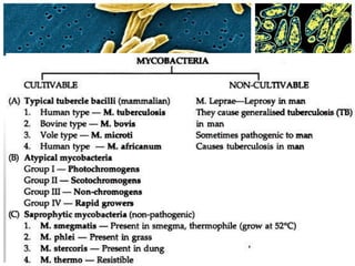
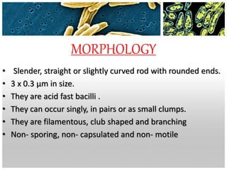
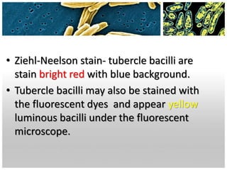
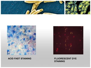
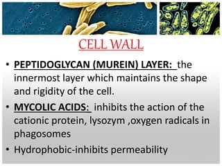
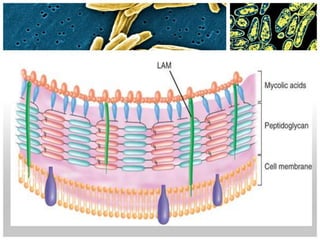
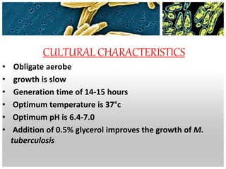
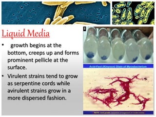
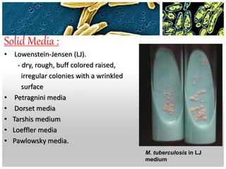
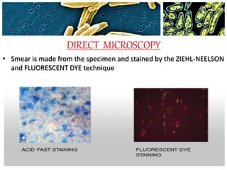
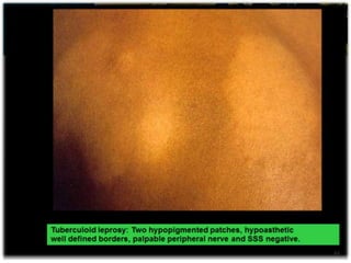
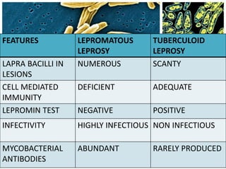
MYCOBACTERIUM TUBERCULOSIS
• M. tuberculosis was discovered in
1882 by Robert Koch.
MORPHOLOGY
• Slender, straight or slightly curved rod with rounded ends.
• 3 x 0.3 μm in size.
• They are acid fast bacilli .
• They can occur singly, in pairs or as small clumps.
• They are filamentous, club shaped and branching
• Non- sporing, non- capsulated and non- motile
Mycobacterium seen under electron microscope
• Ziehl-Neelson stain- tubercle bacilli are
stain bright red with blue background.
• Tubercle bacilli may also be stained with
the fluorescent dyes and appear yellow
luminous bacilli under the fluorescent
microscope.
ACID FAST STAINING FLUORESCENT DYE
STAINING
CELL WALL
• PEPTIDOGLYCAN (MUREIN) LAYER: the
innermost layer which maintains the shape
and rigidity of the cell.
• MYCOLIC ACIDS: inhibits the action of the
cationic protein, lysozym ,oxygen radicals in
phagosomes
• Hydrophobic-inhibits permeability
CULTURAL CHARACTERISTICS
• Obligate aerobe
• growth is slow
• Generation time of 14-15 hours
• Optimum temperature is 37°c
• Optimum pH is 6.4-7.0
• Addition of 0.5% glycerol improves the growth of M.
tuberculosis
Liquid Media
• growth begins at the
bottom, creeps up and forms
prominent pellicle at the
surface.
• Virulent strains tend to grow
as serpentine cords while
avirulent strains grow in a
more dispersed fashion.
Solid Media :
• Lowenstein-Jensen (LJ).
- dry, rough, buff colored raised,
irregular colonies with a wrinkled
surface
• Petragnini media
• Dorset media
• Tarshis medium
• Loeffler media
• Pawlowsky media.
M. tuberculosis in LJ
medium
RESISTANCE
• Not completely heat resistant, being killed at 60°C
for15-20 minutes.
• Resistant to:
1)5% phenol
2)15% sulphuric acid
3)3% nitric acid
4)4% Sodium hydroxide
• Sensitive to :
1)UV ray and sunlight
2) formaldehyde and glutaraldehyde.
3) 80% ethanol for 2 minutes
4)Iodine for 5minutes.
ANTIGENIC PROPERTIES
Two types:
• Cell wall (insoluble)
• Cytoplasmic antigens(soluble)
Cell wall antigens
They constitutes of lipids, proteins,
polysaccharides.
Lipids :
• The cell wall is rich in mycolic acid, which plays an
important role in pathogenesis.
• Mycolic acid is also responsible for the acid
fastness of the bacteria and cellular tissue
reactions of the body.
Proteins :
• Responsible for the tuberculin reaction.
• Induce delayed type hypersensitivity.
Polysaccharides :
• Their role in pathogenesis is not certain.
• They induce delayed type hypersensitivity.
Cytoplasmic antigens:
• Protein antigens used for bacterial typing
MODE OF TRANSMISSION
• infected droplets
• consumption infected milk
• inoculation leading to tonsillar or
intestinal tuberculosis
• trans-placental route
PHATHOGENESIS
 Tubercle bacilli do not contain or secrete a toxin
 Exact basis of their virulence is not understood, but
seems to be related to their ability to survive and
multiply in macrophages
They lyse the host cell, infect other macrophages and
sometimes disseminate to other parts of lung and
elsewhere in the body.
Humoral immunity appears to be irrelevant
The only specific immune mechanism effective is the
CELL MEDIATED IMMUNITY
 The key cell is the activated CD4+ helper T cell which
develops along two different paths: The Th1 and Th2
cells releasing cytokines such as interferon gamma,
interlukins 1 and 2,toxic necrosis factor alpha and
others
TH1 dependent cytokines activate macrophages, to
kill intracellular mycobacteria resulting in protective
immunity and
 TH2 cytokines induce delayed type hypersensitivity
(DTH) tissue destruction and progressive disease
0RAL MANIFESTATION
• include tongue, palate, buccal mucosa, lips,
gingiva and floor of the mouth.
• Irregular painful ulcer, diffuse inflammatory
lesions and fissures.
• Tuberculous osteomyelitis involve maxilla and
mandible
TUBERCULIN TEST
PRINCIPLE:
Delayed or type IV hypersensitivity
REAGENTS:
1. OLD TUBERCULIN (OT)
2. PURIFIED PROTEIN DERIVATIVE (PPD)
METHORD:
1.MANTOUX TEST
2.HEAF TEST
1. MANTOUX TEST
• 0.1 ml of PPD containing 5 TU is injected intradermally into
flexor aspect of forearm.
• A PPD dose of 1TU is used when extreme hypersensitivity is
suspected.
2. HEAF TEST
• This is done with a multiple puncture apparatus that pricks the
skin.
• A drop of undiluted PPD is spread on the area of skin .
• The multiple puncture apparatus is pressed against the skin.
RESULT:
• site of injection is examined after 48-72 hours
• Positive reaction:
• -induration of 10 mm diameter or more surrounded by
erythema at the site of inoculation
• Positive test only confirms past infection with tubercle bacilli
but does not indicate presence of active stage of the disease.
• The test is helpful in children under 5 years for indication of
active infection.
• The test becomes positive 4-6 weeks after infection or BCG
vaccination.
Laboratory diagnosis
• SPECIMEN COLLECTION
• DIRECT MICROSCOPY
• CULTURE
SPECIMEN COLLECTION
• Sputum
• Laryngeal swabs
• Bronchial washings
• Gastric lavage
Pulmonary
tuberculosis
• urine
Renal
tuberculosis
• CSF (develops a spider web clot
on standing)
Tuberculous
meningitis
•Aspirated
fluid
Bone and
joints
tuberculosis
•Biopsy of
tissue
Tissue
DIRECT MICROSCOPY
• Smear is made from the specimen and stained by the ZIEHL-NEELSON
and FLUORESCENT DYE technique
• To detect the bacilli microscopically, there should be at least 50,000
bacilli per ml of sputum.
• A negative report should not be given till at least 300 fields have been
examined.
• Grading of smears is done according to number of bacilli seenNo. of AFB seen in oil Immersion
field
Report
0/300 FIELDS AFB NOT SEEN
1-2/300 FIELDS DOUBTFUL REPEAT THE SMEAR
1-9/100 FIELDS 1+
1-9/10 FIELDS 2+
1-9/FIELD 3+
10 OR MORE/FIELD 4+
CULTURE
 LOWENSTEIN- JENSEN medium
 The culture media are incubated at 37 ̊C in the dark and in light
 Cultures are examined first after 4 days and then weekly till 8
weeks
 The tubercle bacilli usually grow in 2 to 8 weeks
 In a positive culture characteristic colonies
(dry, rough, raised, irregular colonies )
appears on the culture medium
TREATMENT
The antitubercular drugs:
bactericidal : RIFAMPICIN (R), ISONIAZID (H) ,
PYRAZINAMIDE(Z) , STREPTOMYCIN(S)
bacteriostatic: ETHAMBUTOL, CYCLOSERINE, THIACETAZONE
• Use multi-drug therapy
• 1st 2nd months ISONIAZID,RIFAMPICIN AND
PYRAZINAMIDE
• Next 4 months –ISONIAZID AND RIFAMPACIN
• If drugs are resistance, have to add
STREPTOMYCIN/ETHAMBUTOL
PROPYLAXIS DOSE AND ADMINISTRATION
BCG vaccine:
 vaccine is given intradermally in a dose of
0.1ml
 BCG vaccine should be given soon after birth /
any time during the first year of life.
39
Leprosy
Hansen 1868  Identified First
microorganism
40
Mycobacterium leprae
• Appears as slender, straight or curved rods
• Size is 1 – 8 µm x 0.2-0.5 µm
• Acid fast
• Live bacilli - appear as solid uniform structure.
• Dead bacilli- appear as fragmented with granules.
41
• Seen singly and in groups
• Intracellularly or lying free outside the
cells
• Frequently appears Parallel rows of bacilli
in the globi present a cigar like
appearance
• In tissue sections the bacilli are arranged
in clumps resembling cigarette ends 42
Cultivation
• Not possible in artificial medium
• Can be propagated in Foot pads of Mice (SHEPARD 1960)
• Granulomas develop at the site of inoculation.
• Nine banded armadillo highly susceptible.
• Identified in Chimpanzees too
• Average Generation time 12 -13 days.
• it may vary 8- 42 days.
43
Important Experimental
Animal
44
45
PATHOGENESIS
• A chronic granulomatous disease of humans
• Involves Skin, Peripheral nerves, Nasal mucosa,
capable of affecting any tissues and organs
• Incubation period is very long and variable.
• Sources of infection are nasal discharge and skin
lesions of patients.
• Prolonged close contact with patients is necessary
for transmission of the disease.
MODE OF TRANSMISSION
• Leprosy is a slow communicable disease and the
incubation period varies from 2 to 20 years
• Direct contact
• Maternofoetal transmission
• Milk of infected mother to infant.
Classification ( Madrid 1953)
1) Lepromatous
2) Tuberculoid
3) Dimorphus
4) Intermediate.
51
LEPROMATOUS LEPROSY
• Patient develops nodular skin lesions on face, ear lobes,
hands, feet and less commonly on trunk.
• Slow and symmetric thickening of peripheral nerves and
anaesthesia.
• As a result of loss of sensation nodular skin lesions ulcerate
with repeated trauma
• Skin lesions contain many macrophages packed with bacilli.
• Lepra bacilli are present in large number in skin lesions and
in mucosa of nose, mouth and upper respiratory tract.
52
Tuberculoid leprosy
• Skin lesions are few and consist of non elevated
hypo or hyper pigmented macular patches
involving the face limbs and trunk.
• Local nerve trunks are involved in early stages and
gradually extends into bigger nerve trunks which
become thickened, hard or tender.
• This may lead to deformities of hands and feet.
54
FEATURES LEPROMATOUS
LEPROSY
TUBERCULOID
LEPROSY
LAPRA BACILLI IN
LESIONS
NUMEROUS SCANTY
CELL MEDIATED
IMMUNITY
DEFICIENT ADEQUATE
LEPROMIN TEST NEGATIVE POSITIVE
INFECTIVITY HIGHLY INFECTIOUS NON INFECTIOUS
MYCOBACTERIAL
ANTIBODIES
ABUNDANT RARELY PRODUCED
ORAL MANIFESTATIONS
• develops on the tongue ,lip or hard palate.
• lesions contains tumour like mass called lepromas.
• intraoral nodules tends to ulcerate.
• In the facial regions, skeletal changes such as atrophy of
anterior nasal spine, saddle nose and pre-maxillary bone
recession are frequently found with or without tooth loss.
WHO classification
• Two Groups
1. Paucibacillary
2. Multibacillary
Paucibacillary (PB): the number of M. leprae in
the body is small (less than 1 million) and a skin
smear test is negative. The patient presents fewer
skin lesions. all cases of tuberculoid leprosy are PB.
58
2 Multibacillary
• M. leprae can multiply in the body and thus
present in high numbers.
• The bacillus has likely spread to almost all areas of
skin and peripheral nerves.
• A skin smear test is positive and the patient
presents more than five skin lesions.
• all cases of lepromatous type
59
Immunity
• Innate Immunity exists in human beings
• Infection induces Humoral & Cell mediated
immunity
• CMI destroys the bacilli and determines the
recovery.
• Good CMI -Tuberculoid leprosy.
• Deficient CMI - lepromatous type
• Patient exhibits delayed hypersensitivity to the
lepra bacillus protein 60
Lepromin Test
• Lepromin test first described by MITSUDA in
1919.
• lepromin was boiled emulsified lepromatous
tissue –rich in lepra bacilli
• lepromins used as antigens can be of
Human origin - lepromin H
Armadillo origin – lepromin A
61
1.INTEGRAL LEPROMIN:
• Mitsuda’s crude antigen is called the integral
antigen.
• It is now increasingly being prepared from
armadillo-derived M.leprae (lepromin –A)
2.BACILLARY LEPROMIN:
• This contains more of bacillary components with
less of tissue.
• An antigen commonly employed is Dharmendra
antigen.
PROCEDURE
• Intradermal injection of 0.1 ml of lepromin
• Response is biphasic
• Events in the reaction , Biphasic reaction
1. Early reaction of Fernandez
• Induration and erythema developing in 24 – 48 hrs
• remains for 3 – 5 days,
• serous exudate with lymphocytic infiltration
• analogous to tuberculin reaction. 63
2.Late reaction of Mitsuda
• reaction occurs after 1 – 2 weeks.
prominent after 4 weeks
• Consists of indurated skin lesion which may
ulcerate
• Infiltration with Lymphocytes , Epitheloid
cells, and giant cells,
• Positive reaction indicates resistance to
leprosy
64
USES
• 1. CLASSIFICATION OF LEPROSY --:
• positive- tuberculoid
• negative -lepromatous
• 2. ASSESSMENT OF PROGNOSIS:
• Positive-good prognosis
• Negative – bad prognosis
• 3.ASSESSMENT OF RESISTANCE
• Resistance is indicated by positive lepromin test
65
LABORATORY DIAGNOSIS
OVERVIEW :
1. Specimens
2. Acid fast staining
3. Skin and nerve biopsy
4. Lepromin test
66
SPECIMEN
Nasal mucosa, skin lesions, ear lobules.
Skin and nerve biopsy.
67
• Specimens from skin are collected by slit
and scrape method.
• Samples are collected from the edges of
the lesion.
• Skin is cleaned, pinched up tight ,cut
about 5mm long is made with a scalpel
deep enough into the infiltrated layers.
• After wiping off blood or lymph that may have exuded the
blade of the scalpel is turned at right angle to the cut.
• The bottom and the sides of the slit are scraped with the
point of the blade so as to obtain a little tissue pulp which
is smeared uniformly on a slide.
• About six different areas should be sampled, including the
skin over the buttocks, chin, cheek, forehead and ears.
• Smears from the nose are made by scraping a little
material from the nasal septum with a small blade knife.
• Skin biopsy is collected from active edges of the patches
and nerve biopsy from thickened nerves.
ACID FAST STAINING
• stained by ZIEHL-NEELSEN method
using 5% sulphuric acid as
decolourising agent.
• Acid fast bacilli arranged in parallel
bundles within macrophages (lepra
cells) confirms lepromatous
leprosy.
• Viable bacilli stain uniformly and
the dead bacilli are fragmented
irregular or granular.
69
SKIN AND NERVE BIOPSY
it is done when AFB cannot be demonstrated in direct smear.
• Skin biopsy is also useful in diagnosis and accurate
classification of leprosy lesion.
70
LEPROMIN TEST
• Not a diagnostic test but is used to assess the
resistance of patient to M.leprae infection.
• Delayed type of hypersensitive reaction.
71
Treatment of Leprosy
MULTIDRUG REGIME
• For paucibacillary cases:
Rifampicin 600mg/once a month
Dapsone 100mg/daily for 6 months
• For multibacillary
Rifampicin 600 mg / once month
Dapsone 100/day
Clofazimine 50 mg/daily for 2 years
until skin smears are negative
Other Drugs for Leprosy
1.Ethionamide
2.Prothionamide. 72
Prevention Of Leprosy
• Early Diagnosis and treatment.
• Health education
• active surveillance
• vaccination
Field trails with different vaccine
1. BCG
2.Armadillo-derived killed M.laprae
3.BCG + killed leprae bacilli
4. ICRC bacillus
5.Mycobacterium w 74
Dr.T.V.Rao MD 75
Mycobacterium tuberculosis

Mycobacterium tuberculosis

  • 3.
    MYCOBACTERIUM TUBERCULOSIS • M.tuberculosis was discovered in 1882 by Robert Koch.
  • 5.
    MORPHOLOGY • Slender, straightor slightly curved rod with rounded ends. • 3 x 0.3 μm in size. • They are acid fast bacilli . • They can occur singly, in pairs or as small clumps. • They are filamentous, club shaped and branching • Non- sporing, non- capsulated and non- motile
  • 6.
    Mycobacterium seen underelectron microscope
  • 7.
    • Ziehl-Neelson stain-tubercle bacilli are stain bright red with blue background. • Tubercle bacilli may also be stained with the fluorescent dyes and appear yellow luminous bacilli under the fluorescent microscope.
  • 8.
    ACID FAST STAININGFLUORESCENT DYE STAINING
  • 9.
    CELL WALL • PEPTIDOGLYCAN(MUREIN) LAYER: the innermost layer which maintains the shape and rigidity of the cell. • MYCOLIC ACIDS: inhibits the action of the cationic protein, lysozym ,oxygen radicals in phagosomes • Hydrophobic-inhibits permeability
  • 11.
    CULTURAL CHARACTERISTICS • Obligateaerobe • growth is slow • Generation time of 14-15 hours • Optimum temperature is 37°c • Optimum pH is 6.4-7.0 • Addition of 0.5% glycerol improves the growth of M. tuberculosis
  • 12.
    Liquid Media • growthbegins at the bottom, creeps up and forms prominent pellicle at the surface. • Virulent strains tend to grow as serpentine cords while avirulent strains grow in a more dispersed fashion.
  • 13.
    Solid Media : •Lowenstein-Jensen (LJ). - dry, rough, buff colored raised, irregular colonies with a wrinkled surface • Petragnini media • Dorset media • Tarshis medium • Loeffler media • Pawlowsky media. M. tuberculosis in LJ medium
  • 14.
    RESISTANCE • Not completelyheat resistant, being killed at 60°C for15-20 minutes. • Resistant to: 1)5% phenol 2)15% sulphuric acid 3)3% nitric acid 4)4% Sodium hydroxide
  • 15.
    • Sensitive to: 1)UV ray and sunlight 2) formaldehyde and glutaraldehyde. 3) 80% ethanol for 2 minutes 4)Iodine for 5minutes.
  • 16.
    ANTIGENIC PROPERTIES Two types: •Cell wall (insoluble) • Cytoplasmic antigens(soluble)
  • 17.
    Cell wall antigens Theyconstitutes of lipids, proteins, polysaccharides. Lipids : • The cell wall is rich in mycolic acid, which plays an important role in pathogenesis. • Mycolic acid is also responsible for the acid fastness of the bacteria and cellular tissue reactions of the body.
  • 18.
    Proteins : • Responsiblefor the tuberculin reaction. • Induce delayed type hypersensitivity. Polysaccharides : • Their role in pathogenesis is not certain. • They induce delayed type hypersensitivity. Cytoplasmic antigens: • Protein antigens used for bacterial typing
  • 19.
    MODE OF TRANSMISSION •infected droplets • consumption infected milk • inoculation leading to tonsillar or intestinal tuberculosis • trans-placental route
  • 20.
    PHATHOGENESIS  Tubercle bacillido not contain or secrete a toxin  Exact basis of their virulence is not understood, but seems to be related to their ability to survive and multiply in macrophages They lyse the host cell, infect other macrophages and sometimes disseminate to other parts of lung and elsewhere in the body. Humoral immunity appears to be irrelevant The only specific immune mechanism effective is the CELL MEDIATED IMMUNITY
  • 21.
     The keycell is the activated CD4+ helper T cell which develops along two different paths: The Th1 and Th2 cells releasing cytokines such as interferon gamma, interlukins 1 and 2,toxic necrosis factor alpha and others TH1 dependent cytokines activate macrophages, to kill intracellular mycobacteria resulting in protective immunity and  TH2 cytokines induce delayed type hypersensitivity (DTH) tissue destruction and progressive disease
  • 24.
    0RAL MANIFESTATION • includetongue, palate, buccal mucosa, lips, gingiva and floor of the mouth. • Irregular painful ulcer, diffuse inflammatory lesions and fissures. • Tuberculous osteomyelitis involve maxilla and mandible
  • 26.
    TUBERCULIN TEST PRINCIPLE: Delayed ortype IV hypersensitivity REAGENTS: 1. OLD TUBERCULIN (OT) 2. PURIFIED PROTEIN DERIVATIVE (PPD) METHORD: 1.MANTOUX TEST 2.HEAF TEST
  • 27.
    1. MANTOUX TEST •0.1 ml of PPD containing 5 TU is injected intradermally into flexor aspect of forearm. • A PPD dose of 1TU is used when extreme hypersensitivity is suspected. 2. HEAF TEST • This is done with a multiple puncture apparatus that pricks the skin. • A drop of undiluted PPD is spread on the area of skin . • The multiple puncture apparatus is pressed against the skin.
  • 28.
    RESULT: • site ofinjection is examined after 48-72 hours • Positive reaction: • -induration of 10 mm diameter or more surrounded by erythema at the site of inoculation • Positive test only confirms past infection with tubercle bacilli but does not indicate presence of active stage of the disease. • The test is helpful in children under 5 years for indication of active infection. • The test becomes positive 4-6 weeks after infection or BCG vaccination.
  • 30.
    Laboratory diagnosis • SPECIMENCOLLECTION • DIRECT MICROSCOPY • CULTURE
  • 31.
    SPECIMEN COLLECTION • Sputum •Laryngeal swabs • Bronchial washings • Gastric lavage Pulmonary tuberculosis • urine Renal tuberculosis • CSF (develops a spider web clot on standing) Tuberculous meningitis
  • 32.
  • 33.
    DIRECT MICROSCOPY • Smearis made from the specimen and stained by the ZIEHL-NEELSON and FLUORESCENT DYE technique
  • 34.
    • To detectthe bacilli microscopically, there should be at least 50,000 bacilli per ml of sputum. • A negative report should not be given till at least 300 fields have been examined. • Grading of smears is done according to number of bacilli seenNo. of AFB seen in oil Immersion field Report 0/300 FIELDS AFB NOT SEEN 1-2/300 FIELDS DOUBTFUL REPEAT THE SMEAR 1-9/100 FIELDS 1+ 1-9/10 FIELDS 2+ 1-9/FIELD 3+ 10 OR MORE/FIELD 4+
  • 35.
    CULTURE  LOWENSTEIN- JENSENmedium  The culture media are incubated at 37 ̊C in the dark and in light  Cultures are examined first after 4 days and then weekly till 8 weeks  The tubercle bacilli usually grow in 2 to 8 weeks  In a positive culture characteristic colonies (dry, rough, raised, irregular colonies ) appears on the culture medium
  • 36.
    TREATMENT The antitubercular drugs: bactericidal: RIFAMPICIN (R), ISONIAZID (H) , PYRAZINAMIDE(Z) , STREPTOMYCIN(S) bacteriostatic: ETHAMBUTOL, CYCLOSERINE, THIACETAZONE • Use multi-drug therapy • 1st 2nd months ISONIAZID,RIFAMPICIN AND PYRAZINAMIDE • Next 4 months –ISONIAZID AND RIFAMPACIN • If drugs are resistance, have to add STREPTOMYCIN/ETHAMBUTOL
  • 37.
    PROPYLAXIS DOSE ANDADMINISTRATION BCG vaccine:  vaccine is given intradermally in a dose of 0.1ml  BCG vaccine should be given soon after birth / any time during the first year of life.
  • 39.
  • 40.
    Leprosy Hansen 1868 Identified First microorganism 40
  • 41.
    Mycobacterium leprae • Appearsas slender, straight or curved rods • Size is 1 – 8 µm x 0.2-0.5 µm • Acid fast • Live bacilli - appear as solid uniform structure. • Dead bacilli- appear as fragmented with granules. 41
  • 42.
    • Seen singlyand in groups • Intracellularly or lying free outside the cells • Frequently appears Parallel rows of bacilli in the globi present a cigar like appearance • In tissue sections the bacilli are arranged in clumps resembling cigarette ends 42
  • 43.
    Cultivation • Not possiblein artificial medium • Can be propagated in Foot pads of Mice (SHEPARD 1960) • Granulomas develop at the site of inoculation. • Nine banded armadillo highly susceptible. • Identified in Chimpanzees too • Average Generation time 12 -13 days. • it may vary 8- 42 days. 43
  • 44.
  • 45.
  • 46.
    PATHOGENESIS • A chronicgranulomatous disease of humans • Involves Skin, Peripheral nerves, Nasal mucosa, capable of affecting any tissues and organs • Incubation period is very long and variable. • Sources of infection are nasal discharge and skin lesions of patients. • Prolonged close contact with patients is necessary for transmission of the disease.
  • 50.
    MODE OF TRANSMISSION •Leprosy is a slow communicable disease and the incubation period varies from 2 to 20 years • Direct contact • Maternofoetal transmission • Milk of infected mother to infant.
  • 51.
    Classification ( Madrid1953) 1) Lepromatous 2) Tuberculoid 3) Dimorphus 4) Intermediate. 51
  • 52.
    LEPROMATOUS LEPROSY • Patientdevelops nodular skin lesions on face, ear lobes, hands, feet and less commonly on trunk. • Slow and symmetric thickening of peripheral nerves and anaesthesia. • As a result of loss of sensation nodular skin lesions ulcerate with repeated trauma • Skin lesions contain many macrophages packed with bacilli. • Lepra bacilli are present in large number in skin lesions and in mucosa of nose, mouth and upper respiratory tract. 52
  • 54.
    Tuberculoid leprosy • Skinlesions are few and consist of non elevated hypo or hyper pigmented macular patches involving the face limbs and trunk. • Local nerve trunks are involved in early stages and gradually extends into bigger nerve trunks which become thickened, hard or tender. • This may lead to deformities of hands and feet. 54
  • 56.
    FEATURES LEPROMATOUS LEPROSY TUBERCULOID LEPROSY LAPRA BACILLIIN LESIONS NUMEROUS SCANTY CELL MEDIATED IMMUNITY DEFICIENT ADEQUATE LEPROMIN TEST NEGATIVE POSITIVE INFECTIVITY HIGHLY INFECTIOUS NON INFECTIOUS MYCOBACTERIAL ANTIBODIES ABUNDANT RARELY PRODUCED
  • 57.
    ORAL MANIFESTATIONS • developson the tongue ,lip or hard palate. • lesions contains tumour like mass called lepromas. • intraoral nodules tends to ulcerate. • In the facial regions, skeletal changes such as atrophy of anterior nasal spine, saddle nose and pre-maxillary bone recession are frequently found with or without tooth loss.
  • 58.
    WHO classification • TwoGroups 1. Paucibacillary 2. Multibacillary Paucibacillary (PB): the number of M. leprae in the body is small (less than 1 million) and a skin smear test is negative. The patient presents fewer skin lesions. all cases of tuberculoid leprosy are PB. 58
  • 59.
    2 Multibacillary • M.leprae can multiply in the body and thus present in high numbers. • The bacillus has likely spread to almost all areas of skin and peripheral nerves. • A skin smear test is positive and the patient presents more than five skin lesions. • all cases of lepromatous type 59
  • 60.
    Immunity • Innate Immunityexists in human beings • Infection induces Humoral & Cell mediated immunity • CMI destroys the bacilli and determines the recovery. • Good CMI -Tuberculoid leprosy. • Deficient CMI - lepromatous type • Patient exhibits delayed hypersensitivity to the lepra bacillus protein 60
  • 61.
    Lepromin Test • Lepromintest first described by MITSUDA in 1919. • lepromin was boiled emulsified lepromatous tissue –rich in lepra bacilli • lepromins used as antigens can be of Human origin - lepromin H Armadillo origin – lepromin A 61
  • 62.
    1.INTEGRAL LEPROMIN: • Mitsuda’scrude antigen is called the integral antigen. • It is now increasingly being prepared from armadillo-derived M.leprae (lepromin –A) 2.BACILLARY LEPROMIN: • This contains more of bacillary components with less of tissue. • An antigen commonly employed is Dharmendra antigen.
  • 63.
    PROCEDURE • Intradermal injectionof 0.1 ml of lepromin • Response is biphasic • Events in the reaction , Biphasic reaction 1. Early reaction of Fernandez • Induration and erythema developing in 24 – 48 hrs • remains for 3 – 5 days, • serous exudate with lymphocytic infiltration • analogous to tuberculin reaction. 63
  • 64.
    2.Late reaction ofMitsuda • reaction occurs after 1 – 2 weeks. prominent after 4 weeks • Consists of indurated skin lesion which may ulcerate • Infiltration with Lymphocytes , Epitheloid cells, and giant cells, • Positive reaction indicates resistance to leprosy 64
  • 65.
    USES • 1. CLASSIFICATIONOF LEPROSY --: • positive- tuberculoid • negative -lepromatous • 2. ASSESSMENT OF PROGNOSIS: • Positive-good prognosis • Negative – bad prognosis • 3.ASSESSMENT OF RESISTANCE • Resistance is indicated by positive lepromin test 65
  • 66.
    LABORATORY DIAGNOSIS OVERVIEW : 1.Specimens 2. Acid fast staining 3. Skin and nerve biopsy 4. Lepromin test 66
  • 67.
    SPECIMEN Nasal mucosa, skinlesions, ear lobules. Skin and nerve biopsy. 67 • Specimens from skin are collected by slit and scrape method. • Samples are collected from the edges of the lesion. • Skin is cleaned, pinched up tight ,cut about 5mm long is made with a scalpel deep enough into the infiltrated layers.
  • 68.
    • After wipingoff blood or lymph that may have exuded the blade of the scalpel is turned at right angle to the cut. • The bottom and the sides of the slit are scraped with the point of the blade so as to obtain a little tissue pulp which is smeared uniformly on a slide. • About six different areas should be sampled, including the skin over the buttocks, chin, cheek, forehead and ears. • Smears from the nose are made by scraping a little material from the nasal septum with a small blade knife. • Skin biopsy is collected from active edges of the patches and nerve biopsy from thickened nerves.
  • 69.
    ACID FAST STAINING •stained by ZIEHL-NEELSEN method using 5% sulphuric acid as decolourising agent. • Acid fast bacilli arranged in parallel bundles within macrophages (lepra cells) confirms lepromatous leprosy. • Viable bacilli stain uniformly and the dead bacilli are fragmented irregular or granular. 69
  • 70.
    SKIN AND NERVEBIOPSY it is done when AFB cannot be demonstrated in direct smear. • Skin biopsy is also useful in diagnosis and accurate classification of leprosy lesion. 70
  • 71.
    LEPROMIN TEST • Nota diagnostic test but is used to assess the resistance of patient to M.leprae infection. • Delayed type of hypersensitive reaction. 71
  • 72.
    Treatment of Leprosy MULTIDRUGREGIME • For paucibacillary cases: Rifampicin 600mg/once a month Dapsone 100mg/daily for 6 months • For multibacillary Rifampicin 600 mg / once month Dapsone 100/day Clofazimine 50 mg/daily for 2 years until skin smears are negative Other Drugs for Leprosy 1.Ethionamide 2.Prothionamide. 72
  • 74.
    Prevention Of Leprosy •Early Diagnosis and treatment. • Health education • active surveillance • vaccination Field trails with different vaccine 1. BCG 2.Armadillo-derived killed M.laprae 3.BCG + killed leprae bacilli 4. ICRC bacillus 5.Mycobacterium w 74
  • 75.