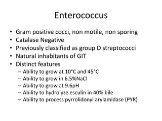
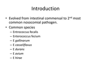
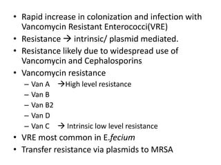
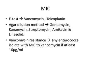
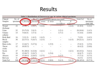
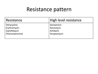
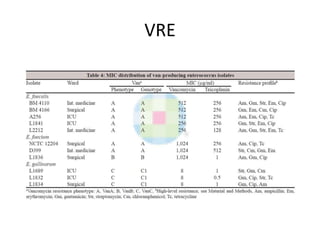
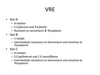
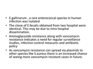
Enterococci are Gram-positive cocci that are natural inhabitants of the gastrointestinal tract. They have become important nosocomial pathogens due to their intrinsic and acquired antibiotic resistance. This study found that Enterococcus faecalis was the most common species isolated from clinical specimens in two Saudi hospitals. Many isolates showed resistance to tetracycline, ciprofloxacin, and chloramphenicol. Vancomycin resistance was observed in 3.9% of isolates, with the VanA phenotype being most common. Pulsed-field gel electrophoresis identified identical clones of E. faecalis isolated from different hospital wards, suggesting intra-hospital transmission. The high resistance rates indicate a need for improved infection control and antibiotic steward
Enterococcus
• Gram positive cocci, non motile, non sporing
• Catalase Negative
• Previously classified as group D streptococci
• Natural inhabitants of GIT
• Distinct features
– Ability to grow at 10°C and 45°C
– Ability to grow in 6.5%NaCl
– Ability to grow at 9.6pH
– Ability to hydrolyze esculin in 40% bile
– Ability to process pyrrolidonyl arylamidase (PYR)
3.
Presumptive identification of
Streptococci & Enterococcci
Hemolysis
Hippurate
Bacitracin
Organism
6.5%NaCl
Optochin
solubility
esculin
Camp
PYR
Bile
Bile
SXT
Lap
Group A ß S R - - + + - - R -
streptococci
Group B ß, none R R + + + - - V R -
streptococci
Group C,G & F ß V S - - + - - - R -
streptococci
Group D α, ß, R R - V + + + + R -
Enterococcci none
Other Group D α, R S - - + - + - R -
streptococci none
Viridans α, none V S - V + - V - R -
streptococci
Pneumococci α V S - - + - - - S +
4.
Classification (based onphenotypic characters in
clinical isolates)
Group 1 Group 3 Group 5
E.avium E.dispar E.columbae
E.gilvus E.durans E.canis
E.malodartus E.hirae E.moraviensis
E.pallens E.ratti
E. pseudoavium E.villorum
E.raffinosus
E.sacchrolyticus
Group 2 Group 4
E.fecalis E.asini
E.fecium E.cecorum
E.casseliflavus E.sulfures
E.gallinarum E.phoeniculicola
E.mundtii Enterococcus sp
E.hemoperoxidus
Enterococcus sp
Koneman textbook of diagnostic microbiology
5.
MANNITOL
Species
PYRUVATE
RAFFINOSE
SORBOSE
ARABINOS
GLUCOSE
SUCROSE
6.5%Nacl
SORBITOL
MGP
10°C
45°C
ADH
PYR
LAP
HIP
E
E.fecalis + + + + + + + + + - - + - + + -
E.fecium + + + + + + - + + - + V V + - -
E.casselifl + NA + + + + - + + - + V + + V +
avus
E.gallinar + + + + + + + + + - + - + + - +
um
E.durans + NA + + + + V + - - - - - - - -
E.hirae + + + + + + - + - - - - + + - -
E.avium + NA + + + - V + + + + + - + + V
LAP-leucine aminopeptidase, PYR- pyrrolidonyl arylamidase, ADH- arginine dihydrolase, HIP – hippurate, MGP-
methyl α-D-glucopyranoside
Intrinsic resistance
• Resistanceto ß-lactams Low affinity of
penicillin binding protein
• Low level resistance to aminoglycosides
Low uptake of these agents
Acquired resistance (Cont’d)
•Chloramphenicol mediated by chloramphenicol acetyl
transferase, prevents binding to 50S ribosome
• Erythromycin resistance occurs as a part of Macrolide-
Lincosamide-streptogramin B resistance phenotype.
– Transferred by resistance deretminant ermB carried on Tn917.
– Leads to methylation of adenosine residue in 23S rRNA.
– Also confers high level resistance to clindamycin
• Tetracycline resistance transferred by plasmid pAMα1
– Promotes active efflux of tetracycline from cells
– Protects ribosomes from inhibition by tetracycline
• Aminoglycosides
– Cell membrane bound inactivating enzymes
– Decreased affinity of ribosomal proteins to aminoglycoside (mutation)
– Decreased uptake of drug
12.
Vancomycin resistance
• Resistanceto glycopeptides is due to alteration of peptidoglycan precursor
D-Ala –D- Ala to D-Ala – D-Lactate / D-Ala-D-Ser
• Genes involved are VanS/VanR
• VanS gene activates D-Lac/D-Ser peptidoglycan precursor
• D-Lac has 1000 times less affinity to vancomycin
• D-Ser exhibits 7fold less affinity to vancomycin
• Six gene clusters have been identified
– VanA, VanB, VanC, VanD, VanE & VanG
• VanA is acquired by Transposon Tn1546
• vanB is acquired by Transposon Tn1547/Tn5382
• Genetic material transferred via conjugation involving a pheromone
induced system.
13.
Prevalence and antimicrobialresistance
pattern of multidrug –resistant
enterococci isolated from clinical
specimens
MM Salem-Bekhit et al
Indian Journal Of Medical Microbiology
(2012) 30(1): 44-51
14.
Introduction
• Evolved fromintestinal commensal to 2nd most
common nosocomial pathogen.
• Common species
– Entrococcus fecalis
– Enterococcus fecium
– E gallinarum
– E casseliflavus
– E durans
– E avium
– E hirae
15.
• Rapid increasein colonization and infection with
Vancomycin Resistant Enterococci(VRE)
• Resistance intrinsic/ plasmid mediated.
• Resistance likely due to widespread use of
Vancomycin and Cephalosporins
• Vancomycin resistance
– Van A High level resistance
– Van B
– Van B2
– Van D
– Van C Intrinsic low level resistance
• VRE most common in E.fecium
• Transfer resistance via plasmids to MRSA
16.
Materials & Methods
Distribution of samples
• Period of study January 2009 – March 2010
• Place of study
• King Khaled University Hospital (140samples)
Urine 32
• King Saud Medical City Hospital (100 Samples)
Blood 30
• Ethical Burns
committee approval 17
• Approval from both hospital ethical committee & from Saudi ministry
Throat swabs
of Health 12
• Isolation
Bed sore 11
– Urine, sterile body fluids & wounds
Devices 14
• Trypticase soy agar with 5% sheep blood
– Stool samples
Stool samples 47
• Inoculated into enterococcal37
Pus broth , incubated overnight at 35°C
• Subcultured to BHIA with 6µg vancomycin/ml & BHIA without
Body fluids
Vancomycin 16
Wound discharge 24
17.
• Presumptive identification
– Growth characteristics on blood agar
– Gram staining morphology,
– Catalase reaction,
– Ability to grow in 6.5%Nacl,
– Bile esculin hydrolysis &
– Biochemicals using API Strep system.
• Enterococcal samples stored in 16%glycerol at
-70°C
MIC
• E-test Vancomycin , Teicoplanin
• Agar dilution method Gentamycin,
Kanamycin, Streptomycin, Amikacin &
Linezolid.
• Vancomycin resistance any enterococcal
isolate with MIC to vancomycin if atleast
16µg/ml
20.
ß-Lactamase production &inhibition
tests
• Nitrocefin 5µl
• Amoxicillin-clavulanic acid, Ampicillin-
sulbactum on disc-agar diffusion method.
21.
Further analysis
• DNAisolation
• Detection of vancomycin determinants
– Denaturation at 94°C for 3 min
– Anneling at 60°C for 45seconds
– Extension at 72°C for 1min
– Final extension at 72°C for 2min
– Amplicons analysed by electrophoresis on 1% agarose
gel containing ethidium bromide
• Restriction fragment length analysis by pulse field
gel electrophoresis
VRE
• Van A
– 8 isolates
– 5 E.faecium and 3 E.fecalis
– Resistant to vancomycin & Teicoplanin
• Van B
– 1 isolate
– Intermediate resistance to Vancomycin and sensitive to
Teicoplanin
• Van C
– 4 isolates
– 3 E.gallinarum and 1 E.casseliflavus
– Intermediate resistance to Vancomycin and sensitive to
Teicoplanin
27.
PGFE of VRE
•8 isolates tested
• 5 isolates had identical profile
• Isolates obtained from patients admitted in
different wards in same hospital
28.
Discussion
• E.fecalis wasthe predominant pathogen
• Many isolates were resistant to Tetracycline, Ciprofloxacin &
Chloramphenicol
• Resistance to Erythromycin was lower compared to other countries
• Nosocomial VRE reported is 3.9% (0.3% in 1989 – 11% in 1996)
• Isolates of VRE in this study were from immunocompromised
patients or with h/o Nosocomial infection
• Vancomycin resistant phenotype Van A was predominant and
resulted in high level resistance
• Van B phenotype showed moderate vancomycin resistance
• Being the mainstay of treatment, ampicillin resistance causes
concern
29.
• E.gallinarum ,a rare enterococcal species in human
infection was isolated
• The clone of E.fecalis obtained from two hospital were
identical. This may be due to intra hospital
dissemination.
• Aminoglycoside resistance along with vancomycin
resistance indicates a need for regular surveillance
studies, infection control measures and antibiotic
policy
• As vancomycin resistance can spread via plasmids to
other species like S.aureus there is an increased chance
of seeing more vancomycin resistant cases in future.
30.
REFERENCES
• Koneman TextbookOf Diagnostic Microbiology
• Kater fisher et al . The ecology, epidemiology and
virulence of enterococcus Microbiology(2009),
155,1749-1757
• PM Giridhara upadhyaya et al. Review of
virulence factors of enterococcus: an emerging
nosocomial pathogen IJMM (2009) 27(4): 301-5
• Barbara E.Murray. The life and times of
enterococcus Clinical microbiology reviews jan
1990. p 46-65