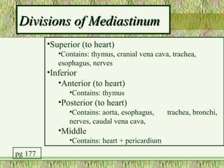
The Thoracic Cavity

   Boundaries of and Structures Within
Remove frame


       Body Cavities
     • Dorsal body cavity
     • Ventral body cavity
          – Thoracic
                • 2 Pleural
                • Mediastinum
          – Divided by Diaphragm
          – Abdominopelvic
                • Abdominal
                • Pelvic


   www.newworldencyclopedia.org/entry/Body_cavity
Serous membrane = Serosa
• Simple squamous epithelium + areolar connective tissue
• 2 Layers
   – Outer layer = PARIETAL serosa
   – Inner layer = VISCERAL serosa
• Between them = Serous Cavity containing Serous Fluid
   – Serous fluid is blood filtrate + secretions by 2 layers of membrane
   – Allows movement of organs with reduced friction
• Types of Serous Membranes
   – Pleural = surrounds lungs
   – Pericardium = surrounds heart, slightly modified
   – Peritoneal = surrounds some abdominal organs
Pleural Cavities
 • Surround the lungs
 • Pleural fluid secreted by pleural
   membranes
    – Holds layers together
    – Reduces friction of organs
 • Benefit of Compartmentalization


                                       pg 159
Pleural Cavities

• 2 Layers
  – Visceral pleura (inner)
     • root of lungs marks transition
     • external surface of lungs
  – Parietal pleura (outer)
     • inner surface of thoracic wall
     • superior surface of diaphragm
     • lateral surface of mediastinum

                                        pg 161
Pleural Abnormalities

• Pleural Effusion
   – Excess fluid in the pleural cavity
   – More than 20X
      • Usually less than 1 ml of fluid
• Pneumothorax
   – Air located in pleural space


                                          Pg 238
Divisions of Mediastinum
         •Superior (to heart)
            •Contains: thymus, cranial vena cava, trachea,
            esophagus, nerves
         •Inferior
            •Anterior (to heart)
               •Contains: thymus
            •Posterior (to heart)
               •Contains: aorta, esophagus,     trachea, bronchi,
               nerves, caudal vena cava,
            •Middle
               •Contains: heart + pericardium
pg 177
Boundaries of Mediastinum
• Lateral
   – parietal pleura of lungs
• Anterior
   – ventral parietal pleura
• Posterior
   – dorsal parietal pleura
• Superior
   – dome of the neck
• Inferior
   – diaphragmatic pleura       pg 159
Respiratory Tract
• Upper Respiratory Tract
  – Superior to Larynx


• Lower Respiratory Tract
  –   Larynx
  –   Trachea
  –   Primary Bronchi
  –   Secondary Bronchi
  –   Rest of Bronchial Tree                pg 992
  –   Lungs
                                            pg 168
Trachea = windpipe

•   Starts at Larynx and travels through mediastinum
•   Located Anterior to Esophagus
•   Trachea terminates into 2 primary bronchi entering lungs
•   Walls contain 16-20 “C” shaped rings Hyaline Cartilage
•   Trachealis Muscle (smooth muscle and soft CT)
•   Layers (deep to superficial)
    – Mucosa = Ciliated Psuedostratified Epithelium
    – Submucosa- contains seromucous glands
    – Adventitia – made of connective tissue, contains cartilage rings

                                                                         pg 966
Bronchial Tree
• Primary (main) Bronchi
   –   Bifurcation of trachea
   –   Basically the same structure
   –   Cartilage rings
   –   Posterior to pulmonary vessels
   –   Right is wider, vertical, shorter
• Secondary (lobar) Bronchi
   – Each primary bronchi divides
   – Same structure as primary bronchi
   – Right lung has 3, Left has 2
• Tertiary (segmental) Bronchi
• Up to 23 divisions
                                           pg 168
Bronchial Tree (continued)
• Bronchioles
  – further divisions, < 1 mm diameter
• Terminal Bronchioles
  – further divisions, 0.5 mm diameter
• Respiratory Zone
  – Respiratory Bronchioles
  – Alveolar Ducts
  – Alveolar Sacs
     • Terminal bunches of Alveoli
     • Respiratory exchange chamber
     • Among alveoli are blood vessels, nerves, lymphatics
                                       www.nlm.nih.gov/.../ency/imagepages/1103.htm
Respiratory Zone (continued)
 • Lining the Walls of Alveoli
    – Respiratory Membrane
       • Type I cells = simple squamous epithelial cells
       • Basal lamina and fine areolar CT
       • Covered with capillaries and elastic fibers
    – Type II cells = cuboidal epithelial cells
       • Secrete fluid containing surfactant
    – Dust Cells (macrophages)
 • Gas exchange
    – Oxygen into blood
    – Carbon Dioxide into alveoli
Throughout Bronchial Tree

• Psuedostratified columnar changes to simple columnar to
  simple cuboidal
• Cartilage rings replaced by cartilage plates once bronchi
  enter the lungs
• Smooth muscle and Elastic fibers remain important
• In Bronchioles
   – Ciliated mucosa disappears, replaced by macrophages in alveoli
   – Cartilage disappears
   – Smooth muscle forms bands around smallest bronchi and
     bronchioles (not found around alveoli)
LUNGS (continued)
•   Located in Pleural Compartments
•   Lateral to Mediastinum
•   Location
     – Apex posterior to clavicle
     – Base lays on Diaphragm
     – Costal Surface = Ant, Lat, Post surfaces contact ribs
•   Left Lung = 2 lobes
     –   Upper
     –   Lower
     –   Oblique Fissure
     –   Cardiac Notch
•   Right Lung = 3 lobes
     –   Upper
     –   Middle
     –   Lower
     –   Oblique fissure
     –   Horizontal fissure                                    pg 168
LUNGS
• Hilus- medial indentation
• Root of Lung = structures enter each lung
   – 2 Pulmonary Veins = carries O2-rich blood from each
     lung to heart
   – 1 Pulmonary Artery = carries O2-poor blood to each
     lung
   – Primary Bronchus
   – Nerve plexus
   – Lymph Vessels
                                                           pg 164
Lung Lobes

• Lobes are anatomically + functionally separate
• Lung lobes divided into Lobules
   – Functionally separate
   – Separated by dense CT
   – Vary in size
• Stroma = lung tissue
   – Areolar CT
   – Many elastic fibers

                                                   pg 178
Esophagus
•   Esophagus
     – Pharynx to Stomach
     – Passes thru diaphragm at esophageal hiatus
     – Anterior to vertebrae, Posterior to trachea
•   Layers of Esophagus (deep to superficial)
     – Mucosa
         • Stratified squamous epithelium
         • Lamina propria (loose CT)
         • Muscularis mucosae
     – Submucosa
         • Loose connective tissue
         • Secretes mucus
     – Muscularis Externa
         • Circular/Longitudinal layers
         • Skeletal m, Mix, then Smooth m
     – Adventitia
         • Fibrous CT
                                                     pg 212
The Diaphragm
         •   Skeletal Muscle
         •   Dome-shaped (relaxed)
         •   Flattens (contracts)
         •   Divides thoracic & abdominopelvic cavities
         •   Attachments
             – O: Inferior Internal rib cage, Lumbar vertebrae (by
               crura)
             – I: Central tendon

pg 136   • Innervated by right + left PHRENIC Nerves
Action of the Diaphragm
         • Primary muscle of respiration (involuntary)
            – Contraction during inspiration
               • Increases volume of thoracic cavity
               • Decreases pressure of thoracic cavity
               • Air moves into lungs (highlow pressure)
         • Forced contraction (voluntary)
            – Used for defecation, urination, labor
               • Decreases volume of abdominal cavity
               • Increases pressure in abdominal cavity
               • Pushes on abdominal organs to move contents out

pg 136
Thoracic Cavity Capacity is Increased by:

          • Contraction of diaphragm
          • Intercostal muscles elevate ribs
          • Rib elevation causes the sternum to move
            anteriorly


 pg 135
Openings of Diaphragm

• PosteriorAnterior
• Aortic Hiatus
  – Aorta
  – Azygos vein
  – Thoracic duct
• Esophageal Hiatus
  – Esophagus
  – Vagus nerves
• Caval Opening
  – Inferior Vena Cava
  – Right Phrenic Nerve     pg 157
Vena Cava

• Superior Vena Cava              • Inferior Vena Cava
  – in Superior mediastinum,        – in Inferior mediastinum
    right side                        (right side), runs through
                                      abdomen
  – Receives blood from regions
                                    – Returns blood to heart from
    above diaphragm                   regions below diaphragm
  – Formed from Rt + Lft            – Formed from Rt + Lft
    Brachiocephalic Veins             Common Iliac Veins
    cranially                       – Empties into Right Atrium
  – Azygos Vein empties into it     – Widest blood vessel in body
    just superior to heart
  – Empties into Right Atrium
Veins of Thoracic Cavity
• Vena Cavae
• Azygos Vein
  – “unpaired”
  – right side of vertebral bodies (at level of T12)
  –   runs superiorly
  –   empties into Sup. Vena Cava
  –   drains right posterior intercostal veins
  –   Connects to hemiazygos and accessory hemiazygos
      that drain left side
                                                        pg 153
Thymus Gland
         • Lymphatic Organ
         • 2-lobed w/lobules
         • Sits on heart and great vessels
         • Immature lymphocytes mature into T-lymphocytes
         • Secretes Thymic Hormones: help T-lymphocytes gain
           immunocompetence
         • Decreases in size w/age
             – Functional tissue is replaced with fatty tissue
         • Contains lobes and lobules
pg 206       – Capsule
             – Cortex
             – Medulla

4+-+Thoracic+Cavity+Grays.ppt

  • 1.
    The Thoracic Cavity Boundaries of and Structures Within
  • 2.
    Remove frame Body Cavities • Dorsal body cavity • Ventral body cavity – Thoracic • 2 Pleural • Mediastinum – Divided by Diaphragm – Abdominopelvic • Abdominal • Pelvic www.newworldencyclopedia.org/entry/Body_cavity
  • 3.
    Serous membrane =Serosa • Simple squamous epithelium + areolar connective tissue • 2 Layers – Outer layer = PARIETAL serosa – Inner layer = VISCERAL serosa • Between them = Serous Cavity containing Serous Fluid – Serous fluid is blood filtrate + secretions by 2 layers of membrane – Allows movement of organs with reduced friction • Types of Serous Membranes – Pleural = surrounds lungs – Pericardium = surrounds heart, slightly modified – Peritoneal = surrounds some abdominal organs
  • 4.
    Pleural Cavities •Surround the lungs • Pleural fluid secreted by pleural membranes – Holds layers together – Reduces friction of organs • Benefit of Compartmentalization pg 159
  • 5.
    Pleural Cavities • 2Layers – Visceral pleura (inner) • root of lungs marks transition • external surface of lungs – Parietal pleura (outer) • inner surface of thoracic wall • superior surface of diaphragm • lateral surface of mediastinum pg 161
  • 6.
    Pleural Abnormalities • PleuralEffusion – Excess fluid in the pleural cavity – More than 20X • Usually less than 1 ml of fluid • Pneumothorax – Air located in pleural space Pg 238
  • 7.
    Divisions of Mediastinum •Superior (to heart) •Contains: thymus, cranial vena cava, trachea, esophagus, nerves •Inferior •Anterior (to heart) •Contains: thymus •Posterior (to heart) •Contains: aorta, esophagus, trachea, bronchi, nerves, caudal vena cava, •Middle •Contains: heart + pericardium pg 177
  • 8.
    Boundaries of Mediastinum •Lateral – parietal pleura of lungs • Anterior – ventral parietal pleura • Posterior – dorsal parietal pleura • Superior – dome of the neck • Inferior – diaphragmatic pleura pg 159
  • 9.
    Respiratory Tract • UpperRespiratory Tract – Superior to Larynx • Lower Respiratory Tract – Larynx – Trachea – Primary Bronchi – Secondary Bronchi – Rest of Bronchial Tree pg 992 – Lungs pg 168
  • 10.
    Trachea = windpipe • Starts at Larynx and travels through mediastinum • Located Anterior to Esophagus • Trachea terminates into 2 primary bronchi entering lungs • Walls contain 16-20 “C” shaped rings Hyaline Cartilage • Trachealis Muscle (smooth muscle and soft CT) • Layers (deep to superficial) – Mucosa = Ciliated Psuedostratified Epithelium – Submucosa- contains seromucous glands – Adventitia – made of connective tissue, contains cartilage rings pg 966
  • 11.
    Bronchial Tree • Primary(main) Bronchi – Bifurcation of trachea – Basically the same structure – Cartilage rings – Posterior to pulmonary vessels – Right is wider, vertical, shorter • Secondary (lobar) Bronchi – Each primary bronchi divides – Same structure as primary bronchi – Right lung has 3, Left has 2 • Tertiary (segmental) Bronchi • Up to 23 divisions pg 168
  • 12.
    Bronchial Tree (continued) •Bronchioles – further divisions, < 1 mm diameter • Terminal Bronchioles – further divisions, 0.5 mm diameter • Respiratory Zone – Respiratory Bronchioles – Alveolar Ducts – Alveolar Sacs • Terminal bunches of Alveoli • Respiratory exchange chamber • Among alveoli are blood vessels, nerves, lymphatics www.nlm.nih.gov/.../ency/imagepages/1103.htm
  • 13.
    Respiratory Zone (continued) • Lining the Walls of Alveoli – Respiratory Membrane • Type I cells = simple squamous epithelial cells • Basal lamina and fine areolar CT • Covered with capillaries and elastic fibers – Type II cells = cuboidal epithelial cells • Secrete fluid containing surfactant – Dust Cells (macrophages) • Gas exchange – Oxygen into blood – Carbon Dioxide into alveoli
  • 14.
    Throughout Bronchial Tree •Psuedostratified columnar changes to simple columnar to simple cuboidal • Cartilage rings replaced by cartilage plates once bronchi enter the lungs • Smooth muscle and Elastic fibers remain important • In Bronchioles – Ciliated mucosa disappears, replaced by macrophages in alveoli – Cartilage disappears – Smooth muscle forms bands around smallest bronchi and bronchioles (not found around alveoli)
  • 15.
    LUNGS (continued) • Located in Pleural Compartments • Lateral to Mediastinum • Location – Apex posterior to clavicle – Base lays on Diaphragm – Costal Surface = Ant, Lat, Post surfaces contact ribs • Left Lung = 2 lobes – Upper – Lower – Oblique Fissure – Cardiac Notch • Right Lung = 3 lobes – Upper – Middle – Lower – Oblique fissure – Horizontal fissure pg 168
  • 16.
    LUNGS • Hilus- medialindentation • Root of Lung = structures enter each lung – 2 Pulmonary Veins = carries O2-rich blood from each lung to heart – 1 Pulmonary Artery = carries O2-poor blood to each lung – Primary Bronchus – Nerve plexus – Lymph Vessels pg 164
  • 17.
    Lung Lobes • Lobesare anatomically + functionally separate • Lung lobes divided into Lobules – Functionally separate – Separated by dense CT – Vary in size • Stroma = lung tissue – Areolar CT – Many elastic fibers pg 178
  • 18.
    Esophagus • Esophagus – Pharynx to Stomach – Passes thru diaphragm at esophageal hiatus – Anterior to vertebrae, Posterior to trachea • Layers of Esophagus (deep to superficial) – Mucosa • Stratified squamous epithelium • Lamina propria (loose CT) • Muscularis mucosae – Submucosa • Loose connective tissue • Secretes mucus – Muscularis Externa • Circular/Longitudinal layers • Skeletal m, Mix, then Smooth m – Adventitia • Fibrous CT pg 212
  • 19.
    The Diaphragm • Skeletal Muscle • Dome-shaped (relaxed) • Flattens (contracts) • Divides thoracic & abdominopelvic cavities • Attachments – O: Inferior Internal rib cage, Lumbar vertebrae (by crura) – I: Central tendon pg 136 • Innervated by right + left PHRENIC Nerves
  • 20.
    Action of theDiaphragm • Primary muscle of respiration (involuntary) – Contraction during inspiration • Increases volume of thoracic cavity • Decreases pressure of thoracic cavity • Air moves into lungs (highlow pressure) • Forced contraction (voluntary) – Used for defecation, urination, labor • Decreases volume of abdominal cavity • Increases pressure in abdominal cavity • Pushes on abdominal organs to move contents out pg 136
  • 21.
    Thoracic Cavity Capacityis Increased by: • Contraction of diaphragm • Intercostal muscles elevate ribs • Rib elevation causes the sternum to move anteriorly pg 135
  • 22.
    Openings of Diaphragm •PosteriorAnterior • Aortic Hiatus – Aorta – Azygos vein – Thoracic duct • Esophageal Hiatus – Esophagus – Vagus nerves • Caval Opening – Inferior Vena Cava – Right Phrenic Nerve pg 157
  • 23.
    Vena Cava • SuperiorVena Cava • Inferior Vena Cava – in Superior mediastinum, – in Inferior mediastinum right side (right side), runs through abdomen – Receives blood from regions – Returns blood to heart from above diaphragm regions below diaphragm – Formed from Rt + Lft – Formed from Rt + Lft Brachiocephalic Veins Common Iliac Veins cranially – Empties into Right Atrium – Azygos Vein empties into it – Widest blood vessel in body just superior to heart – Empties into Right Atrium
  • 24.
    Veins of ThoracicCavity • Vena Cavae • Azygos Vein – “unpaired” – right side of vertebral bodies (at level of T12) – runs superiorly – empties into Sup. Vena Cava – drains right posterior intercostal veins – Connects to hemiazygos and accessory hemiazygos that drain left side pg 153
  • 25.
    Thymus Gland • Lymphatic Organ • 2-lobed w/lobules • Sits on heart and great vessels • Immature lymphocytes mature into T-lymphocytes • Secretes Thymic Hormones: help T-lymphocytes gain immunocompetence • Decreases in size w/age – Functional tissue is replaced with fatty tissue • Contains lobes and lobules pg 206 – Capsule – Cortex – Medulla