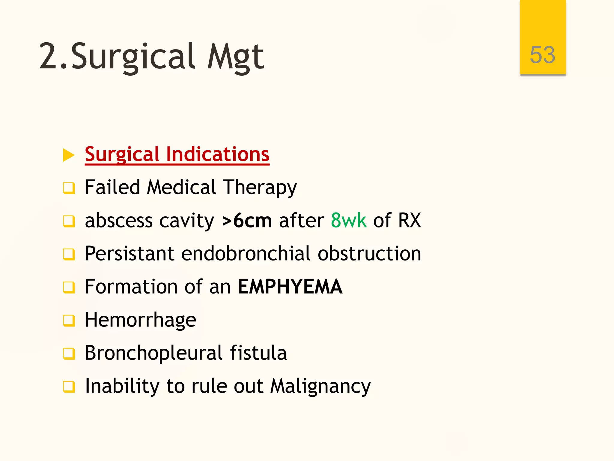
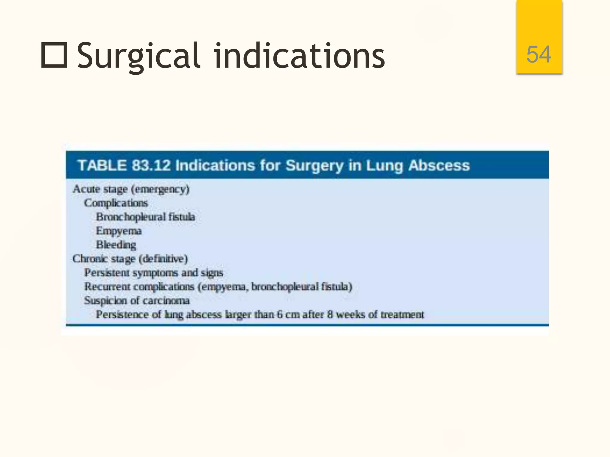
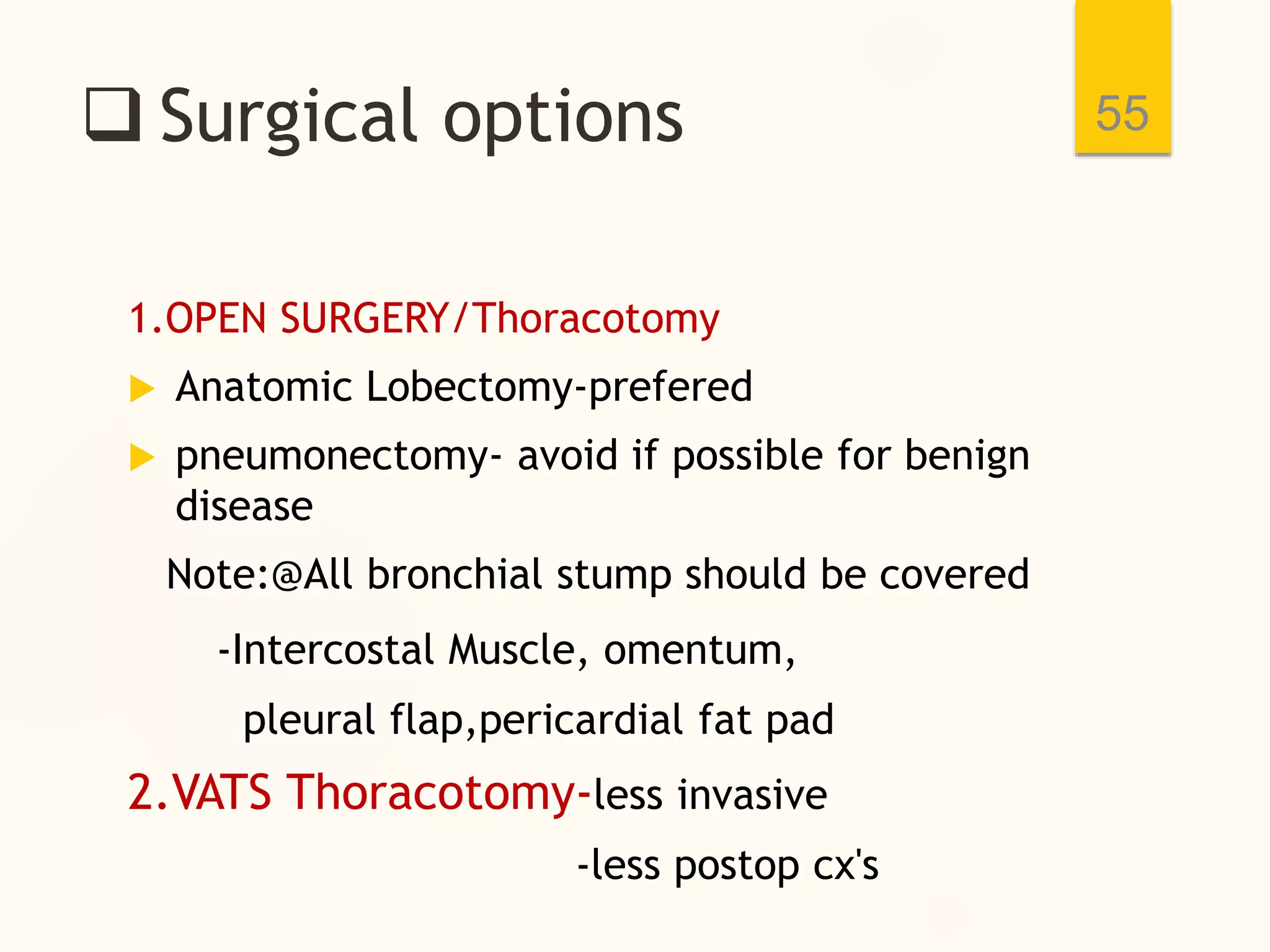
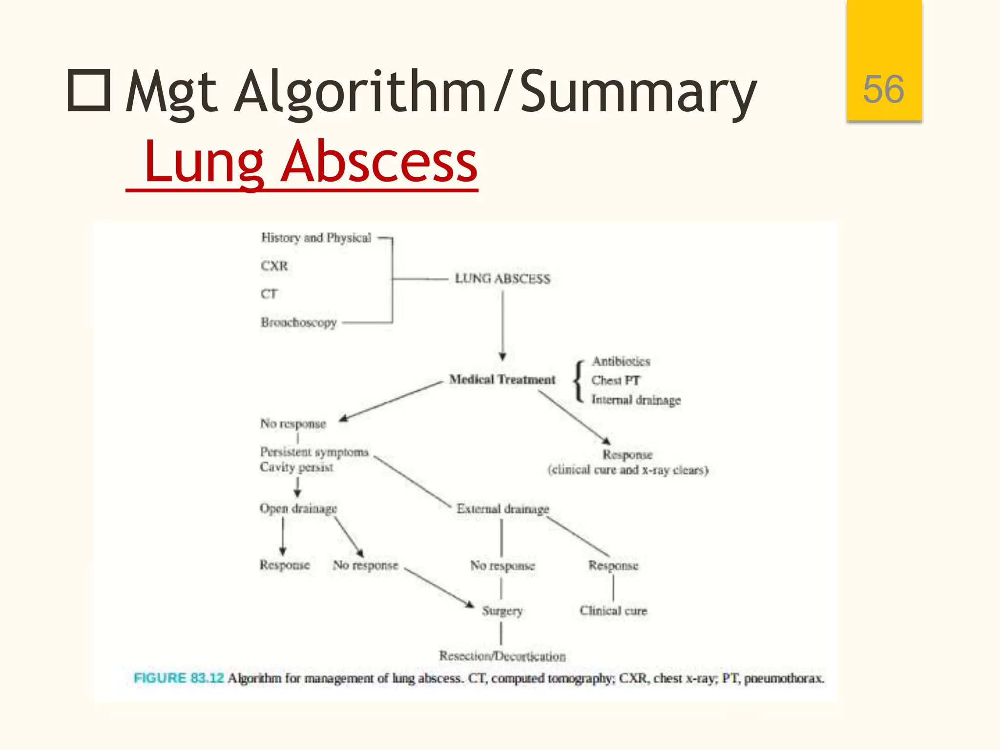
Mgt of suppurative Lung Disease
(Bronchiectasis,lung Abscess)
Addis Ababa University,
black lion hospital
Presentor:- Doti G.( GSRlll)
Moderator:-Dr Samuel(CTSF2)
Oct 5,2021
1
 Outline
 Introduction
 Bronchiectasis
 lung Abscess
2
 Objectives
 To know different types of SLD
 To describe the etiology & pathogenesis of
each ds
 To understand clinical features & DX of each
SLD
 To discuss the management modalities of
each SLD
3
 Introduction
Anatomy of lungs
4
 Bronchopulmonary
segment-largest fxnal unit of anatomic
lobe
5
10 BPS 8-9 BPS
-main bronchus
-lobar bronchus
-segmental bronchus
 Intro---
SLD=Definition
 Suppuration:- ''pus forming''
 Suppurative lung diseases:-are disorders
associated with puss formation within the
lungs & are named according to site;
 Bronchi---------------Bronchiectasis
 lung parenchyma--- Lung abscess
 pleural space--------Empyema Thoracis
6
 Bronchiectasis
 DEFINITION
 EPIDEMIOLOGY
 CLASSIFICATION
 AETIOLOGY
 PATHOPHYSIOLOGY
 CLINICAL FEATURES
 DIAGNOSIS/INVESTIGATIONS
 DDX
 MANAGEMENT OPTIONS
 CX'S & PROGNOSIS
7
 Bronchiectasis
 This is an abnormal irreversible dilatation
of one or more bronchi or bronchioles,large
or small sized airways,with destruction of
their elastic and muscular components
 pseudobronchiectasis-temporary(wks-mo)
cylinderical dilatation of bronchi due acute
pneumonic process=No surgical implications
 Usually due to acute or chronic infection
resulting in recurent inflammation, airflow
obstruction & impaired clearance of secretions
 classified as an obstructive airway disease
 May be focal or diffused
8
 EPIDEMIOLOGY
 Currently there are no estimates of
incidence but prevalence varies by
country,region and underlying aetiology.
 recent study in USA shows:-HRCT identified
bronchiectasis in 50% of pt Dx to have Chronic
Bronchitis or COPD by GP previously
 There is decline in its prevalence in
developed countries with emergence of
vaccines & antibiotics in the 20th century.
 still prevalent in developing country due to
emergency of drug-resistant microrg/MDR.TB
 Overall it increases with age
9
 CLASSIFICATION
Pathological/radiologic
classification
1.cylinderical/Tubular
-Bronchi is dilated &
non-tapering
-Bronchi directly
communicate with lung
parenchymawithout tapering
-usually post TB infection
10
 CLASSIFICATION----
2.Cystic/ Saccular
 Progressive dilation of
the airway which end in
saclike Cystic structure
 Resemble cluster of
grapes
 common in bacterial
pulmonary infection,
FB obstruction &
bronchial stricture
11
 CLASSIFI------
 3.Varicose /Mixed
Bronchiectasis
-Areas of Dilatation
& Constriction
-Resembles
varicose vein
12
 AETIOLOGY-
BRONCHIECTASIS
- in 50% of cases there is no initiating events
13
 Congenital causes
 Cystic Fibrosis-chronic
resp.infection
 primary Ciliary
Dyskinesia
 alpha1-antitrypsin
deficiency
 Hypogammaglobunemia
 Tendency to have
Diffuse & Bilateral
distribution
 Acquired causes
 post infections
 Bacterial
-H.infleunza=1/3
-Mycobacterial TB-scarring
-MAC
 Viral
-measles,influenza
 AETIOLOGY-
BRONCHIECTASIS----
 fungi- Aspergillosis
 Mechanica obstruction
 intrinsic-FB Aspiration,tumor,mucus plug
 Extrinsic-lymph node,tumor
 Immune mediated diseases- post transplant
 Autoimmune /rheumatologic ds
 Toxic gas exposure-chlorine,ammonia gas
14
 Pathogenesis
1.viscious cycle hypothesis
15
-start with impaired defense Mzm
Bronchopulmonary
manifestations
2.Atelectasis
-repeated inflammation increased mucus pxn
airway compression lung collapse airway obst-
Bronchopulmonary manifestations
16
AIRWAY OBSTRUCTION Bronchopulmonary symptoms
 clinical presentations
 A typical patient has classical symptoms of
daily purulent, mucopurulent or mucoid
sputum discharge, cough, fatigue,
 Haemoptysis
 Recurrent pneumonia
 Dyspnea,exercise intolerance
 fever
 wheezing
 pleuritic chest pain
17
 clinical features---
 Non-specific signs
 pallor
 clubbing- Chronic hypoxia
 cyanosis
 tachycardia
 crackles/rales
 cor-pulmonale
 respiratory failure
18
 Diagnosis
Imaging- CXR
 first imaging examination
 posterobasal segment of
lower lobes is mostly
involved
 bronchial wall thickening
 lobar collapse
 airway dilatation
 tram-track sign-parallel line
opacities-Cylinderical BE
 Heart displacement
 normal-mild cases
19
 HRCT
20
 Gold standard modality
 Confirms all cases
 97%sensitivity
 93-100% specificity
 airway dilatations
 signet ring( airway diameter
>1.5 times adjacent vessels)
 mediastinal shifting-the
same side
BRONCHOSCOPE
21
 not routine, there
are indications
 Rigid bronchoscope
is prefered
-allows large sample
bronchography
22
-Nowdays largely
replaced by HRCT
-Disadv=Contrast
associated S/E
=Cost
 Other investigations:-
to identify underlying causes
 PFT=FEV1 should 1.5L for lobectomy
 CBC
 Sweat chloride test->60meq/l & genetic
testing= CFTR protein-chloride channel-CysticFibrosis
 Sputum- Gram stain & culture for AFB,
bacteria & Fungi growth
 immunological tests
 Quantitative IgA,IgG & IgM levels
 Quantitative alpha1-antitrypsin levels
 Autoimmune serologic markers
23
 Mgt- Bronchiectasis
MDT Approach
surgeon,pediatrician,physiotherapist involved
 Goals:-
 Eradication of infection
 RX of underlying causes
 Improve secretion clearance
 Relieving airway obstruction
 Reducing complications
 prevent exacerbations & improve quality of
life
 curative- in strictly localized ds
24
 Mgt-Bronchiectasis
 Medical mgt
1.Antibiotics:- for post infectious causes
 Imperical broad spectrum against most
suspicious pathogens
 start with B-lactam ABC's-Amoxacilline
 can be modified according to CST
 7-14days IV ABC's for acute exacerbations
-Monitor clinical & radiologic response
 Anti-TB =mycobacterial tuberculosis
25
 Mgt----
2. Bronchial Hygiene/Airway clearance
-Hydration-improves mucociliary activities
-Mucolytics- eg Guafinosine
-bronchodilators:-alph2 agonist
-Hyperosmolar agents-eg.saline salt
-chest physiotherapy
-postural drainage
-active breath cycles-ballon inflation
-manual techniques-eg chest clapping/blow
26
 Mgt-----
3.Supportive Measures
-Anti-inflammatory drugs-Halt ds progression
-Smoking cessation=improve mucociliary fxn
-Nutritional supplements=eg,vitamines
-Oxygen therapy for severe cases
 Allergic Bronchopulmonary Aspergillosis
-oral corticosteroid + AZOLE antifungal
 -Immunoglobulin def-= IV Immunoglobulin
27
 Surgical mgt
 options:-
1. Thoracoscopic/ VATS Lung Resection
2. Open Thoracotomy + Lung resection
(lobectomy or segementectomy)
indications:-
 refractory to optimal medical therapy
 Massive Haemoptsis- eg,post TB
bronchiectasis( pulmonary artery laceration)
 unilateral-segmental or lobar distribution
 bilateral-not more than one lobe in each lung
28
 Surgical techniques
 Preoperative planning
 Po Antibiotics for 10 days before surgery
 chest physiotherapy should started 10d pre-op
-Smokers- should stop smoking for 6-8wks
- FEV1 >1.5L for lobectomy
> 2l for pneumonectomy
 position:- lateral decubitus position
29
 Surgical techniques
Left lower Lobectomy
 Posterolateral incision
prefered:-
-Bcoz it provide bird's
eye view of fissure b/n
lul & lower lobe
Disadv=muscle
resection
landmarks-
30
4th-6th ICS
 surgical steps
 The open LL lobectomy for bronchiectasis
involves 3 main steps:
 in contrast to lung ca lobectomy( vein 1st
principle), pA ligation preceeds vein.
 (1) ligation of the interlobar basal
pulmonary artery,
 (2) ligation of the inferior pulmonary vein
(IPV),
 (3) division of the lower lobe bronchus.
31
 step1:- Division of
interlobar basal PA
32
Step2:- Division of IPV 33
IPV
 step3:- Division of LLLB 34
postop care
 Aim:-To prevent common postop cx's
such as pain, atelectasis & pneumonia
 postop Analgesia
-essential to prevent postop resp cx's
-facilitate pt cough & walk ability
35
 postop care----
 options
 Thoracic Epidural Anesthasia
-mixture of LA & Narcotics used
-Gold standard
 paravertabral catheter-LA
 paravertaberal blocks
 NSAID
 Narcotics-
 Neuroleptic- Gabapentin
- if intercostal nerve bundle compression is likely
36
 Respiratory physiotherapy
 Lung expansion techniques :-includes
 incentive spirometry,
 deep breathing exercises,
 postural drainage, percussion & vibration,
coughing, suctioning,
 continuous positive airway pressure (CPAP).
 DVT prophylaxis-UFH/LMWH after 24hrs
 GI prophylaxis- stress gastritis= IV cimitidine
37
 mgt----last option
lung Transplantation
-End stage ds-Disseminated BE
Bilateral, all lobes bronchiectasis
38
 prognosis(PX)
poor pxtic factors includes
 life threatening haemoptysis
 Bilateral lung involvement
 Associated lung abscess
 Malnutrition
 Chronic anemia
 frequency of exacerbation
 Specific pathogens:-
p.aeroginosa,Aspergillus ,MAC
39
 LUNG ABSCESS
 INTRODUCTION
 CLASSIFICATION
 AETIOLOGY
 PATHOGENESIS
 CF
 DX
 MGT
40
 Lung Abscess
introduction
 lung Abscess is necrosis of pulmonary tissue
& formation of cavities( >2cm) containing
necrotic debris or pus caused by microbial
infection
 formation of multiple small(<2cm) abscesses
is called Necrotizing pneuminia or lung
gangrene
 More common in Male & older age
 in the pre-antibiotic era 1/3 of pt died
 Nowdays most of uncomplicated LA respond
to prolonged Antibiotic therapy
41
 Classification
lung Abscess
 Duration:- Acute(<6wks),subacute(6-12wk)or
chronic(>12wks)
 Origins:primary LA=aspiration or pneumonia
Secondary LA- obstruction(FB,tumor)
-Bronchiectasis,BEF
-spread from Extra p.site
eg.perforated Appendix
 setup:-Community Acquired LA= Anaerobic oral flora
-Hospital Acquired LA= aerobic organisms. eg P.aerginosa
42
 Aetiology
based on pathophysiology
 Aspiration pneumonia from oropharyngeal
flora
 Dental/periodontal sepsis
 paranasal sinus infection
 Depressed conscious level
-Alcohol/sedative abuse
-Anaesthesia
-Epilepsy
-Head injury/Coma
43
 Aetiology----
 Impaired Larygeal Closure
-prolonged ET intubation
-recurrent laryngeal nerve palsy
 Disturbance of swallowing
-oesophageal stricture,Achalasia, GERD
 Haematogenous spread from a distal site
-UTI, Abdominal sepsis,Amebic liver Abscess
44
 pathognesis
bacteria reach lower airway
( not cleared by DEFENSE MZM)-mucociliary dfs
ASPIRATION PNEUMONITIS
7-14 days later
TISSUE NECROSIS EMPYEMA( BPF).
LUNG ABSCESS
45
Bacteremia(
haematogenous/septic emboli)
Impaired host defense mzm
Distant focus
eg.abdominal sepsis
 CF-LUNG ABSCESS
 The typical pt presents with a hx of an
antecedent event suspicious for aspiration,
pulmonary infection, or pneumonia.
 productive cough, foul breath,fever
rigors,fatigue,,dyspnea,hemoptysis
 absence of fever or leucocytosis suggest
non-infectious cause of LA
 95% of LA occur in posterior segment of Upper lobe
& superior segment of Lower lobes.
 DX is by IMAGING
 CXR,BRONCHOSCOPE & HRCT scan
46
 CXR
 Abscess is often
unilateral & single
involving posterior
segments of the
upper lobes & the
apical segment of
lower lobe due to
gravitywhile lying
down
 Air-fluid level
47
Air fluid level
CT-lung Abscess
48
 Absecss in lt upper
lobe thick & irregular
wall air -fluid level
 Does not cross
fissures
A)Multiple cavitation,Disseminated
infiltration & B)fistulization
49
bronchopleural fistula
causing empyema &
pneumothorax
sagital view
Axialview
 Microbiologic studies
 Samples: sputum, BAL,Transtracheal
aspiration,transthoracic needle aspiration
under CT guidance
 All above specimens for:-
-Gram stain
-CST
-cytology
 Bronchoscope- R/O obstructive lesions
- Bronchial drainage(Rx)
 CBC,ESR,CRP- non-specific
50
 Mgt
1.Medical Mgt
 ANTIBIOTICS
-Targeted & prolonged (6-8wks)ABC is Mainstay of Rx
- start with abc against suspected pathogens
-comminity Acquired LA- clindamycine/metr
-Hospt Acquired= ciprofloxacilline
@ 90% of pt (aerobic LA)respond to ABC alone
 Chest physiotherapy-aggressive
 Internal drainage- bronchoscopic lavage
51
if pt not respond to above
measures-sx's/cavity persist
 External drainage( Acute stage)
 Closed- tube thoracostomy
 CT-guided catheter drainage
 pneumonostomy(cavernostomy)
 open drainage(chronic stage)
-thoracotomy + drainage
 if no response to above measures
about 10% of pts require surgical
intervention as last option
52
2.Surgical Mgt
 Surgical Indications
 Failed Medical Therapy
 abscess cavity >6cm after 8wk of RX
 Persistant endobronchial obstruction
 Formation of an EMPHYEMA
 Hemorrhage
 Bronchopleural fistula
 Inability to rule out Malignancy
53
 Surgical indications 54
 Surgical options
1.OPEN SURGERY/Thoracotomy
 Anatomic Lobectomy-prefered
 pneumonectomy- avoid if possible for benign
disease
Note:@All bronchial stump should be covered
-Intercostal Muscle, omentum,
pleural flap,pericardial fat pad
2.VATS Thoracotomy-less invasive
-less postop cx's
55
 Mgt Algorithm/Summary
Lung Abscess
56
References 57
MEDSCAPE.COM
THANK YOU
For your Attention
58

Suppurative lung disease.ppt

  • 1.
    Mgt of suppurativeLung Disease (Bronchiectasis,lung Abscess) Addis Ababa University, black lion hospital Presentor:- Doti G.( GSRlll) Moderator:-Dr Samuel(CTSF2) Oct 5,2021 1
  • 2.
     Outline  Introduction Bronchiectasis  lung Abscess 2
  • 3.
     Objectives  Toknow different types of SLD  To describe the etiology & pathogenesis of each ds  To understand clinical features & DX of each SLD  To discuss the management modalities of each SLD 3
  • 4.
  • 5.
     Bronchopulmonary segment-largest fxnalunit of anatomic lobe 5 10 BPS 8-9 BPS -main bronchus -lobar bronchus -segmental bronchus
  • 6.
     Intro--- SLD=Definition  Suppuration:-''pus forming''  Suppurative lung diseases:-are disorders associated with puss formation within the lungs & are named according to site;  Bronchi---------------Bronchiectasis  lung parenchyma--- Lung abscess  pleural space--------Empyema Thoracis 6
  • 7.
     Bronchiectasis  DEFINITION EPIDEMIOLOGY  CLASSIFICATION  AETIOLOGY  PATHOPHYSIOLOGY  CLINICAL FEATURES  DIAGNOSIS/INVESTIGATIONS  DDX  MANAGEMENT OPTIONS  CX'S & PROGNOSIS 7
  • 8.
     Bronchiectasis  Thisis an abnormal irreversible dilatation of one or more bronchi or bronchioles,large or small sized airways,with destruction of their elastic and muscular components  pseudobronchiectasis-temporary(wks-mo) cylinderical dilatation of bronchi due acute pneumonic process=No surgical implications  Usually due to acute or chronic infection resulting in recurent inflammation, airflow obstruction & impaired clearance of secretions  classified as an obstructive airway disease  May be focal or diffused 8
  • 9.
     EPIDEMIOLOGY  Currentlythere are no estimates of incidence but prevalence varies by country,region and underlying aetiology.  recent study in USA shows:-HRCT identified bronchiectasis in 50% of pt Dx to have Chronic Bronchitis or COPD by GP previously  There is decline in its prevalence in developed countries with emergence of vaccines & antibiotics in the 20th century.  still prevalent in developing country due to emergency of drug-resistant microrg/MDR.TB  Overall it increases with age 9
  • 10.
     CLASSIFICATION Pathological/radiologic classification 1.cylinderical/Tubular -Bronchi isdilated & non-tapering -Bronchi directly communicate with lung parenchymawithout tapering -usually post TB infection 10
  • 11.
     CLASSIFICATION---- 2.Cystic/ Saccular Progressive dilation of the airway which end in saclike Cystic structure  Resemble cluster of grapes  common in bacterial pulmonary infection, FB obstruction & bronchial stricture 11
  • 12.
     CLASSIFI------  3.Varicose/Mixed Bronchiectasis -Areas of Dilatation & Constriction -Resembles varicose vein 12
  • 13.
     AETIOLOGY- BRONCHIECTASIS - in50% of cases there is no initiating events 13  Congenital causes  Cystic Fibrosis-chronic resp.infection  primary Ciliary Dyskinesia  alpha1-antitrypsin deficiency  Hypogammaglobunemia  Tendency to have Diffuse & Bilateral distribution  Acquired causes  post infections  Bacterial -H.infleunza=1/3 -Mycobacterial TB-scarring -MAC  Viral -measles,influenza
  • 14.
     AETIOLOGY- BRONCHIECTASIS----  fungi-Aspergillosis  Mechanica obstruction  intrinsic-FB Aspiration,tumor,mucus plug  Extrinsic-lymph node,tumor  Immune mediated diseases- post transplant  Autoimmune /rheumatologic ds  Toxic gas exposure-chlorine,ammonia gas 14
  • 15.
     Pathogenesis 1.viscious cyclehypothesis 15 -start with impaired defense Mzm Bronchopulmonary manifestations
  • 16.
    2.Atelectasis -repeated inflammation increasedmucus pxn airway compression lung collapse airway obst- Bronchopulmonary manifestations 16 AIRWAY OBSTRUCTION Bronchopulmonary symptoms
  • 17.
     clinical presentations A typical patient has classical symptoms of daily purulent, mucopurulent or mucoid sputum discharge, cough, fatigue,  Haemoptysis  Recurrent pneumonia  Dyspnea,exercise intolerance  fever  wheezing  pleuritic chest pain 17
  • 18.
     clinical features--- Non-specific signs  pallor  clubbing- Chronic hypoxia  cyanosis  tachycardia  crackles/rales  cor-pulmonale  respiratory failure 18
  • 19.
     Diagnosis Imaging- CXR first imaging examination  posterobasal segment of lower lobes is mostly involved  bronchial wall thickening  lobar collapse  airway dilatation  tram-track sign-parallel line opacities-Cylinderical BE  Heart displacement  normal-mild cases 19
  • 20.
     HRCT 20  Goldstandard modality  Confirms all cases  97%sensitivity  93-100% specificity  airway dilatations  signet ring( airway diameter >1.5 times adjacent vessels)  mediastinal shifting-the same side
  • 21.
    BRONCHOSCOPE 21  not routine,there are indications  Rigid bronchoscope is prefered -allows large sample
  • 22.
    bronchography 22 -Nowdays largely replaced byHRCT -Disadv=Contrast associated S/E =Cost
  • 23.
     Other investigations:- toidentify underlying causes  PFT=FEV1 should 1.5L for lobectomy  CBC  Sweat chloride test->60meq/l & genetic testing= CFTR protein-chloride channel-CysticFibrosis  Sputum- Gram stain & culture for AFB, bacteria & Fungi growth  immunological tests  Quantitative IgA,IgG & IgM levels  Quantitative alpha1-antitrypsin levels  Autoimmune serologic markers 23
  • 24.
     Mgt- Bronchiectasis MDTApproach surgeon,pediatrician,physiotherapist involved  Goals:-  Eradication of infection  RX of underlying causes  Improve secretion clearance  Relieving airway obstruction  Reducing complications  prevent exacerbations & improve quality of life  curative- in strictly localized ds 24
  • 25.
     Mgt-Bronchiectasis  Medicalmgt 1.Antibiotics:- for post infectious causes  Imperical broad spectrum against most suspicious pathogens  start with B-lactam ABC's-Amoxacilline  can be modified according to CST  7-14days IV ABC's for acute exacerbations -Monitor clinical & radiologic response  Anti-TB =mycobacterial tuberculosis 25
  • 26.
     Mgt---- 2. BronchialHygiene/Airway clearance -Hydration-improves mucociliary activities -Mucolytics- eg Guafinosine -bronchodilators:-alph2 agonist -Hyperosmolar agents-eg.saline salt -chest physiotherapy -postural drainage -active breath cycles-ballon inflation -manual techniques-eg chest clapping/blow 26
  • 27.
     Mgt----- 3.Supportive Measures -Anti-inflammatorydrugs-Halt ds progression -Smoking cessation=improve mucociliary fxn -Nutritional supplements=eg,vitamines -Oxygen therapy for severe cases  Allergic Bronchopulmonary Aspergillosis -oral corticosteroid + AZOLE antifungal  -Immunoglobulin def-= IV Immunoglobulin 27
  • 28.
     Surgical mgt options:- 1. Thoracoscopic/ VATS Lung Resection 2. Open Thoracotomy + Lung resection (lobectomy or segementectomy) indications:-  refractory to optimal medical therapy  Massive Haemoptsis- eg,post TB bronchiectasis( pulmonary artery laceration)  unilateral-segmental or lobar distribution  bilateral-not more than one lobe in each lung 28
  • 29.
     Surgical techniques Preoperative planning  Po Antibiotics for 10 days before surgery  chest physiotherapy should started 10d pre-op -Smokers- should stop smoking for 6-8wks - FEV1 >1.5L for lobectomy > 2l for pneumonectomy  position:- lateral decubitus position 29
  • 30.
     Surgical techniques Leftlower Lobectomy  Posterolateral incision prefered:- -Bcoz it provide bird's eye view of fissure b/n lul & lower lobe Disadv=muscle resection landmarks- 30 4th-6th ICS
  • 31.
     surgical steps The open LL lobectomy for bronchiectasis involves 3 main steps:  in contrast to lung ca lobectomy( vein 1st principle), pA ligation preceeds vein.  (1) ligation of the interlobar basal pulmonary artery,  (2) ligation of the inferior pulmonary vein (IPV),  (3) division of the lower lobe bronchus. 31
  • 32.
     step1:- Divisionof interlobar basal PA 32
  • 33.
  • 34.
  • 35.
    postop care  Aim:-Toprevent common postop cx's such as pain, atelectasis & pneumonia  postop Analgesia -essential to prevent postop resp cx's -facilitate pt cough & walk ability 35
  • 36.
     postop care---- options  Thoracic Epidural Anesthasia -mixture of LA & Narcotics used -Gold standard  paravertabral catheter-LA  paravertaberal blocks  NSAID  Narcotics-  Neuroleptic- Gabapentin - if intercostal nerve bundle compression is likely 36
  • 37.
     Respiratory physiotherapy Lung expansion techniques :-includes  incentive spirometry,  deep breathing exercises,  postural drainage, percussion & vibration, coughing, suctioning,  continuous positive airway pressure (CPAP).  DVT prophylaxis-UFH/LMWH after 24hrs  GI prophylaxis- stress gastritis= IV cimitidine 37
  • 38.
     mgt----last option lungTransplantation -End stage ds-Disseminated BE Bilateral, all lobes bronchiectasis 38
  • 39.
     prognosis(PX) poor pxticfactors includes  life threatening haemoptysis  Bilateral lung involvement  Associated lung abscess  Malnutrition  Chronic anemia  frequency of exacerbation  Specific pathogens:- p.aeroginosa,Aspergillus ,MAC 39
  • 40.
     LUNG ABSCESS INTRODUCTION  CLASSIFICATION  AETIOLOGY  PATHOGENESIS  CF  DX  MGT 40
  • 41.
     Lung Abscess introduction lung Abscess is necrosis of pulmonary tissue & formation of cavities( >2cm) containing necrotic debris or pus caused by microbial infection  formation of multiple small(<2cm) abscesses is called Necrotizing pneuminia or lung gangrene  More common in Male & older age  in the pre-antibiotic era 1/3 of pt died  Nowdays most of uncomplicated LA respond to prolonged Antibiotic therapy 41
  • 42.
     Classification lung Abscess Duration:- Acute(<6wks),subacute(6-12wk)or chronic(>12wks)  Origins:primary LA=aspiration or pneumonia Secondary LA- obstruction(FB,tumor) -Bronchiectasis,BEF -spread from Extra p.site eg.perforated Appendix  setup:-Community Acquired LA= Anaerobic oral flora -Hospital Acquired LA= aerobic organisms. eg P.aerginosa 42
  • 43.
     Aetiology based onpathophysiology  Aspiration pneumonia from oropharyngeal flora  Dental/periodontal sepsis  paranasal sinus infection  Depressed conscious level -Alcohol/sedative abuse -Anaesthesia -Epilepsy -Head injury/Coma 43
  • 44.
     Aetiology----  ImpairedLarygeal Closure -prolonged ET intubation -recurrent laryngeal nerve palsy  Disturbance of swallowing -oesophageal stricture,Achalasia, GERD  Haematogenous spread from a distal site -UTI, Abdominal sepsis,Amebic liver Abscess 44
  • 45.
     pathognesis bacteria reachlower airway ( not cleared by DEFENSE MZM)-mucociliary dfs ASPIRATION PNEUMONITIS 7-14 days later TISSUE NECROSIS EMPYEMA( BPF). LUNG ABSCESS 45 Bacteremia( haematogenous/septic emboli) Impaired host defense mzm Distant focus eg.abdominal sepsis
  • 46.
     CF-LUNG ABSCESS The typical pt presents with a hx of an antecedent event suspicious for aspiration, pulmonary infection, or pneumonia.  productive cough, foul breath,fever rigors,fatigue,,dyspnea,hemoptysis  absence of fever or leucocytosis suggest non-infectious cause of LA  95% of LA occur in posterior segment of Upper lobe & superior segment of Lower lobes.  DX is by IMAGING  CXR,BRONCHOSCOPE & HRCT scan 46
  • 47.
     CXR  Abscessis often unilateral & single involving posterior segments of the upper lobes & the apical segment of lower lobe due to gravitywhile lying down  Air-fluid level 47 Air fluid level
  • 48.
    CT-lung Abscess 48  Absecssin lt upper lobe thick & irregular wall air -fluid level  Does not cross fissures
  • 49.
    A)Multiple cavitation,Disseminated infiltration &B)fistulization 49 bronchopleural fistula causing empyema & pneumothorax sagital view Axialview
  • 50.
     Microbiologic studies Samples: sputum, BAL,Transtracheal aspiration,transthoracic needle aspiration under CT guidance  All above specimens for:- -Gram stain -CST -cytology  Bronchoscope- R/O obstructive lesions - Bronchial drainage(Rx)  CBC,ESR,CRP- non-specific 50
  • 51.
     Mgt 1.Medical Mgt ANTIBIOTICS -Targeted & prolonged (6-8wks)ABC is Mainstay of Rx - start with abc against suspected pathogens -comminity Acquired LA- clindamycine/metr -Hospt Acquired= ciprofloxacilline @ 90% of pt (aerobic LA)respond to ABC alone  Chest physiotherapy-aggressive  Internal drainage- bronchoscopic lavage 51
  • 52.
    if pt notrespond to above measures-sx's/cavity persist  External drainage( Acute stage)  Closed- tube thoracostomy  CT-guided catheter drainage  pneumonostomy(cavernostomy)  open drainage(chronic stage) -thoracotomy + drainage  if no response to above measures about 10% of pts require surgical intervention as last option 52
  • 53.
    2.Surgical Mgt  SurgicalIndications  Failed Medical Therapy  abscess cavity >6cm after 8wk of RX  Persistant endobronchial obstruction  Formation of an EMPHYEMA  Hemorrhage  Bronchopleural fistula  Inability to rule out Malignancy 53
  • 54.
  • 55.
     Surgical options 1.OPENSURGERY/Thoracotomy  Anatomic Lobectomy-prefered  pneumonectomy- avoid if possible for benign disease Note:@All bronchial stump should be covered -Intercostal Muscle, omentum, pleural flap,pericardial fat pad 2.VATS Thoracotomy-less invasive -less postop cx's 55
  • 56.
  • 57.
  • 58.
    THANK YOU For yourAttention 58

Editor's Notes

  • #10 previously,in USA the incidence in adult population is 52 per 100,000 populations
  • #14 Alpha1-antitrypsin is a protein that protect the lung and liver.AATD is autosomal dominal genetic inherited disorder that can result in COPD,Bronchiectasis anf CLD(Cirrhosis,HCC)
  • #24 CF =There is defect in cystic fibrosis gene w/c code for cystic fibrosis transmembrane regulator(CFTR) which fxn as chloride channel