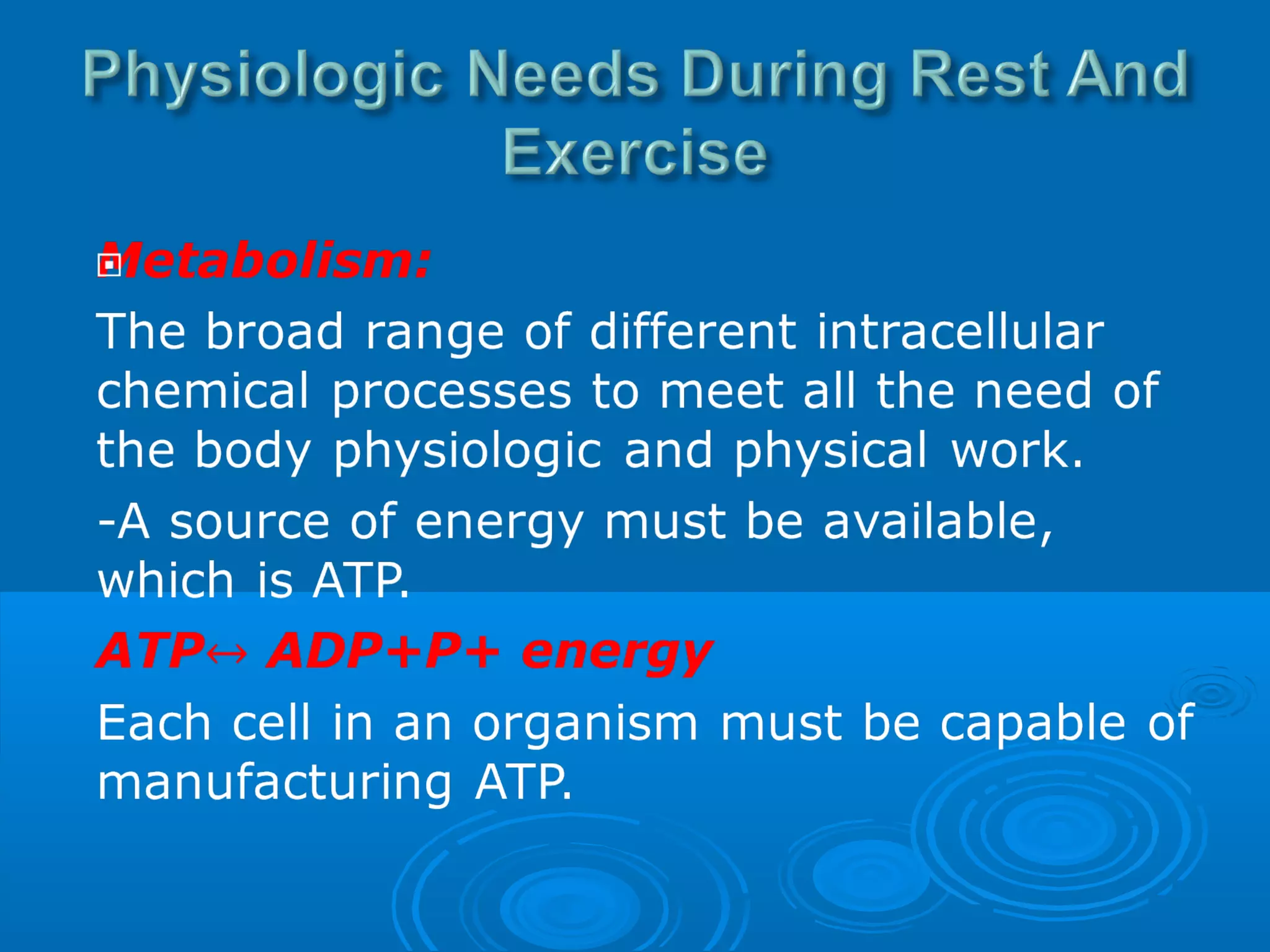
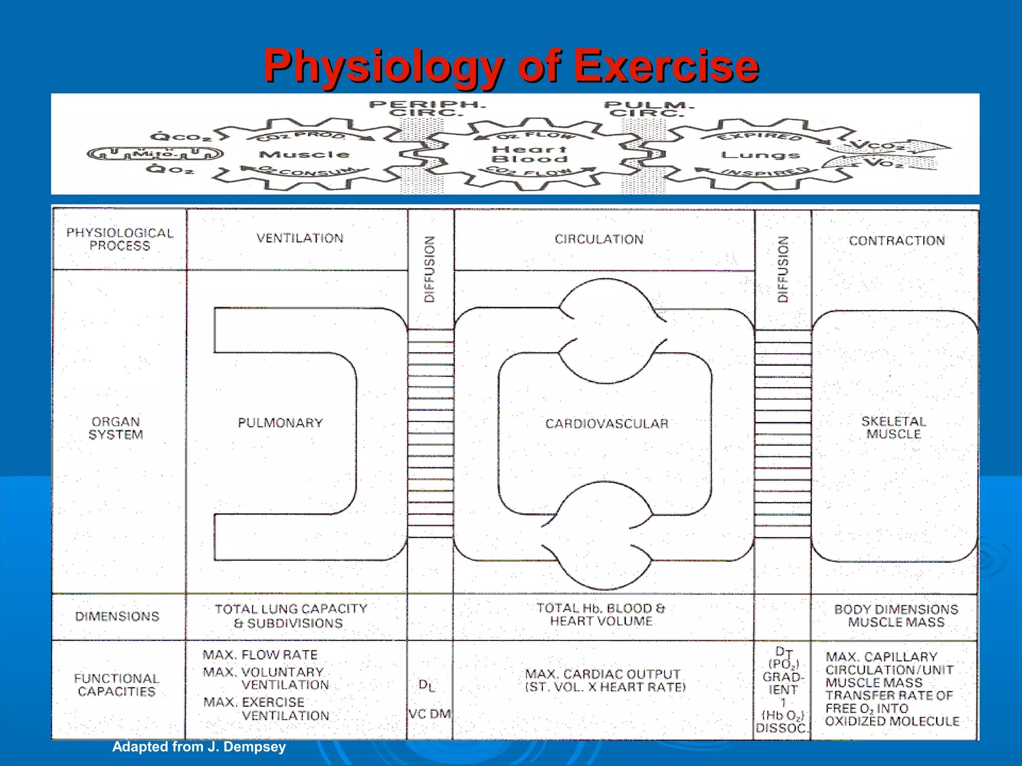
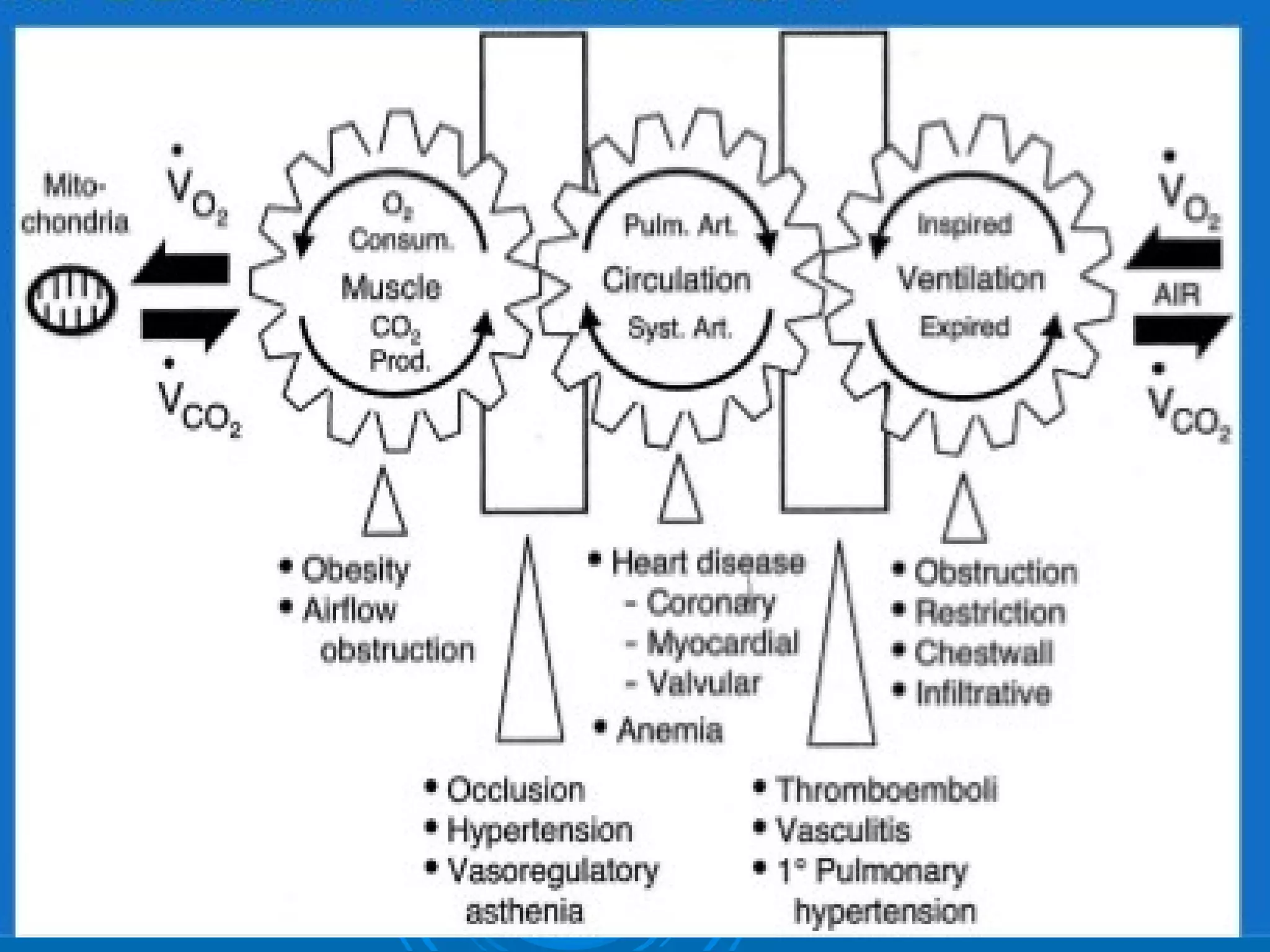
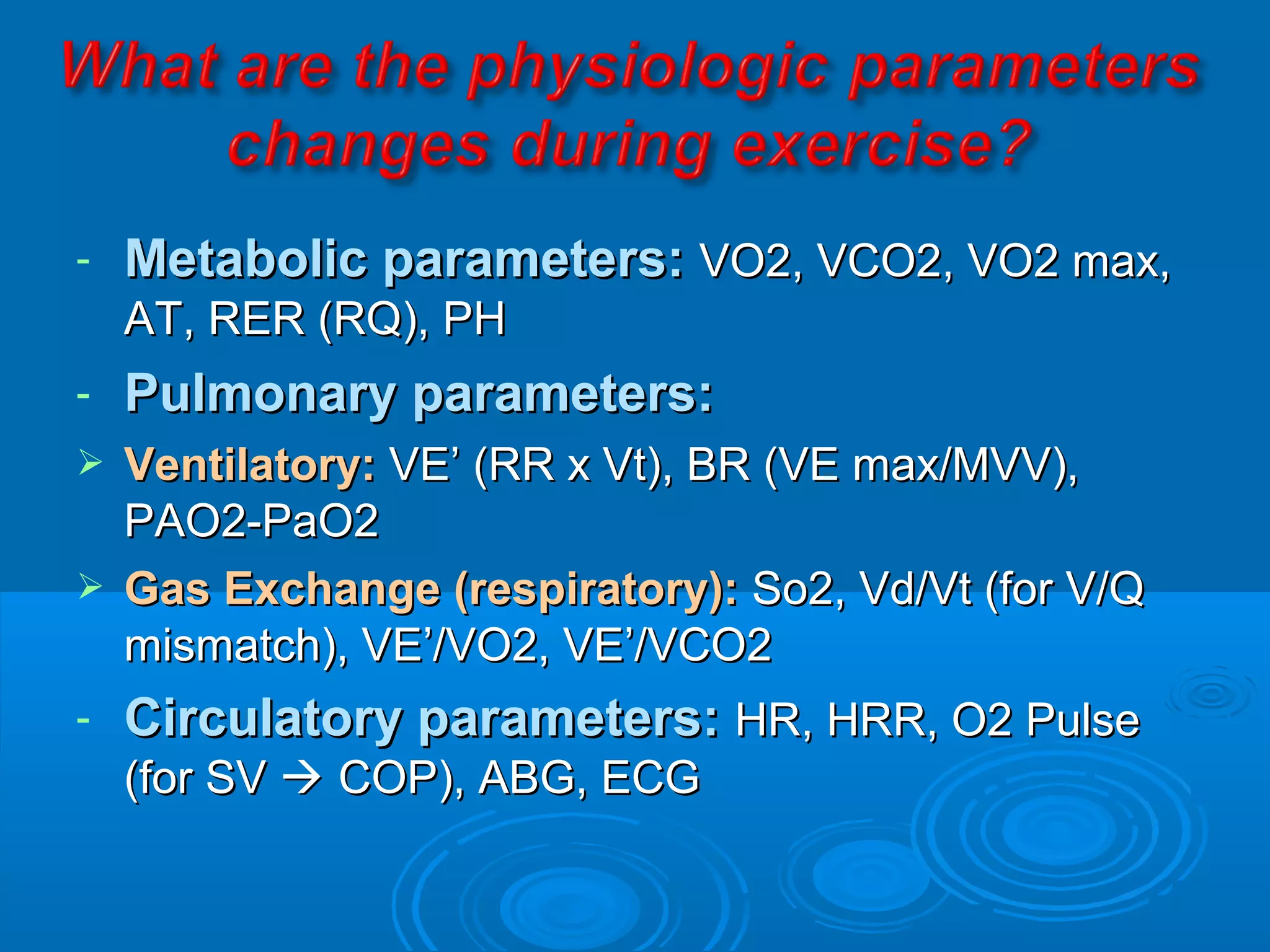
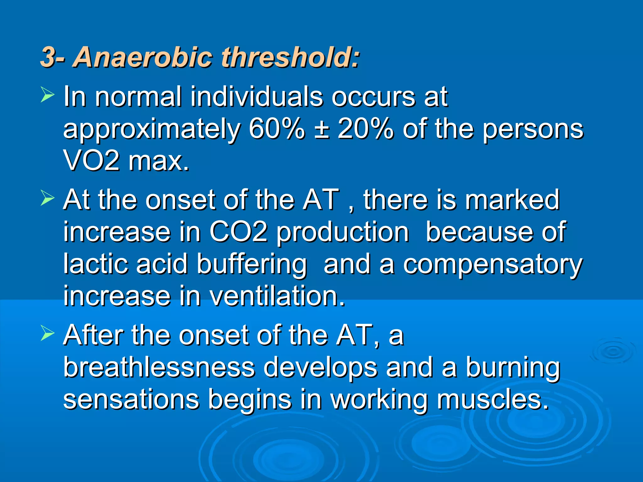
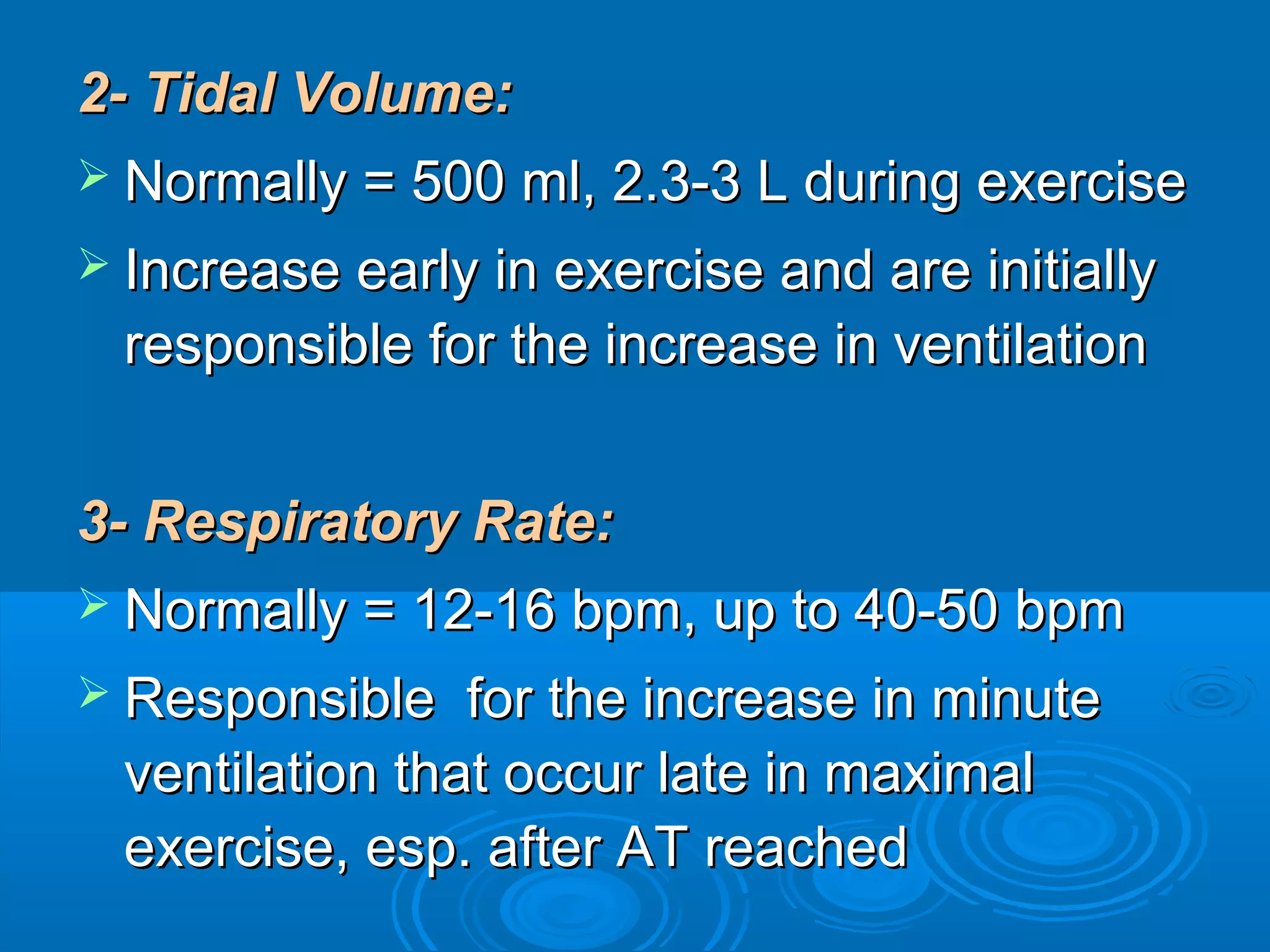
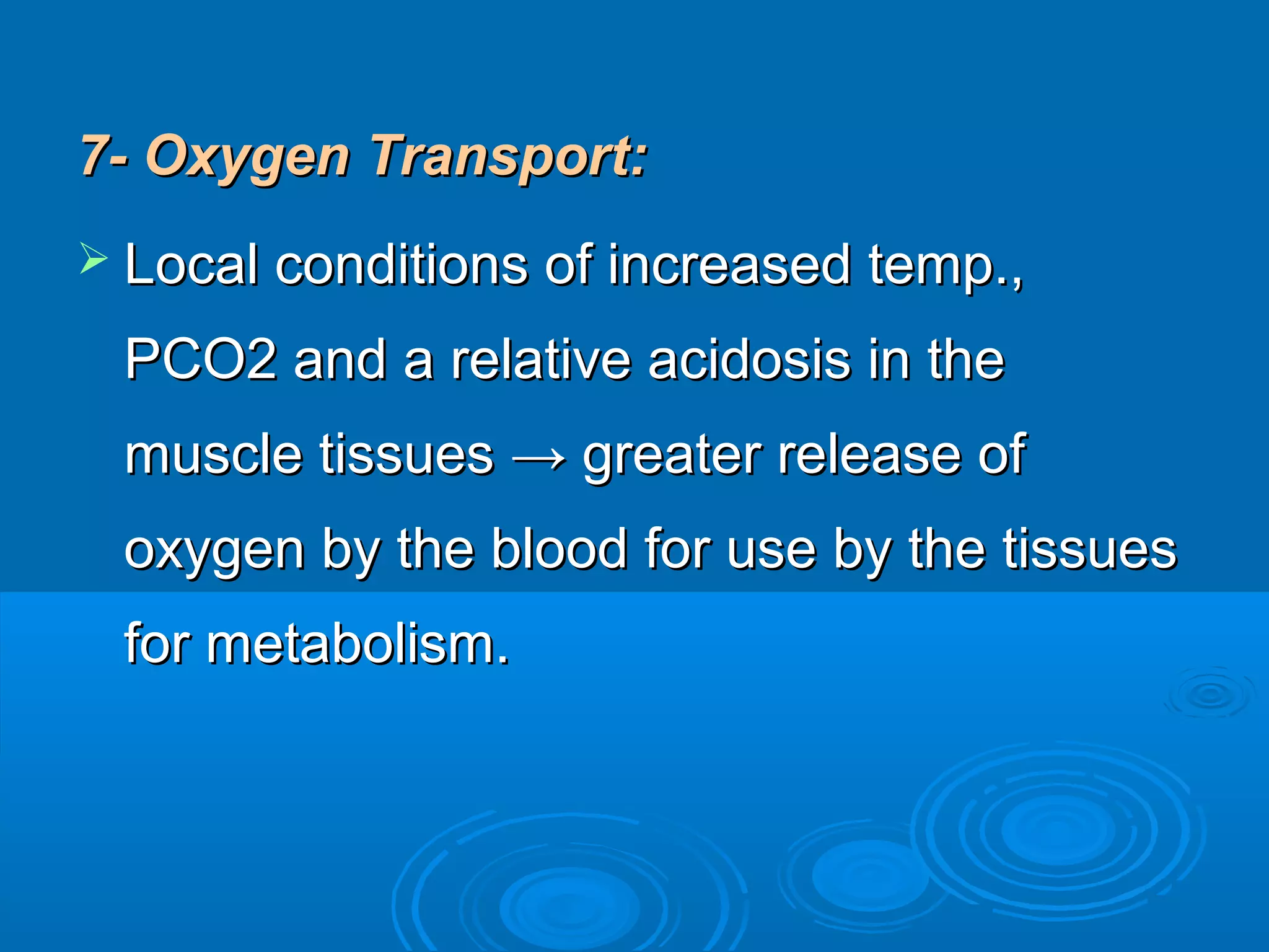
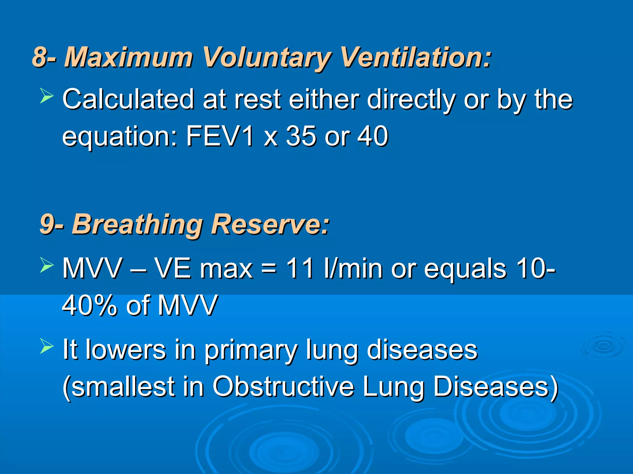
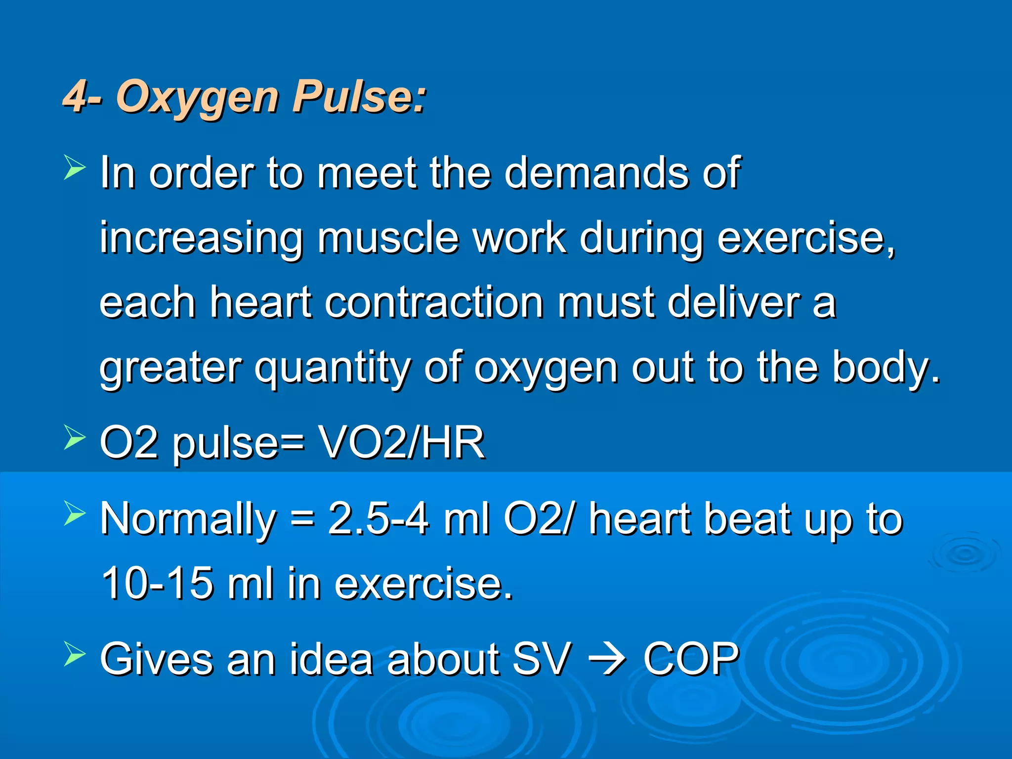
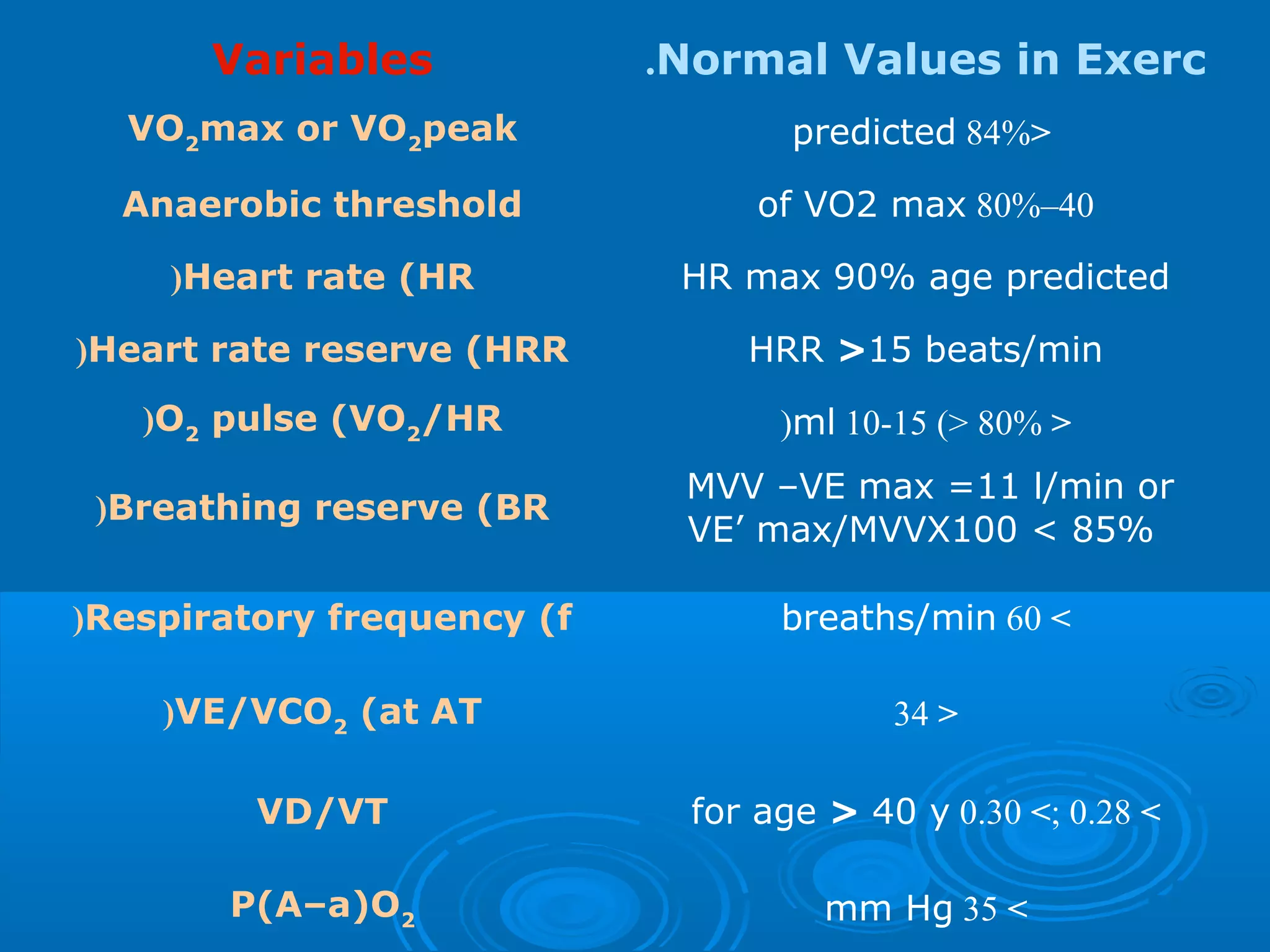
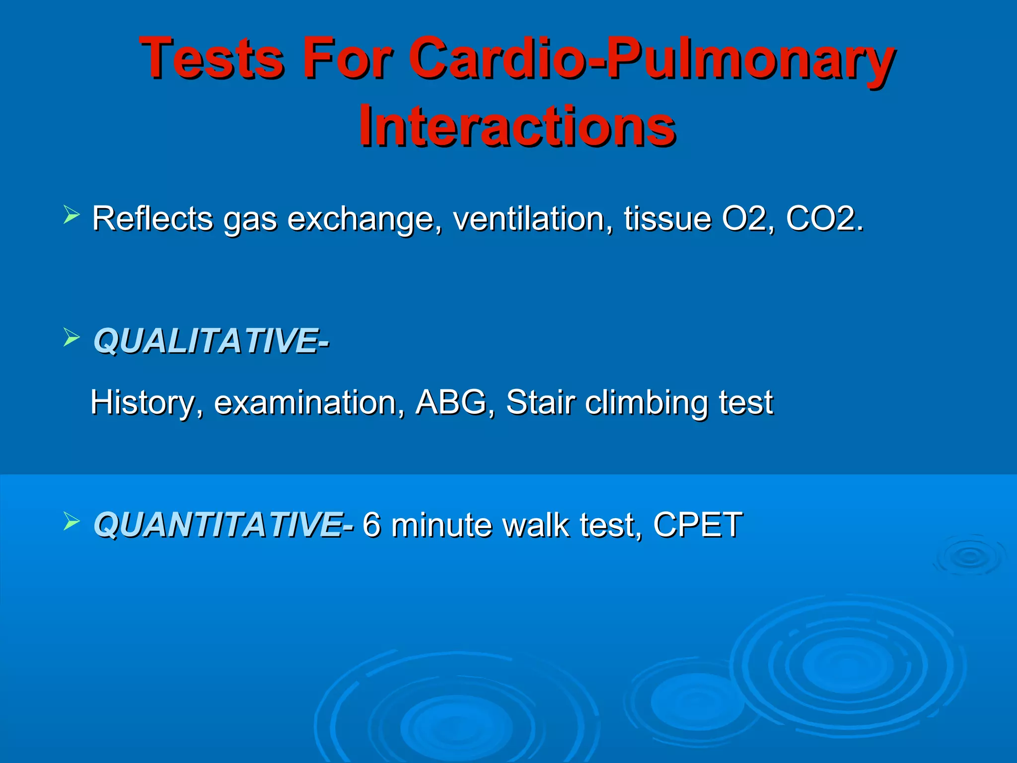
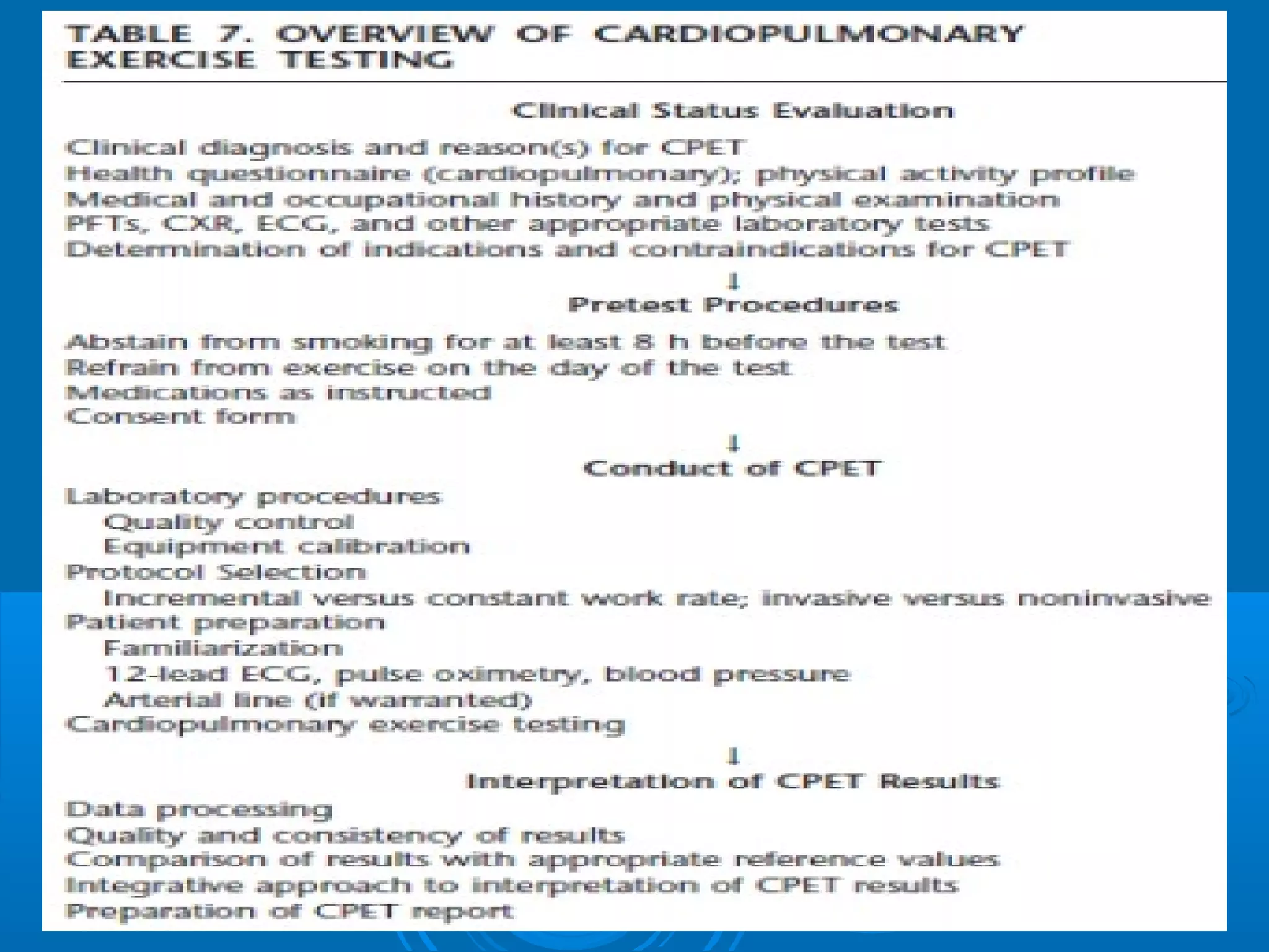
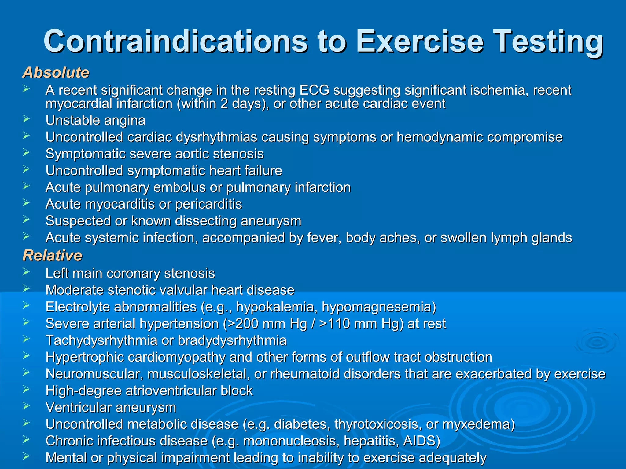
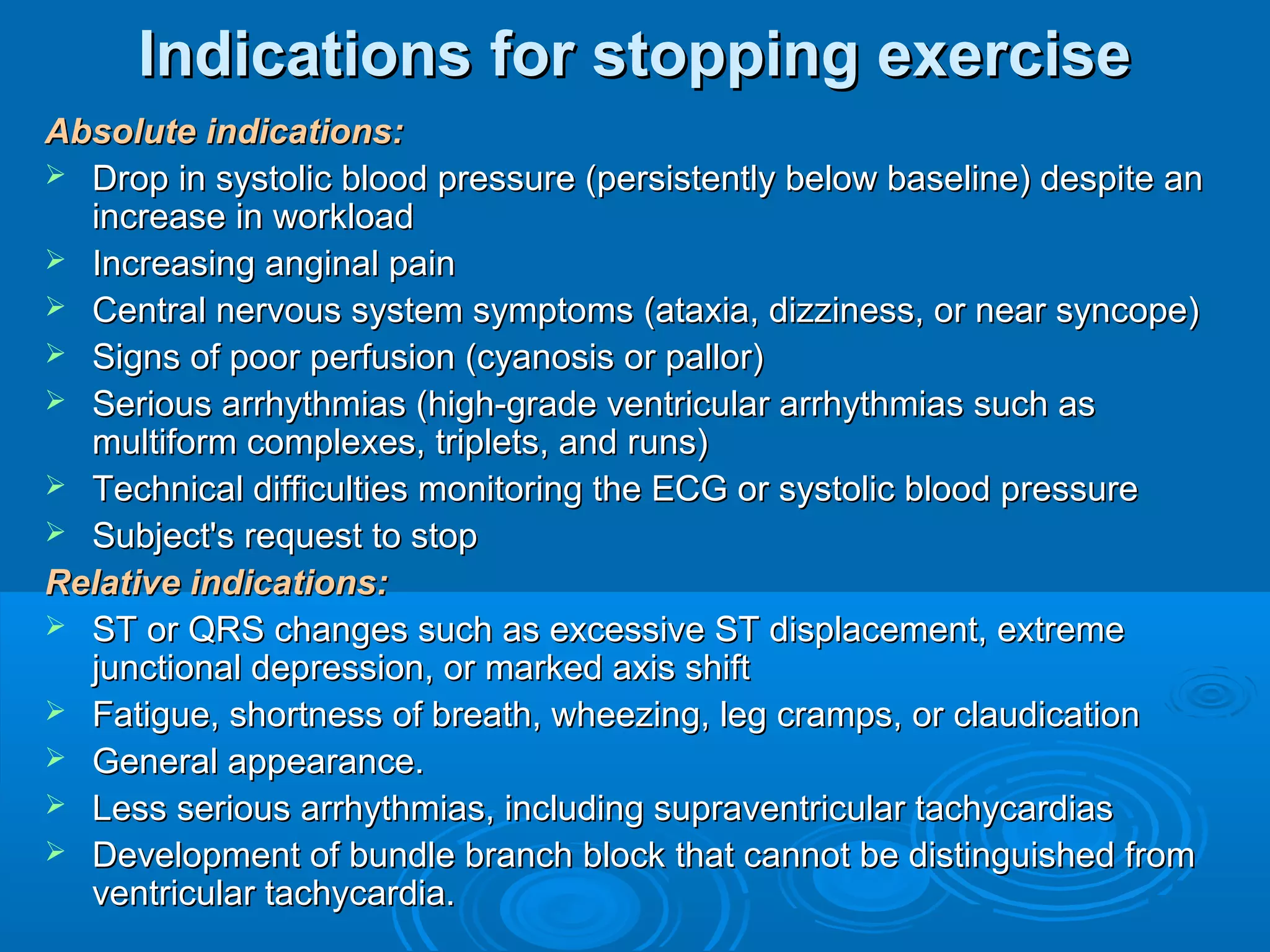
ByBy
Riham Hazem RaafatRiham Hazem Raafat
Lecturer of Chest DiseasesLecturer of Chest Diseases
Ainshams UniversityAinshams University
Exercise Adaptation &Exercise Adaptation &
Cardio-Pulmonary ExerciseCardio-Pulmonary Exercise
TestingTesting
Physiology of ExercisePhysiology of Exercise
Adapted from J. Dempsey
 Takes place by means of diffusion ofTakes place by means of diffusion of
substances.substances.
External respiration:External respiration: (within the lungs)(within the lungs)
Respiratory exchange ratio (R) is the ratio ofRespiratory exchange ratio (R) is the ratio of
this exchange within the lungs betweenthis exchange within the lungs between
oxygen and carbon dioxideoxygen and carbon dioxide..
R= 200/250=0.8R= 200/250=0.8
Internal respiration:Internal respiration: (within the muscles)(within the muscles)
Gas exchange and nutrients.Gas exchange and nutrients.
 Oxygen is consumed in the production ofOxygen is consumed in the production of
ATP and that co2 is produced as wasteATP and that co2 is produced as waste
product.product.
Respiratory quotient (RQ)Respiratory quotient (RQ) this ratio of o2this ratio of o2
consumption to co2 production.consumption to co2 production.
 It equals 0.8It equals 0.8
- Metabolic parameters:Metabolic parameters: VO2, VCO2, VO2 max,VO2, VCO2, VO2 max,
AT, RER (RQ), PHAT, RER (RQ), PH
- Pulmonary parameters:Pulmonary parameters:
 Ventilatory:Ventilatory: VE’ (RR x Vt), BR (VE max/MVV),VE’ (RR x Vt), BR (VE max/MVV),
PAO2-PaO2PAO2-PaO2
 Gas Exchange (respiratory):Gas Exchange (respiratory): So2, Vd/Vt (for V/QSo2, Vd/Vt (for V/Q
mismatch), VE’/VO2, VE’/VCO2mismatch), VE’/VO2, VE’/VCO2
- Circulatory parameters:Circulatory parameters: HR, HRR, O2 PulseHR, HRR, O2 Pulse
(for SV(for SV  COP), ABG, ECGCOP), ABG, ECG
- Metabolic parameters:- Metabolic parameters:
1- Oxygen consumption:1- Oxygen consumption:
-Normally at rest average 250 ml/min (3.5-4-Normally at rest average 250 ml/min (3.5-4
ml/min/kg), with exercise it will increaseml/min/kg), with exercise it will increase
directly with the level of muscular work, VO2directly with the level of muscular work, VO2
increase until exhaustion occurs andincrease until exhaustion occurs and
maximum level of O2consumption(VO2max)maximum level of O2consumption(VO2max)
is reached.is reached.
-- VO2 max is a reproducible , well definedVO2 max is a reproducible , well defined
physiologic end point so it is used as aphysiologic end point so it is used as a
definitive indicator of an individual s musculardefinitive indicator of an individual s muscular
work capacity. Normally range from 1700-work capacity. Normally range from 1700-
5800 ml/ min5800 ml/ min
2- CO2 production:2- CO2 production:
 Normally 200ml/min (2.8 ml/min/kg)Normally 200ml/min (2.8 ml/min/kg)
 During the initial phase of exercise itDuring the initial phase of exercise it
increase at a rate similar to VO2, once theincrease at a rate similar to VO2, once the
anaerobic threshold has been reached ,anaerobic threshold has been reached ,
VCO2 increases at a faster rate than VO2.VCO2 increases at a faster rate than VO2.
the faster rate is the result of additional CO2the faster rate is the result of additional CO2
production from HCO3/CO2 bufferingproduction from HCO3/CO2 buffering
mechanism.mechanism.
 PET CO2PET CO2 (end tidal pressure of CO2): is an index(end tidal pressure of CO2): is an index
of pulmonary blood flowof pulmonary blood flow
3- Anaerobic threshold:3- Anaerobic threshold:
 In normal individuals occurs atIn normal individuals occurs at
approximately 60% ± 20% of the personsapproximately 60% ± 20% of the persons
VO2 max.VO2 max.
 At the onset of the AT , there is markedAt the onset of the AT , there is marked
increase in CO2 production because ofincrease in CO2 production because of
lactic acid buffering and a compensatorylactic acid buffering and a compensatory
increase in ventilation.increase in ventilation.
 After the onset of the AT, aAfter the onset of the AT, a
breathlessness develops and a burningbreathlessness develops and a burning
sensations begins in working muscles.sensations begins in working muscles.
4- Respiratory Quotient:4- Respiratory Quotient:
 CO2 production ↑ during exercise esp. afterCO2 production ↑ during exercise esp. after
the AT has been achieved →↑RQ fromthe AT has been achieved →↑RQ from
resting levels of 0.8 to beyond 1.0resting levels of 0.8 to beyond 1.0
 The subject will be able to continue exerciseThe subject will be able to continue exercise
for a short period of time as much as 1.5for a short period of time as much as 1.5
5- Blood PH:5- Blood PH:
 Remains relatively unchanged till the onset ofRemains relatively unchanged till the onset of
AT → the blood gradually becomes moreAT → the blood gradually becomes more
acidotic as the body is less able to buffer theacidotic as the body is less able to buffer the
excessive acid (H) produced by anaerobicexcessive acid (H) produced by anaerobic
metabolism .. It’s a direct way to measure ATmetabolism .. It’s a direct way to measure AT
with Lactate Level.with Lactate Level.
- Pulmonary Parameters:- Pulmonary Parameters:
1-Minute Ventilation (VE’):1-Minute Ventilation (VE’):
 Normally = 5-6 l/min,100 l/min in maximal exercise.Normally = 5-6 l/min,100 l/min in maximal exercise.
 At the very start of an exercise → vent. ↑(d.t. resp.At the very start of an exercise → vent. ↑(d.t. resp.
centers stimulation by the brain motor cortex andcenters stimulation by the brain motor cortex and
joint proprioceptors)joint proprioceptors)
 Humeral factors (chemoreceptors) do the fine tuningHumeral factors (chemoreceptors) do the fine tuning
of vent.of vent.
 The level of vent. continue increasingThe level of vent. continue increasing
correspondingly with the increase of the workload tillcorrespondingly with the increase of the workload till
AT reached → vent. ↑ in a rate greater than the rateAT reached → vent. ↑ in a rate greater than the rate
of workload ↑to compensate for the additional C02of workload ↑to compensate for the additional C02
produced during anaerobic metabolism.produced during anaerobic metabolism.
2- Tidal Volume:2- Tidal Volume:
 Normally = 500 ml, 2.3-3 L during exerciseNormally = 500 ml, 2.3-3 L during exercise
 Increase early in exercise and are initiallyIncrease early in exercise and are initially
responsible for the increase in ventilationresponsible for the increase in ventilation
3- Respiratory Rate:3- Respiratory Rate:
 Normally = 12-16 bpm, up to 40-50 bpmNormally = 12-16 bpm, up to 40-50 bpm
 Responsible for the increase in minuteResponsible for the increase in minute
ventilation that occur late in maximalventilation that occur late in maximal
exercise, esp. after AT reachedexercise, esp. after AT reached
4- Dead space/ Tidal volume Ratio:4- Dead space/ Tidal volume Ratio:
 Normally = 0.20-0.40, ↓ during exerciseNormally = 0.20-0.40, ↓ during exercise
0.04-0.280.04-0.28
 ↓↓ significantly during exercise d.t ↑ in tidalsignificantly during exercise d.t ↑ in tidal
volume with constant dead spacevolume with constant dead space
5- Pulmonary capillary blood transit time:5- Pulmonary capillary blood transit time:
 Normally= 0.75 second, ↓ 0.38 second d.tNormally= 0.75 second, ↓ 0.38 second d.t
↑ C.O.↑ C.O.
6- Alveolar-Arterial Oxygen Difference:6- Alveolar-Arterial Oxygen Difference:
 Normally= 10 mmHg changes little until aNormally= 10 mmHg changes little until a
heavy workload is achieved . However, itheavy workload is achieved . However, it
can increase to 20-30 mmHg.can increase to 20-30 mmHg.
 PAO2 = (FiO2- PaCO2) = (150- PaCO2)PAO2 = (FiO2- PaCO2) = (150- PaCO2)
PAO2-PaO2 = {150- (PaCO2 ÷ 0.8)}- PaO2PAO2-PaO2 = {150- (PaCO2 ÷ 0.8)}- PaO2
(alveolar air equation)(alveolar air equation)
7- Oxygen Transport:7- Oxygen Transport:
 Local conditions of increased temp.,Local conditions of increased temp.,
PCO2 and a relative acidosis in thePCO2 and a relative acidosis in the
muscle tissues → greater release ofmuscle tissues → greater release of
oxygen by the blood for use by the tissuesoxygen by the blood for use by the tissues
for metabolism.for metabolism.
8- Maximum Voluntary Ventilation:8- Maximum Voluntary Ventilation:
 Calculated at rest either directly or by theCalculated at rest either directly or by the
equation: FEV1 x 35 or 40equation: FEV1 x 35 or 40
9- Breathing Reserve:9- Breathing Reserve:
 MVV – VE max = 11 l/min or equals 10-MVV – VE max = 11 l/min or equals 10-
40% of MVV40% of MVV
 It lowers in primary lung diseasesIt lowers in primary lung diseases
(smallest in Obstructive Lung Diseases)(smallest in Obstructive Lung Diseases)
- Cardiovascular Parameters:- Cardiovascular Parameters:
1- Cardiac Output:1- Cardiac Output:
 Normally = 4-6 L/min up to 20 L/minNormally = 4-6 L/min up to 20 L/min
 Increase linearly with increases in theIncrease linearly with increases in the
workload during exercise till the point ofworkload during exercise till the point of
exhaustion.exhaustion.
 At work levels of up 50% of an individualsAt work levels of up 50% of an individuals
exercise capacity, the ↑ in COP is d.t ↑ inexercise capacity, the ↑ in COP is d.t ↑ in
heart rate and stroke volume together.heart rate and stroke volume together.
After this point, it’ll be d.t. ↑ in heart rate.After this point, it’ll be d.t. ↑ in heart rate.
2- Stroke Volume:2- Stroke Volume:
 Normally = 50-80 ml can double duringNormally = 50-80 ml can double during
exercise.exercise.
 Increase linearly with increase in workloadIncrease linearly with increase in workload
until a maximum value is achieved, ≈ 50%until a maximum value is achieved, ≈ 50%
of an individuals capacity for exercise.of an individuals capacity for exercise.
 After a HR of about 120 bpm , there is littleAfter a HR of about 120 bpm , there is little
additional increase in SV → COPadditional increase in SV → COP
increase based in HR.increase based in HR.
3- Heart Rate:3- Heart Rate:
 Can increase as much as 2.5-4 times theCan increase as much as 2.5-4 times the
resting HR.resting HR.
 HR max is achieved just prior of totalHR max is achieved just prior of total
exhaustion, considered as physiologic endexhaustion, considered as physiologic end
point for each individual.point for each individual.
 HR max (±10bpm)= 210-(0.65 X age)HR max (±10bpm)= 210-(0.65 X age)
 HR max (±10bpm)=220- ageHR max (±10bpm)=220- age
4- Oxygen Pulse:4- Oxygen Pulse:
 In order to meet the demands ofIn order to meet the demands of
increasing muscle work during exercise,increasing muscle work during exercise,
each heart contraction must deliver aeach heart contraction must deliver a
greater quantity of oxygen out to the body.greater quantity of oxygen out to the body.
 O2 pulse= VO2/HRO2 pulse= VO2/HR
 Normally = 2.5-4 ml O2/ heart beat up toNormally = 2.5-4 ml O2/ heart beat up to
10-15 ml in exercise.10-15 ml in exercise.
 Gives an idea about SVGives an idea about SV  COPCOP
5- Blood Pressure:5- Blood Pressure:
 During exerciseDuring exercise →↑systolic blood pressure (up→↑systolic blood pressure (up
to 200mmHg) while diastolic blood pressureto 200mmHg) while diastolic blood pressure
remains relatively stable( may ↑up to 90mmHg)remains relatively stable( may ↑up to 90mmHg)
 Pulse pressure (difference between systolic andPulse pressure (difference between systolic and
diastolic pressure) ↑ during exercise.diastolic pressure) ↑ during exercise.
6- Arterial- Venous O2 Content6- Arterial- Venous O2 Content
Difference:Difference:
 During maximal exercise the difference ↑ 2.5-3During maximal exercise the difference ↑ 2.5-3
times the resting value. (N: 5 vol %)times the resting value. (N: 5 vol %)
 The ↑ is due to the greater amounts of O2 thatThe ↑ is due to the greater amounts of O2 that
are extracted by the working muscle tissue duringare extracted by the working muscle tissue during
exercise.exercise.
7- Distribution of Circulation:7- Distribution of Circulation:
 Circulation to the skeletal musclesCirculation to the skeletal muscles
increases which in turn increase theincreases which in turn increase the
cardiac output.cardiac output.
 Circulation to the heart ↑.Circulation to the heart ↑.
 Skin perfusion ↑ as cooling mechanism forSkin perfusion ↑ as cooling mechanism for
the body but can ↓ at extreme exercise levelsthe body but can ↓ at extreme exercise levels
(as the muscles demand ↑)(as the muscles demand ↑)
Variables Normal Values in Exerc.
VO2
max or VO2
peak <84%predicted
Anaerobic threshold 40–80%of VO2 max
Heart rate (HR( HR max 90% age predicted
Heart rate reserve (HRR( HRR >15 beats/min
O2
pulse (VO2
/HR( <80%)<10-15ml(
Breathing reserve (BR(
MVV –VE max =11 l/min or
VE’ max/MVVX100 < 85%
Respiratory frequency (f( >60breaths/min
VE/VCO2
(at AT( <34
VD/VT >0.28;>0.30for age > 40 y
P(A–a)O2 >35mm Hg
Tests For Cardio-PulmonaryTests For Cardio-Pulmonary
InteractionsInteractions
 Reflects gas exchange, ventilation, tissue O2, CO2.Reflects gas exchange, ventilation, tissue O2, CO2.
 QUALITATIVE-QUALITATIVE-
History, examination, ABG, Stair climbing testHistory, examination, ABG, Stair climbing test
 QUANTITATIVE-QUANTITATIVE- 6 minute walk test, CPET6 minute walk test, CPET
1) Stair Climbing Test1) Stair Climbing Test
 If able to climb 3 flights of stairs without stopping/dyspnea -If able to climb 3 flights of stairs without stopping/dyspnea -
↓ed morbidity & mortality↓ed morbidity & mortality
 If not able to climb 2 flights – high riskIf not able to climb 2 flights – high risk
2) 6MWT or CPET:2) 6MWT or CPET:
- Gold standardGold standard
- C.P. reserve is measured by estimating max. O2 uptakeC.P. reserve is measured by estimating max. O2 uptake
during exerciseduring exercise
- Modified if pt. can’t walk – bicycle/ arm exercisesModified if pt. can’t walk – bicycle/ arm exercises
- If pt. is able to walk for >2000 feet during 6 minIf pt. is able to walk for >2000 feet during 6 min
- VO2 max > 15 ml/kg/minVO2 max > 15 ml/kg/min
- If 1080 feet in 6 mins : VO2 of 12 ml/kg/minIf 1080 feet in 6 mins : VO2 of 12 ml/kg/min
- Simultaneously oximetry is done & if Spo2 falls >4%- highSimultaneously oximetry is done & if Spo2 falls >4%- high
riskrisk
Indications of Exercise TestingIndications of Exercise Testing
A) Evaluate exercise intolerance or level of fitnessA) Evaluate exercise intolerance or level of fitness
B) Document or diagnose exercise limitation as a result of fatigue,B) Document or diagnose exercise limitation as a result of fatigue,
dyspnea, or paindyspnea, or pain
1.1. Cardiovascular diseasesCardiovascular diseases
a. Myocardial ischemia or dyskinesiaa. Myocardial ischemia or dyskinesia
b. Cardiomyopathyb. Cardiomyopathy
c. Congestive heart failurec. Congestive heart failure
d. Peripheral vascular diseased. Peripheral vascular disease
e. Selection for heart transplantatione. Selection for heart transplantation
2.2. Pulmonary diseasesPulmonary diseases
a. Airway obstruction (including cystic fibrosis) or hyperreactivitya. Airway obstruction (including cystic fibrosis) or hyperreactivity
b. Interstitial lung diseaseb. Interstitial lung disease
c. Pulmonary vascular diseasec. Pulmonary vascular disease
3.3. Mixed cardiovascular and pulmonary etiologiesMixed cardiovascular and pulmonary etiologies
4.4. Unexplained dyspneaUnexplained dyspnea
C) Exercise evaluation for cardiac or pulmonary rehabilitationC) Exercise evaluation for cardiac or pulmonary rehabilitation
1. Exercise desaturation/hypoxemia1. Exercise desaturation/hypoxemia
2. Oxygen prescription2. Oxygen prescription
D) Assess preoperative risk, particularly lung resection or reductionD) Assess preoperative risk, particularly lung resection or reduction
E) Assess disability, particularly related to occupational lung diseaseE) Assess disability, particularly related to occupational lung disease
F) Evaluate therapeutic interventions G) Pulmonary RehabilitationF) Evaluate therapeutic interventions G) Pulmonary Rehabilitation
Contraindications to Exercise TestingContraindications to Exercise Testing
AbsoluteAbsolute
 A recent significant change in the resting ECG suggesting significant ischemia, recentA recent significant change in the resting ECG suggesting significant ischemia, recent
myocardial infarction (within 2 days), or other acute cardiac eventmyocardial infarction (within 2 days), or other acute cardiac event
 Unstable anginaUnstable angina
 Uncontrolled cardiac dysrhythmias causing symptoms or hemodynamic compromiseUncontrolled cardiac dysrhythmias causing symptoms or hemodynamic compromise
 Symptomatic severe aortic stenosisSymptomatic severe aortic stenosis
 Uncontrolled symptomatic heart failureUncontrolled symptomatic heart failure
 Acute pulmonary embolus or pulmonary infarctionAcute pulmonary embolus or pulmonary infarction
 Acute myocarditis or pericarditisAcute myocarditis or pericarditis
 Suspected or known dissecting aneurysmSuspected or known dissecting aneurysm
 Acute systemic infection, accompanied by fever, body aches, or swollen lymph glandsAcute systemic infection, accompanied by fever, body aches, or swollen lymph glands
RelativeRelative
 Left main coronary stenosisLeft main coronary stenosis
 Moderate stenotic valvular heart diseaseModerate stenotic valvular heart disease
 Electrolyte abnormalities (e.g., hypokalemia, hypomagnesemia)Electrolyte abnormalities (e.g., hypokalemia, hypomagnesemia)
 Severe arterial hypertension (>200 mm Hg / >110 mm Hg) at restSevere arterial hypertension (>200 mm Hg / >110 mm Hg) at rest
 Tachydysrhythmia or bradydysrhythmiaTachydysrhythmia or bradydysrhythmia
 Hypertrophic cardiomyopathy and other forms of outflow tract obstructionHypertrophic cardiomyopathy and other forms of outflow tract obstruction
 Neuromuscular, musculoskeletal, or rheumatoid disorders that are exacerbated by exerciseNeuromuscular, musculoskeletal, or rheumatoid disorders that are exacerbated by exercise
 High-degree atrioventricular blockHigh-degree atrioventricular block
 Ventricular aneurysmVentricular aneurysm
 Uncontrolled metabolic disease (e.g. diabetes, thyrotoxicosis, or myxedema)Uncontrolled metabolic disease (e.g. diabetes, thyrotoxicosis, or myxedema)
 Chronic infectious disease (e.g. mononucleosis, hepatitis, AIDS)Chronic infectious disease (e.g. mononucleosis, hepatitis, AIDS)
 Mental or physical impairment leading to inability to exercise adequatelyMental or physical impairment leading to inability to exercise adequately
Indications for stopping exerciseIndications for stopping exercise
Absolute indications:Absolute indications:
 Drop in systolic blood pressure (persistently below baseline) despite anDrop in systolic blood pressure (persistently below baseline) despite an
increase in workloadincrease in workload
 Increasing anginal painIncreasing anginal pain
 Central nervous system symptoms (ataxia, dizziness, or near syncope)Central nervous system symptoms (ataxia, dizziness, or near syncope)
 Signs of poor perfusion (cyanosis or pallor)Signs of poor perfusion (cyanosis or pallor)
 Serious arrhythmias (high-grade ventricular arrhythmias such asSerious arrhythmias (high-grade ventricular arrhythmias such as
multiform complexes, triplets, and runs)multiform complexes, triplets, and runs)
 Technical difficulties monitoring the ECG or systolic blood pressureTechnical difficulties monitoring the ECG or systolic blood pressure
 Subject's request to stopSubject's request to stop
Relative indications:Relative indications:
 ST or QRS changes such as excessive ST displacement, extremeST or QRS changes such as excessive ST displacement, extreme
junctional depression, or marked axis shiftjunctional depression, or marked axis shift
 Fatigue, shortness of breath, wheezing, leg cramps, or claudicationFatigue, shortness of breath, wheezing, leg cramps, or claudication
 General appearance.General appearance.
 Less serious arrhythmias, including supraventricular tachycardiasLess serious arrhythmias, including supraventricular tachycardias
 Development of bundle branch block that cannot be distinguished fromDevelopment of bundle branch block that cannot be distinguished from
ventricular tachycardia.ventricular tachycardia.
Nine Panel PlotNine Panel Plot
Patterns of abnormal response toPatterns of abnormal response to
exercise in different diseasesexercise in different diseases
 
COPD ILD PVD Obesity
Deconditio
ning
Heart
failure
V’O2
,peak Reduced Reduced Reduced
Reduced
or Normal
Reduced Reduced
AT
Normal or 
Low
Normal or 
Low
Low Normal Normal Low
BR
Vd / Vt
Reduced
Increased
Reduced
Increased
Normal Normal Normal Normal
HRR
Normal or 
Increa
sed
Normal or 
Increa
sed
Normal Normal Normal Reduced
O2
pulse Normal Normal Reduced Normal Normal Reduced
Fall in
SaO2
Present Present Present Absent Absent Absent
Exercise Adaptation and CPET

Exercise Adaptation and CPET

  • 1.
    ByBy Riham Hazem RaafatRihamHazem Raafat Lecturer of Chest DiseasesLecturer of Chest Diseases Ainshams UniversityAinshams University Exercise Adaptation &Exercise Adaptation & Cardio-Pulmonary ExerciseCardio-Pulmonary Exercise TestingTesting
  • 4.
    Physiology of ExercisePhysiologyof Exercise Adapted from J. Dempsey
  • 7.
     Takes placeby means of diffusion ofTakes place by means of diffusion of substances.substances. External respiration:External respiration: (within the lungs)(within the lungs) Respiratory exchange ratio (R) is the ratio ofRespiratory exchange ratio (R) is the ratio of this exchange within the lungs betweenthis exchange within the lungs between oxygen and carbon dioxideoxygen and carbon dioxide.. R= 200/250=0.8R= 200/250=0.8 Internal respiration:Internal respiration: (within the muscles)(within the muscles) Gas exchange and nutrients.Gas exchange and nutrients.
  • 8.
     Oxygen isconsumed in the production ofOxygen is consumed in the production of ATP and that co2 is produced as wasteATP and that co2 is produced as waste product.product. Respiratory quotient (RQ)Respiratory quotient (RQ) this ratio of o2this ratio of o2 consumption to co2 production.consumption to co2 production.  It equals 0.8It equals 0.8
  • 11.
    - Metabolic parameters:Metabolicparameters: VO2, VCO2, VO2 max,VO2, VCO2, VO2 max, AT, RER (RQ), PHAT, RER (RQ), PH - Pulmonary parameters:Pulmonary parameters:  Ventilatory:Ventilatory: VE’ (RR x Vt), BR (VE max/MVV),VE’ (RR x Vt), BR (VE max/MVV), PAO2-PaO2PAO2-PaO2  Gas Exchange (respiratory):Gas Exchange (respiratory): So2, Vd/Vt (for V/QSo2, Vd/Vt (for V/Q mismatch), VE’/VO2, VE’/VCO2mismatch), VE’/VO2, VE’/VCO2 - Circulatory parameters:Circulatory parameters: HR, HRR, O2 PulseHR, HRR, O2 Pulse (for SV(for SV  COP), ABG, ECGCOP), ABG, ECG
  • 12.
    - Metabolic parameters:-Metabolic parameters: 1- Oxygen consumption:1- Oxygen consumption: -Normally at rest average 250 ml/min (3.5-4-Normally at rest average 250 ml/min (3.5-4 ml/min/kg), with exercise it will increaseml/min/kg), with exercise it will increase directly with the level of muscular work, VO2directly with the level of muscular work, VO2 increase until exhaustion occurs andincrease until exhaustion occurs and maximum level of O2consumption(VO2max)maximum level of O2consumption(VO2max) is reached.is reached. -- VO2 max is a reproducible , well definedVO2 max is a reproducible , well defined physiologic end point so it is used as aphysiologic end point so it is used as a definitive indicator of an individual s musculardefinitive indicator of an individual s muscular work capacity. Normally range from 1700-work capacity. Normally range from 1700- 5800 ml/ min5800 ml/ min
  • 13.
    2- CO2 production:2-CO2 production:  Normally 200ml/min (2.8 ml/min/kg)Normally 200ml/min (2.8 ml/min/kg)  During the initial phase of exercise itDuring the initial phase of exercise it increase at a rate similar to VO2, once theincrease at a rate similar to VO2, once the anaerobic threshold has been reached ,anaerobic threshold has been reached , VCO2 increases at a faster rate than VO2.VCO2 increases at a faster rate than VO2. the faster rate is the result of additional CO2the faster rate is the result of additional CO2 production from HCO3/CO2 bufferingproduction from HCO3/CO2 buffering mechanism.mechanism.  PET CO2PET CO2 (end tidal pressure of CO2): is an index(end tidal pressure of CO2): is an index of pulmonary blood flowof pulmonary blood flow
  • 14.
    3- Anaerobic threshold:3-Anaerobic threshold:  In normal individuals occurs atIn normal individuals occurs at approximately 60% ± 20% of the personsapproximately 60% ± 20% of the persons VO2 max.VO2 max.  At the onset of the AT , there is markedAt the onset of the AT , there is marked increase in CO2 production because ofincrease in CO2 production because of lactic acid buffering and a compensatorylactic acid buffering and a compensatory increase in ventilation.increase in ventilation.  After the onset of the AT, aAfter the onset of the AT, a breathlessness develops and a burningbreathlessness develops and a burning sensations begins in working muscles.sensations begins in working muscles.
  • 16.
    4- Respiratory Quotient:4-Respiratory Quotient:  CO2 production ↑ during exercise esp. afterCO2 production ↑ during exercise esp. after the AT has been achieved →↑RQ fromthe AT has been achieved →↑RQ from resting levels of 0.8 to beyond 1.0resting levels of 0.8 to beyond 1.0  The subject will be able to continue exerciseThe subject will be able to continue exercise for a short period of time as much as 1.5for a short period of time as much as 1.5 5- Blood PH:5- Blood PH:  Remains relatively unchanged till the onset ofRemains relatively unchanged till the onset of AT → the blood gradually becomes moreAT → the blood gradually becomes more acidotic as the body is less able to buffer theacidotic as the body is less able to buffer the excessive acid (H) produced by anaerobicexcessive acid (H) produced by anaerobic metabolism .. It’s a direct way to measure ATmetabolism .. It’s a direct way to measure AT with Lactate Level.with Lactate Level.
  • 17.
    - Pulmonary Parameters:-Pulmonary Parameters: 1-Minute Ventilation (VE’):1-Minute Ventilation (VE’):  Normally = 5-6 l/min,100 l/min in maximal exercise.Normally = 5-6 l/min,100 l/min in maximal exercise.  At the very start of an exercise → vent. ↑(d.t. resp.At the very start of an exercise → vent. ↑(d.t. resp. centers stimulation by the brain motor cortex andcenters stimulation by the brain motor cortex and joint proprioceptors)joint proprioceptors)  Humeral factors (chemoreceptors) do the fine tuningHumeral factors (chemoreceptors) do the fine tuning of vent.of vent.  The level of vent. continue increasingThe level of vent. continue increasing correspondingly with the increase of the workload tillcorrespondingly with the increase of the workload till AT reached → vent. ↑ in a rate greater than the rateAT reached → vent. ↑ in a rate greater than the rate of workload ↑to compensate for the additional C02of workload ↑to compensate for the additional C02 produced during anaerobic metabolism.produced during anaerobic metabolism.
  • 18.
    2- Tidal Volume:2-Tidal Volume:  Normally = 500 ml, 2.3-3 L during exerciseNormally = 500 ml, 2.3-3 L during exercise  Increase early in exercise and are initiallyIncrease early in exercise and are initially responsible for the increase in ventilationresponsible for the increase in ventilation 3- Respiratory Rate:3- Respiratory Rate:  Normally = 12-16 bpm, up to 40-50 bpmNormally = 12-16 bpm, up to 40-50 bpm  Responsible for the increase in minuteResponsible for the increase in minute ventilation that occur late in maximalventilation that occur late in maximal exercise, esp. after AT reachedexercise, esp. after AT reached
  • 19.
    4- Dead space/Tidal volume Ratio:4- Dead space/ Tidal volume Ratio:  Normally = 0.20-0.40, ↓ during exerciseNormally = 0.20-0.40, ↓ during exercise 0.04-0.280.04-0.28  ↓↓ significantly during exercise d.t ↑ in tidalsignificantly during exercise d.t ↑ in tidal volume with constant dead spacevolume with constant dead space 5- Pulmonary capillary blood transit time:5- Pulmonary capillary blood transit time:  Normally= 0.75 second, ↓ 0.38 second d.tNormally= 0.75 second, ↓ 0.38 second d.t ↑ C.O.↑ C.O.
  • 20.
    6- Alveolar-Arterial OxygenDifference:6- Alveolar-Arterial Oxygen Difference:  Normally= 10 mmHg changes little until aNormally= 10 mmHg changes little until a heavy workload is achieved . However, itheavy workload is achieved . However, it can increase to 20-30 mmHg.can increase to 20-30 mmHg.  PAO2 = (FiO2- PaCO2) = (150- PaCO2)PAO2 = (FiO2- PaCO2) = (150- PaCO2) PAO2-PaO2 = {150- (PaCO2 ÷ 0.8)}- PaO2PAO2-PaO2 = {150- (PaCO2 ÷ 0.8)}- PaO2 (alveolar air equation)(alveolar air equation)
  • 21.
    7- Oxygen Transport:7-Oxygen Transport:  Local conditions of increased temp.,Local conditions of increased temp., PCO2 and a relative acidosis in thePCO2 and a relative acidosis in the muscle tissues → greater release ofmuscle tissues → greater release of oxygen by the blood for use by the tissuesoxygen by the blood for use by the tissues for metabolism.for metabolism.
  • 22.
    8- Maximum VoluntaryVentilation:8- Maximum Voluntary Ventilation:  Calculated at rest either directly or by theCalculated at rest either directly or by the equation: FEV1 x 35 or 40equation: FEV1 x 35 or 40 9- Breathing Reserve:9- Breathing Reserve:  MVV – VE max = 11 l/min or equals 10-MVV – VE max = 11 l/min or equals 10- 40% of MVV40% of MVV  It lowers in primary lung diseasesIt lowers in primary lung diseases (smallest in Obstructive Lung Diseases)(smallest in Obstructive Lung Diseases)
  • 23.
    - Cardiovascular Parameters:-Cardiovascular Parameters: 1- Cardiac Output:1- Cardiac Output:  Normally = 4-6 L/min up to 20 L/minNormally = 4-6 L/min up to 20 L/min  Increase linearly with increases in theIncrease linearly with increases in the workload during exercise till the point ofworkload during exercise till the point of exhaustion.exhaustion.  At work levels of up 50% of an individualsAt work levels of up 50% of an individuals exercise capacity, the ↑ in COP is d.t ↑ inexercise capacity, the ↑ in COP is d.t ↑ in heart rate and stroke volume together.heart rate and stroke volume together. After this point, it’ll be d.t. ↑ in heart rate.After this point, it’ll be d.t. ↑ in heart rate.
  • 24.
    2- Stroke Volume:2-Stroke Volume:  Normally = 50-80 ml can double duringNormally = 50-80 ml can double during exercise.exercise.  Increase linearly with increase in workloadIncrease linearly with increase in workload until a maximum value is achieved, ≈ 50%until a maximum value is achieved, ≈ 50% of an individuals capacity for exercise.of an individuals capacity for exercise.  After a HR of about 120 bpm , there is littleAfter a HR of about 120 bpm , there is little additional increase in SV → COPadditional increase in SV → COP increase based in HR.increase based in HR.
  • 25.
    3- Heart Rate:3-Heart Rate:  Can increase as much as 2.5-4 times theCan increase as much as 2.5-4 times the resting HR.resting HR.  HR max is achieved just prior of totalHR max is achieved just prior of total exhaustion, considered as physiologic endexhaustion, considered as physiologic end point for each individual.point for each individual.  HR max (±10bpm)= 210-(0.65 X age)HR max (±10bpm)= 210-(0.65 X age)  HR max (±10bpm)=220- ageHR max (±10bpm)=220- age
  • 26.
    4- Oxygen Pulse:4-Oxygen Pulse:  In order to meet the demands ofIn order to meet the demands of increasing muscle work during exercise,increasing muscle work during exercise, each heart contraction must deliver aeach heart contraction must deliver a greater quantity of oxygen out to the body.greater quantity of oxygen out to the body.  O2 pulse= VO2/HRO2 pulse= VO2/HR  Normally = 2.5-4 ml O2/ heart beat up toNormally = 2.5-4 ml O2/ heart beat up to 10-15 ml in exercise.10-15 ml in exercise.  Gives an idea about SVGives an idea about SV  COPCOP
  • 27.
    5- Blood Pressure:5-Blood Pressure:  During exerciseDuring exercise →↑systolic blood pressure (up→↑systolic blood pressure (up to 200mmHg) while diastolic blood pressureto 200mmHg) while diastolic blood pressure remains relatively stable( may ↑up to 90mmHg)remains relatively stable( may ↑up to 90mmHg)  Pulse pressure (difference between systolic andPulse pressure (difference between systolic and diastolic pressure) ↑ during exercise.diastolic pressure) ↑ during exercise. 6- Arterial- Venous O2 Content6- Arterial- Venous O2 Content Difference:Difference:  During maximal exercise the difference ↑ 2.5-3During maximal exercise the difference ↑ 2.5-3 times the resting value. (N: 5 vol %)times the resting value. (N: 5 vol %)  The ↑ is due to the greater amounts of O2 thatThe ↑ is due to the greater amounts of O2 that are extracted by the working muscle tissue duringare extracted by the working muscle tissue during exercise.exercise.
  • 28.
    7- Distribution ofCirculation:7- Distribution of Circulation:  Circulation to the skeletal musclesCirculation to the skeletal muscles increases which in turn increase theincreases which in turn increase the cardiac output.cardiac output.  Circulation to the heart ↑.Circulation to the heart ↑.  Skin perfusion ↑ as cooling mechanism forSkin perfusion ↑ as cooling mechanism for the body but can ↓ at extreme exercise levelsthe body but can ↓ at extreme exercise levels (as the muscles demand ↑)(as the muscles demand ↑)
  • 29.
    Variables Normal Valuesin Exerc. VO2 max or VO2 peak <84%predicted Anaerobic threshold 40–80%of VO2 max Heart rate (HR( HR max 90% age predicted Heart rate reserve (HRR( HRR >15 beats/min O2 pulse (VO2 /HR( <80%)<10-15ml( Breathing reserve (BR( MVV –VE max =11 l/min or VE’ max/MVVX100 < 85% Respiratory frequency (f( >60breaths/min VE/VCO2 (at AT( <34 VD/VT >0.28;>0.30for age > 40 y P(A–a)O2 >35mm Hg
  • 31.
    Tests For Cardio-PulmonaryTestsFor Cardio-Pulmonary InteractionsInteractions  Reflects gas exchange, ventilation, tissue O2, CO2.Reflects gas exchange, ventilation, tissue O2, CO2.  QUALITATIVE-QUALITATIVE- History, examination, ABG, Stair climbing testHistory, examination, ABG, Stair climbing test  QUANTITATIVE-QUANTITATIVE- 6 minute walk test, CPET6 minute walk test, CPET
  • 32.
    1) Stair ClimbingTest1) Stair Climbing Test  If able to climb 3 flights of stairs without stopping/dyspnea -If able to climb 3 flights of stairs without stopping/dyspnea - ↓ed morbidity & mortality↓ed morbidity & mortality  If not able to climb 2 flights – high riskIf not able to climb 2 flights – high risk 2) 6MWT or CPET:2) 6MWT or CPET: - Gold standardGold standard - C.P. reserve is measured by estimating max. O2 uptakeC.P. reserve is measured by estimating max. O2 uptake during exerciseduring exercise - Modified if pt. can’t walk – bicycle/ arm exercisesModified if pt. can’t walk – bicycle/ arm exercises - If pt. is able to walk for >2000 feet during 6 minIf pt. is able to walk for >2000 feet during 6 min - VO2 max > 15 ml/kg/minVO2 max > 15 ml/kg/min - If 1080 feet in 6 mins : VO2 of 12 ml/kg/minIf 1080 feet in 6 mins : VO2 of 12 ml/kg/min - Simultaneously oximetry is done & if Spo2 falls >4%- highSimultaneously oximetry is done & if Spo2 falls >4%- high riskrisk
  • 36.
    Indications of ExerciseTestingIndications of Exercise Testing A) Evaluate exercise intolerance or level of fitnessA) Evaluate exercise intolerance or level of fitness B) Document or diagnose exercise limitation as a result of fatigue,B) Document or diagnose exercise limitation as a result of fatigue, dyspnea, or paindyspnea, or pain 1.1. Cardiovascular diseasesCardiovascular diseases a. Myocardial ischemia or dyskinesiaa. Myocardial ischemia or dyskinesia b. Cardiomyopathyb. Cardiomyopathy c. Congestive heart failurec. Congestive heart failure d. Peripheral vascular diseased. Peripheral vascular disease e. Selection for heart transplantatione. Selection for heart transplantation 2.2. Pulmonary diseasesPulmonary diseases a. Airway obstruction (including cystic fibrosis) or hyperreactivitya. Airway obstruction (including cystic fibrosis) or hyperreactivity b. Interstitial lung diseaseb. Interstitial lung disease c. Pulmonary vascular diseasec. Pulmonary vascular disease 3.3. Mixed cardiovascular and pulmonary etiologiesMixed cardiovascular and pulmonary etiologies 4.4. Unexplained dyspneaUnexplained dyspnea C) Exercise evaluation for cardiac or pulmonary rehabilitationC) Exercise evaluation for cardiac or pulmonary rehabilitation 1. Exercise desaturation/hypoxemia1. Exercise desaturation/hypoxemia 2. Oxygen prescription2. Oxygen prescription D) Assess preoperative risk, particularly lung resection or reductionD) Assess preoperative risk, particularly lung resection or reduction E) Assess disability, particularly related to occupational lung diseaseE) Assess disability, particularly related to occupational lung disease F) Evaluate therapeutic interventions G) Pulmonary RehabilitationF) Evaluate therapeutic interventions G) Pulmonary Rehabilitation
  • 38.
    Contraindications to ExerciseTestingContraindications to Exercise Testing AbsoluteAbsolute  A recent significant change in the resting ECG suggesting significant ischemia, recentA recent significant change in the resting ECG suggesting significant ischemia, recent myocardial infarction (within 2 days), or other acute cardiac eventmyocardial infarction (within 2 days), or other acute cardiac event  Unstable anginaUnstable angina  Uncontrolled cardiac dysrhythmias causing symptoms or hemodynamic compromiseUncontrolled cardiac dysrhythmias causing symptoms or hemodynamic compromise  Symptomatic severe aortic stenosisSymptomatic severe aortic stenosis  Uncontrolled symptomatic heart failureUncontrolled symptomatic heart failure  Acute pulmonary embolus or pulmonary infarctionAcute pulmonary embolus or pulmonary infarction  Acute myocarditis or pericarditisAcute myocarditis or pericarditis  Suspected or known dissecting aneurysmSuspected or known dissecting aneurysm  Acute systemic infection, accompanied by fever, body aches, or swollen lymph glandsAcute systemic infection, accompanied by fever, body aches, or swollen lymph glands RelativeRelative  Left main coronary stenosisLeft main coronary stenosis  Moderate stenotic valvular heart diseaseModerate stenotic valvular heart disease  Electrolyte abnormalities (e.g., hypokalemia, hypomagnesemia)Electrolyte abnormalities (e.g., hypokalemia, hypomagnesemia)  Severe arterial hypertension (>200 mm Hg / >110 mm Hg) at restSevere arterial hypertension (>200 mm Hg / >110 mm Hg) at rest  Tachydysrhythmia or bradydysrhythmiaTachydysrhythmia or bradydysrhythmia  Hypertrophic cardiomyopathy and other forms of outflow tract obstructionHypertrophic cardiomyopathy and other forms of outflow tract obstruction  Neuromuscular, musculoskeletal, or rheumatoid disorders that are exacerbated by exerciseNeuromuscular, musculoskeletal, or rheumatoid disorders that are exacerbated by exercise  High-degree atrioventricular blockHigh-degree atrioventricular block  Ventricular aneurysmVentricular aneurysm  Uncontrolled metabolic disease (e.g. diabetes, thyrotoxicosis, or myxedema)Uncontrolled metabolic disease (e.g. diabetes, thyrotoxicosis, or myxedema)  Chronic infectious disease (e.g. mononucleosis, hepatitis, AIDS)Chronic infectious disease (e.g. mononucleosis, hepatitis, AIDS)  Mental or physical impairment leading to inability to exercise adequatelyMental or physical impairment leading to inability to exercise adequately
  • 47.
    Indications for stoppingexerciseIndications for stopping exercise Absolute indications:Absolute indications:  Drop in systolic blood pressure (persistently below baseline) despite anDrop in systolic blood pressure (persistently below baseline) despite an increase in workloadincrease in workload  Increasing anginal painIncreasing anginal pain  Central nervous system symptoms (ataxia, dizziness, or near syncope)Central nervous system symptoms (ataxia, dizziness, or near syncope)  Signs of poor perfusion (cyanosis or pallor)Signs of poor perfusion (cyanosis or pallor)  Serious arrhythmias (high-grade ventricular arrhythmias such asSerious arrhythmias (high-grade ventricular arrhythmias such as multiform complexes, triplets, and runs)multiform complexes, triplets, and runs)  Technical difficulties monitoring the ECG or systolic blood pressureTechnical difficulties monitoring the ECG or systolic blood pressure  Subject's request to stopSubject's request to stop Relative indications:Relative indications:  ST or QRS changes such as excessive ST displacement, extremeST or QRS changes such as excessive ST displacement, extreme junctional depression, or marked axis shiftjunctional depression, or marked axis shift  Fatigue, shortness of breath, wheezing, leg cramps, or claudicationFatigue, shortness of breath, wheezing, leg cramps, or claudication  General appearance.General appearance.  Less serious arrhythmias, including supraventricular tachycardiasLess serious arrhythmias, including supraventricular tachycardias  Development of bundle branch block that cannot be distinguished fromDevelopment of bundle branch block that cannot be distinguished from ventricular tachycardia.ventricular tachycardia.
  • 50.
  • 51.
    Patterns of abnormalresponse toPatterns of abnormal response to exercise in different diseasesexercise in different diseases   COPD ILD PVD Obesity Deconditio ning Heart failure V’O2 ,peak Reduced Reduced Reduced Reduced or Normal Reduced Reduced AT Normal or  Low Normal or  Low Low Normal Normal Low BR Vd / Vt Reduced Increased Reduced Increased Normal Normal Normal Normal HRR Normal or  Increa sed Normal or  Increa sed Normal Normal Normal Reduced O2 pulse Normal Normal Reduced Normal Normal Reduced Fall in SaO2 Present Present Present Absent Absent Absent