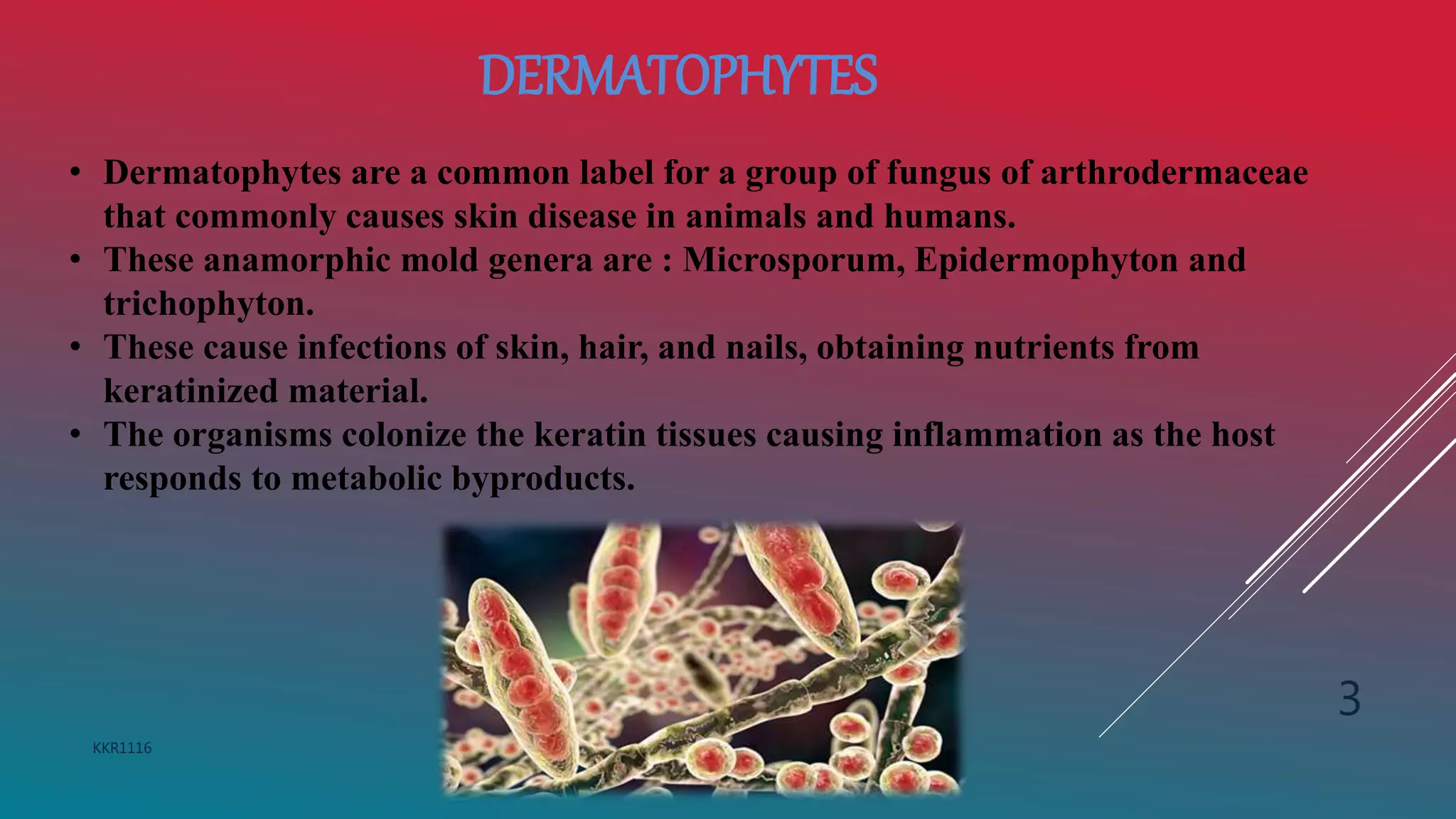
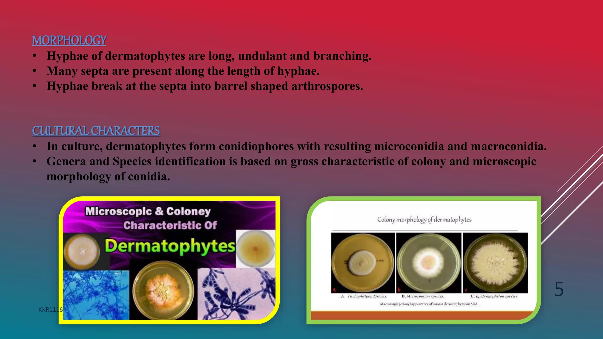
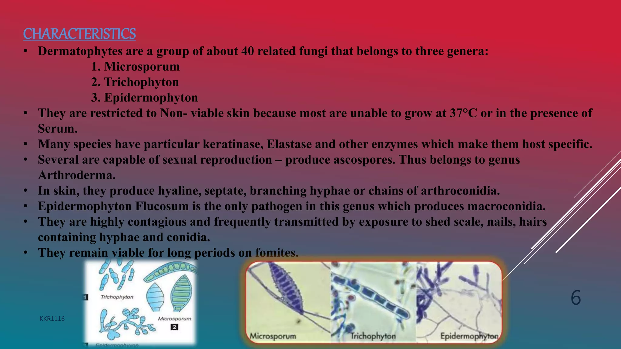
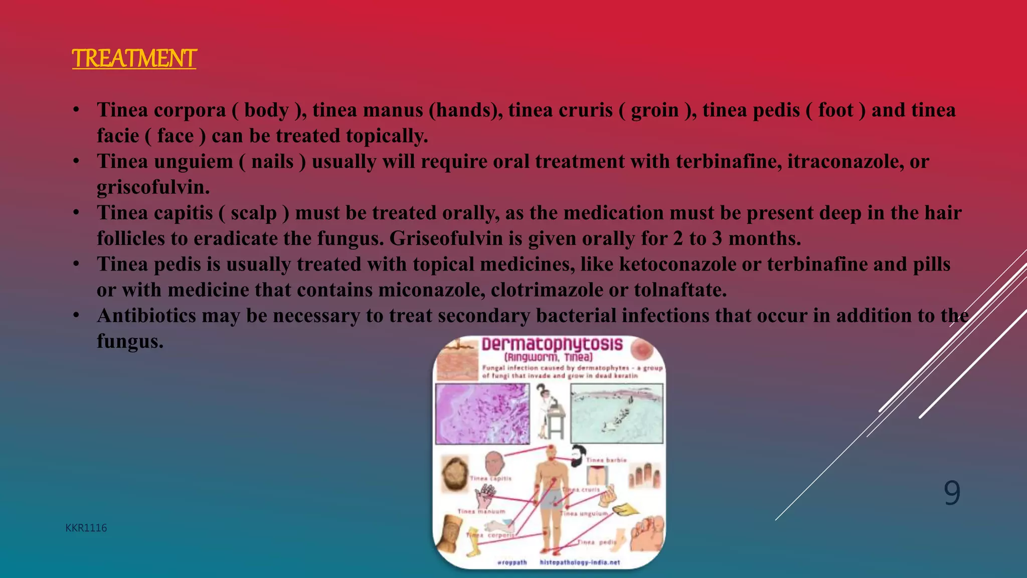
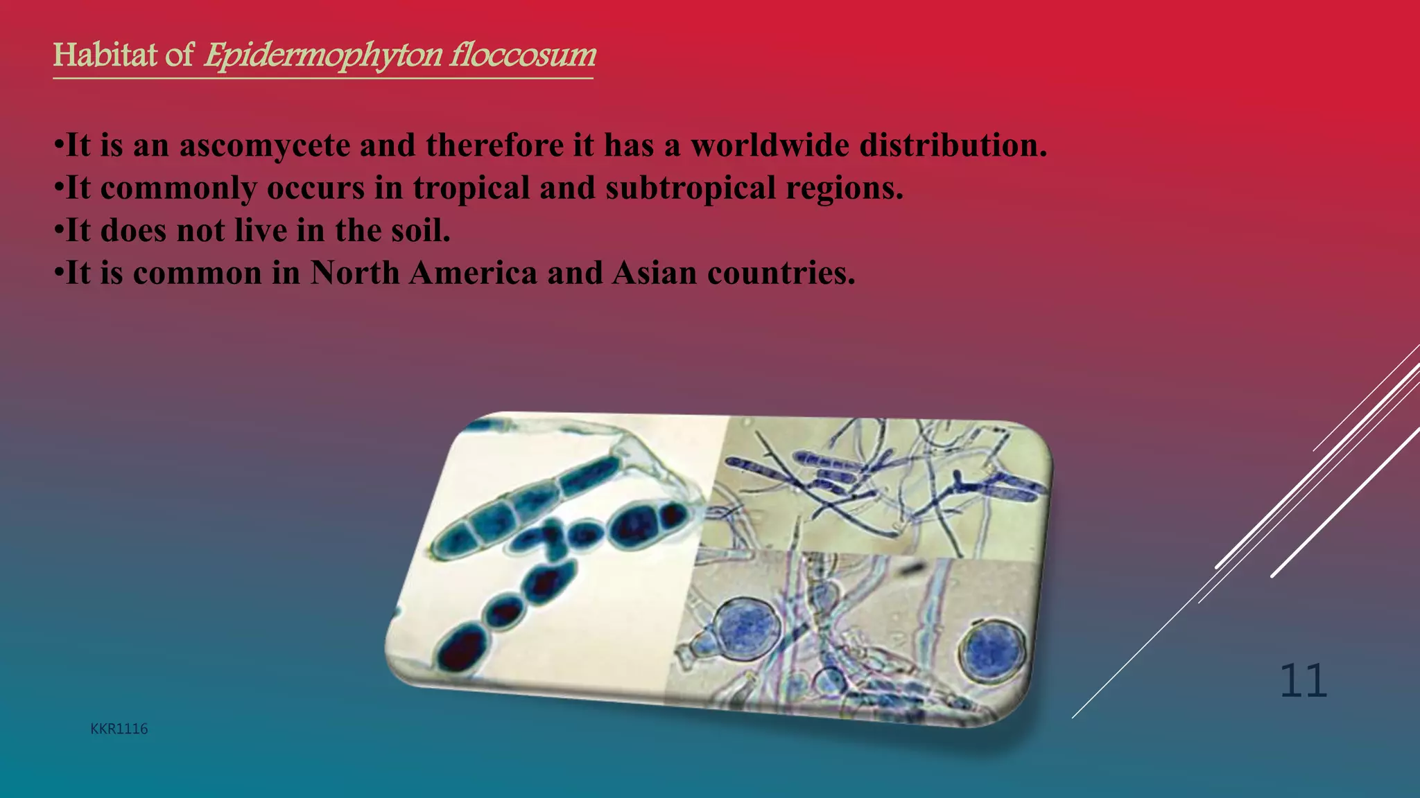
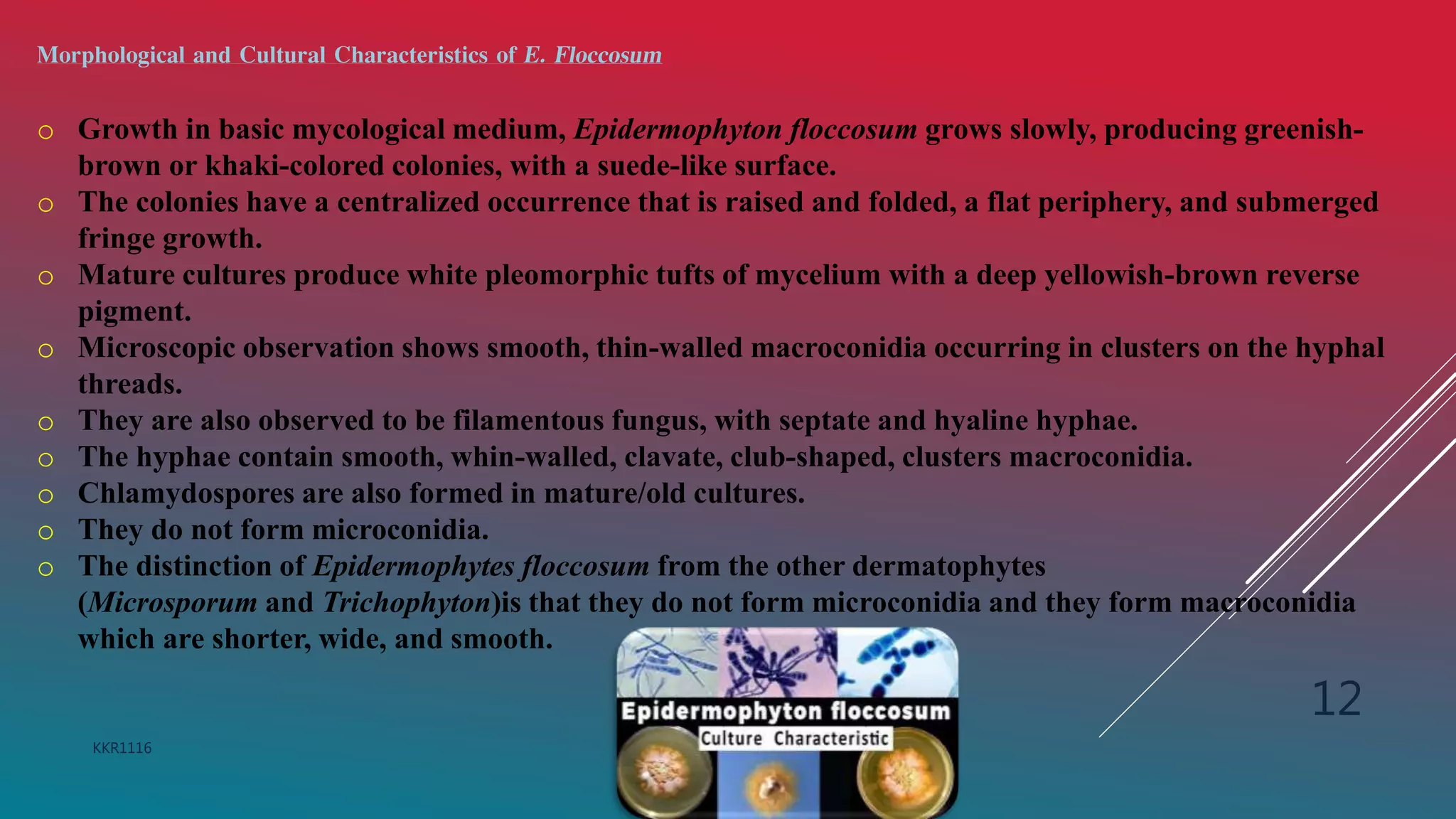
1) Dermatomycosis is a fungal infection of the skin, hair, and nails caused by dermatophytes such as Microsporum, Epidermophyton, and Trichophyton. Common symptoms include a skin rash and nail discoloration.
2) Epidermophyton floccosum is an anthropophilic dermatophyte that causes infections like athlete's foot and ringworm. It produces smooth-walled macroconidia in clusters and grows in culture as greenish-brown colonies.
3) Infections are diagnosed microscopically by viewing macroconidia in skin scrapings or cultures. Topical and oral antifungals
it is based on Harrisons and Davidson text book of internal medicine and Anathanarayanan textbook of microbiology. many clinical pictures have been embeded for better understanding. most common conditions seen in dermatology wards.
Largest viruses that infect vertebrates
Can be seen under light microscope
Poxvirus diseases are characterized by skin lesions – localized or generalized
Important diseases caused by poxviruses are-
Smallpox
Monkeypox
Cowpox
Tanapox
Molluscum contagiosum
Superficial Mycoses Mycology - Tinea Versicolor / Tinea Nigra/Piedra
For Downloading PDF note
As the channel name suggests, our channel will be a perfect lounge for the malayali medicos..we wil be covering videos which will be like lecture classes related to the subjects biochemistry and microbiology in which we are specialised.. It will be a better learning experience for the students especially for those who are not able to understand and follow the normal classes in college..we assure the students that you will get a basic idea regarding the topic and extra reading can be done from the reference textbooks..
Qualification
AHLAD T O
Maneesha M Joseph
MSc MLT (Microbiology)
Assistant Professor
Baby memorial college of allied Health science
Kozhikode
Our Partner Channel
Health & Voyage channel link - https://youtu.be/nzKqRVjlwc0
#Superficial Mycoses Mycology microbiology
#Medical
#Microbiology
#Superficial Mycoses Mycology malayalam lecturer
#Mallu Medicos Lounge
##MalluMedicosLounge
#MLT
#Tinea Versicolor
#Tinea Nigra
#Piedra
Introduction
Disease
Important Properties
Transmission & Epidemiology
Risk factor of reactivation
Pathogenesis
Clinical Findings
Laboratory Diagnosis
Approaches to the diagnosis of latent infections
Treatment
Prevention
Poxviruses are brick or oval-shaped viruses with large double-stranded DNA genomes. Poxviruses exist throughout the world and cause disease in humans and many other types of animals. Poxvirus infections typically result in the formation of lesions, skin nodules, or disseminated rash.
Cryptococcosis also called as Torulosis is a subacute or chronic fungal infection caused by Cryptococcus neoformans. It leads to compications such as fatal meningoencephalitis. It is an opportunistic infection in HIV-infected patients. The PPT discuss on the morphology of the fungus, pathogenesis, laboratory diagnosis and treatment.
Pneumonia is an infection of one or both lungs caused by bacteria, viruses and fungi. An infection of lung that involves the small air alveoli and the tissue around is called pneumonia.
Medical Mycology Black Piedra and White Piedra.pptxDeborahAR1
Black piedra is a fungal infection of the hair shafts. It is also known as Trichomycosis nodosa. The fungal elements are attached to the hair shaft to form nodules along the hair shaft. It predominantly affects scalp hair, although involvement of the beard, mustache and pubic hairs is also known.
White Piedra is a superficial fungal infection of the hair caused by Trichosporon asahii. It is also known as trichomycosis nodosa or trichomycosis nodularis.
Treponema is a genus of spiral-shaped bacteria. The major treponeme species of human pathogens is Treponema pallidum, whose subspecies are responsible for diseases such as syphilis, bejel, and yaws.
Richard's entangled aventures in wonderlandRichard Gill
Since the loophole-free Bell experiments of 2020 and the Nobel prizes in physics of 2022, critics of Bell's work have retreated to the fortress of super-determinism. Now, super-determinism is a derogatory word - it just means "determinism". Palmer, Hance and Hossenfelder argue that quantum mechanics and determinism are not incompatible, using a sophisticated mathematical construction based on a subtle thinning of allowed states and measurements in quantum mechanics, such that what is left appears to make Bell's argument fail, without altering the empirical predictions of quantum mechanics. I think however that it is a smoke screen, and the slogan "lost in math" comes to my mind. I will discuss some other recent disproofs of Bell's theorem using the language of causality based on causal graphs. Causal thinking is also central to law and justice. I will mention surprising connections to my work on serial killer nurse cases, in particular the Dutch case of Lucia de Berk and the current UK case of Lucy Letby.
Multi-source connectivity as the driver of solar wind variability in the heli...Sérgio Sacani
The ambient solar wind that flls the heliosphere originates from multiple
sources in the solar corona and is highly structured. It is often described
as high-speed, relatively homogeneous, plasma streams from coronal
holes and slow-speed, highly variable, streams whose source regions are
under debate. A key goal of ESA/NASA’s Solar Orbiter mission is to identify
solar wind sources and understand what drives the complexity seen in the
heliosphere. By combining magnetic feld modelling and spectroscopic
techniques with high-resolution observations and measurements, we show
that the solar wind variability detected in situ by Solar Orbiter in March
2022 is driven by spatio-temporal changes in the magnetic connectivity to
multiple sources in the solar atmosphere. The magnetic feld footpoints
connected to the spacecraft moved from the boundaries of a coronal hole
to one active region (12961) and then across to another region (12957). This
is refected in the in situ measurements, which show the transition from fast
to highly Alfvénic then to slow solar wind that is disrupted by the arrival of
a coronal mass ejection. Our results describe solar wind variability at 0.5 au
but are applicable to near-Earth observatories.
Slide 1: Title Slide
Extrachromosomal Inheritance
Slide 2: Introduction to Extrachromosomal Inheritance
Definition: Extrachromosomal inheritance refers to the transmission of genetic material that is not found within the nucleus.
Key Components: Involves genes located in mitochondria, chloroplasts, and plasmids.
Slide 3: Mitochondrial Inheritance
Mitochondria: Organelles responsible for energy production.
Mitochondrial DNA (mtDNA): Circular DNA molecule found in mitochondria.
Inheritance Pattern: Maternally inherited, meaning it is passed from mothers to all their offspring.
Diseases: Examples include Leber’s hereditary optic neuropathy (LHON) and mitochondrial myopathy.
Slide 4: Chloroplast Inheritance
Chloroplasts: Organelles responsible for photosynthesis in plants.
Chloroplast DNA (cpDNA): Circular DNA molecule found in chloroplasts.
Inheritance Pattern: Often maternally inherited in most plants, but can vary in some species.
Examples: Variegation in plants, where leaf color patterns are determined by chloroplast DNA.
Slide 5: Plasmid Inheritance
Plasmids: Small, circular DNA molecules found in bacteria and some eukaryotes.
Features: Can carry antibiotic resistance genes and can be transferred between cells through processes like conjugation.
Significance: Important in biotechnology for gene cloning and genetic engineering.
Slide 6: Mechanisms of Extrachromosomal Inheritance
Non-Mendelian Patterns: Do not follow Mendel’s laws of inheritance.
Cytoplasmic Segregation: During cell division, organelles like mitochondria and chloroplasts are randomly distributed to daughter cells.
Heteroplasmy: Presence of more than one type of organellar genome within a cell, leading to variation in expression.
Slide 7: Examples of Extrachromosomal Inheritance
Four O’clock Plant (Mirabilis jalapa): Shows variegated leaves due to different cpDNA in leaf cells.
Petite Mutants in Yeast: Result from mutations in mitochondrial DNA affecting respiration.
Slide 8: Importance of Extrachromosomal Inheritance
Evolution: Provides insight into the evolution of eukaryotic cells.
Medicine: Understanding mitochondrial inheritance helps in diagnosing and treating mitochondrial diseases.
Agriculture: Chloroplast inheritance can be used in plant breeding and genetic modification.
Slide 9: Recent Research and Advances
Gene Editing: Techniques like CRISPR-Cas9 are being used to edit mitochondrial and chloroplast DNA.
Therapies: Development of mitochondrial replacement therapy (MRT) for preventing mitochondrial diseases.
Slide 10: Conclusion
Summary: Extrachromosomal inheritance involves the transmission of genetic material outside the nucleus and plays a crucial role in genetics, medicine, and biotechnology.
Future Directions: Continued research and technological advancements hold promise for new treatments and applications.
Slide 11: Questions and Discussion
Invite Audience: Open the floor for any questions or further discussion on the topic.
Seminar of U.V. Spectroscopy by SAMIR PANDASAMIR PANDA
Spectroscopy is a branch of science dealing the study of interaction of electromagnetic radiation with matter.
Ultraviolet-visible spectroscopy refers to absorption spectroscopy or reflect spectroscopy in the UV-VIS spectral region.
Ultraviolet-visible spectroscopy is an analytical method that can measure the amount of light received by the analyte.
This pdf is about the Schizophrenia.
For more details visit on YouTube; @SELF-EXPLANATORY;
https://www.youtube.com/channel/UCAiarMZDNhe1A3Rnpr_WkzA/videos
Thanks...!
THE IMPORTANCE OF MARTIAN ATMOSPHERE SAMPLE RETURN.Sérgio Sacani
The return of a sample of near-surface atmosphere from Mars would facilitate answers to several first-order science questions surrounding the formation and evolution of the planet. One of the important aspects of terrestrial planet formation in general is the role that primary atmospheres played in influencing the chemistry and structure of the planets and their antecedents. Studies of the martian atmosphere can be used to investigate the role of a primary atmosphere in its history. Atmosphere samples would also inform our understanding of the near-surface chemistry of the planet, and ultimately the prospects for life. High-precision isotopic analyses of constituent gases are needed to address these questions, requiring that the analyses are made on returned samples rather than in situ.
Deep Behavioral Phenotyping in Systems Neuroscience for Functional Atlasing a...Ana Luísa Pinho
Functional Magnetic Resonance Imaging (fMRI) provides means to characterize brain activations in response to behavior. However, cognitive neuroscience has been limited to group-level effects referring to the performance of specific tasks. To obtain the functional profile of elementary cognitive mechanisms, the combination of brain responses to many tasks is required. Yet, to date, both structural atlases and parcellation-based activations do not fully account for cognitive function and still present several limitations. Further, they do not adapt overall to individual characteristics. In this talk, I will give an account of deep-behavioral phenotyping strategies, namely data-driven methods in large task-fMRI datasets, to optimize functional brain-data collection and improve inference of effects-of-interest related to mental processes. Key to this approach is the employment of fast multi-functional paradigms rich on features that can be well parametrized and, consequently, facilitate the creation of psycho-physiological constructs to be modelled with imaging data. Particular emphasis will be given to music stimuli when studying high-order cognitive mechanisms, due to their ecological nature and quality to enable complex behavior compounded by discrete entities. I will also discuss how deep-behavioral phenotyping and individualized models applied to neuroimaging data can better account for the subject-specific organization of domain-general cognitive systems in the human brain. Finally, the accumulation of functional brain signatures brings the possibility to clarify relationships among tasks and create a univocal link between brain systems and mental functions through: (1) the development of ontologies proposing an organization of cognitive processes; and (2) brain-network taxonomies describing functional specialization. To this end, tools to improve commensurability in cognitive science are necessary, such as public repositories, ontology-based platforms and automated meta-analysis tools. I will thus discuss some brain-atlasing resources currently under development, and their applicability in cognitive as well as clinical neuroscience.
1. KARTHIK REDDY C A
MSC 3rd SEM MICROBIOLOGY
Reg No – NP20AL61
NRUPATHUNGA UNIVERSITY
BANGALORE - 01
KKR1116
1
2. INTRODUCTION
Dermatomycosis is one of the most frequent fungal
infections of skin and skin appendages, which
encompass nails and hair.
It is a mycotic diseases of skin caused by a few mycetes :
Dermatophytes and some opportunistic fungi.
These fungal infections impair superficial layers of the
skin, hair and nails.
Dermatomycosis mainly caused by filamentous fungi.
KKR1116
2
3. DERMATOPHYTES
• Dermatophytes are a common label for a group of fungus of arthrodermaceae
that commonly causes skin disease in animals and humans.
• These anamorphic mold genera are : Microsporum, Epidermophyton and
trichophyton.
• These cause infections of skin, hair, and nails, obtaining nutrients from
keratinized material.
• The organisms colonize the keratin tissues causing inflammation as the host
responds to metabolic byproducts.
KKR1116
3
4. EPIDERMIOLOGY
Dermatophyte fungi are the most common fungal infections world wide.
Dermatophytosis was first described by David Gruby, a Hungarian
physician in 1841.
Before Gruby, various scientist described lesions which were ring like and
were thought to be infective.
The description of lesions dates back to the Roman era.
Around 1890, Raimond sabouraud advance knowledge of
dermatomycology by studying extensively into the taxonomy, morphology
and treatment of dermatophytes even classifying these fungal agents into 4
genera.
Dermatophytosis has been prevalent since early 1900’S at which time
ringworm was treated with compounds of mercury or sometimes sulfur or
iodine.
KKR1116
4
5. MORPHOLOGY
• Hyphae of dermatophytes are long, undulant and branching.
• Many septa are present along the length of hyphae.
• Hyphae break at the septa into barrel shaped arthrospores.
CULTURALCHARACTERS
• In culture, dermatophytes form conidiophores with resulting microconidia and macroconidia.
• Genera and Species identification is based on gross characteristic of colony and microscopic
morphology of conidia.
KKR1116
5
6. CHARACTERISTICS
• Dermatophytes are a group of about 40 related fungi that belongs to three genera:
1. Microsporum
2. Trichophyton
3. Epidermophyton
• They are restricted to Non- viable skin because most are unable to grow at 37°C or in the presence of
Serum.
• Many species have particular keratinase, Elastase and other enzymes which make them host specific.
• Several are capable of sexual reproduction – produce ascospores. Thus belongs to genus
Arthroderma.
• In skin, they produce hyaline, septate, branching hyphae or chains of arthroconidia.
• Epidermophyton Flucosum is the only pathogen in this genus which produces macroconidia.
• They are highly contagious and frequently transmitted by exposure to shed scale, nails, hairs
containing hyphae and conidia.
• They remain viable for long periods on fomites.
KKR1116
6
7. SYMPTOMS
o The first symptom is a distinctive skin rash on the face, eyelids,
chest, nails, cuticle areas, knees or elbows.
o The rash is patchy and usually a bluish to purple colour.
o May also get muscle weakness that gets work over weeks or
months. This muscle weakness usually starts in neck, arms, or
hips and can be felt on both sides of body.
o Other symptoms are –
o Muscle pain and tenderness
o Swallowing problems and lung problems
o Hard calcium deposits underneath, the skin which is mostly seen
in children.
o Fatigue, unintentional weight loss, fever.
There is a subtype of dermatomyositis that includes the rash but
not muscle weakness. This is know as amyopathic dermatomyositis.
KKR1116
7
8. LABORATORYDIAGNOSIS
• MRI is used to observe abnormal muscles.
• Electromyography ( EMG ) to record electrical impulses that control
muscles.
• Blood analysis to check the levels of muscle enzymes and
autoantibodies, which are antibodies that attack normal cells.
• A muscle biopsy to look for inflammation and other problems
associated with the disease in a sample of muscle tissue.
• A skin biopsy to look for changes caused by the disease in a skin
sample.
KKR1116
8
9. TREATMENT
• Tinea corpora ( body ), tinea manus (hands), tinea cruris ( groin ), tinea pedis ( foot ) and tinea
facie ( face ) can be treated topically.
• Tinea unguiem ( nails ) usually will require oral treatment with terbinafine, itraconazole, or
griscofulvin.
• Tinea capitis ( scalp ) must be treated orally, as the medication must be present deep in the hair
follicles to eradicate the fungus. Griseofulvin is given orally for 2 to 3 months.
• Tinea pedis is usually treated with topical medicines, like ketoconazole or terbinafine and pills
or with medicine that contains miconazole, clotrimazole or tolnaftate.
• Antibiotics may be necessary to treat secondary bacterial infections that occur in addition to the
fungus.
KKR1116
9
10. Epidermophyton
• Epidermophyton floccosum is a filamentous dermatophytic fungus known to cause
skin and nail infections in humans.
• It is an anthropophilic dermatophyte hence it is transmissible from one individual to
another.
• Common infections caused by dermatophytes include tinea pedis (athlete’s foot),
tinea crusis, tinea corporis, and onychomycosis.
• It is the third most common cause of Tinea pedis (athletes foot).
KKR1116
10
11. Habitat of Epidermophyton floccosum
•It is an ascomycete and therefore it has a worldwide distribution.
•It commonly occurs in tropical and subtropical regions.
•It does not live in the soil.
•It is common in North America and Asian countries.
KKR1116
11
12. Morphological and Cultural Characteristics of E. Floccosum
o Growth in basic mycological medium, Epidermophyton floccosum grows slowly, producing greenish-
brown or khaki-colored colonies, with a suede-like surface.
o The colonies have a centralized occurrence that is raised and folded, a flat periphery, and submerged
fringe growth.
o Mature cultures produce white pleomorphic tufts of mycelium with a deep yellowish-brown reverse
pigment.
o Microscopic observation shows smooth, thin-walled macroconidia occurring in clusters on the hyphal
threads.
o They are also observed to be filamentous fungus, with septate and hyaline hyphae.
o The hyphae contain smooth, whin-walled, clavate, club-shaped, clusters macroconidia.
o Chlamydospores are also formed in mature/old cultures.
o They do not form microconidia.
o The distinction of Epidermophytes floccosum from the other dermatophytes
(Microsporum and Trichophyton)is that they do not form microconidia and they form macroconidia
which are shorter, wide, and smooth.
KKR1116
12
13. Figure: Pleomorphism of E. floccosum. A: Overview of the Wild-Type surface; B: Revise
side of wild-type; C: Overview of pleomorphic E. floccosum; D: Revise side of
pleomorphic E. floccosum.
Image Source: Creative Biolabs
KKR1116
13
14. Pathogenesis and clinical manifestations of Epidermophyton floccosum
o Epidermophyton floccosum is transmitted from person-to-person via skin contact, causing cutaneous
and subcutaneous infections on the skin, and nails.
o As a dermatophyte, it causes dermatophytosis affecting the keratinized regions in the body including
hair, skin, and nails.
o Symptoms vary depending on the site of the body affected, such as:
o Hair infections are Tinea capitis, Tinea barbae characterized by invasion of hair follicles (without
perforation), hair loss (ectothrix), and/or hair breakage (endothrix).
o On the skin, the formation of lesions is commonly characterized by circular or annular ringworm
infections (tinea corporis).
o As an anthropophilic fungus, it does not cause inflammations or hypersensitivity.
o They have also been associated with causing keratitis of the eye.
o Infection of the nails is commonly characterized by discoloration, dystrophy, hyperkeratosis, and
onycholysis.
o Manifestations occur several weeks after contracting the fungus.
o Disseminated and invasive disease infections are rare.
o However, invasive Epidermophyton floccosum infection has been documented in persons with Behcet’s
syndrome.
KKR1116
14
15. Laboratory Diagnosis of Epidermophytonfloccosum
Specimen: Skin scrapings, pus/lesion biopsies, hair scarpings
Microscopic Examination
10-20% KOH Wet mount for observation of smooth, thin-walled, wide macroconidia.
Cultural Examination
Growth in Sabouraud Dextrose Agar with cycloheximide and chloramphenicol to suppress mold and
bacterial growth and observation of growth in 3 weeks. The colonies formed are greenish-brown or
khaki-colored with a suede-like surface.
The colonies also appear raised and folded at the center with a flattened periphery.
Extended growth produces white colonies showing pleomorphism in mycelial growth, producing deep
yellow to brown mycelium on reverse pigmentation.
KKR1116
15
16. Molecularcharacterization and differentiation
Use of PCR-Restriction Fragment Length Plormophism (PCR-RFLP) is used to distinguish the 12
species that cause dermatophytosis infections.
Real-time PCR is also used to identify Epidermophyton floccosum after fungal lysis
Treatment of Epidermophytonfloccosuminfections
Tinea capitis is tretaed with griseofulvin or oral azoles (ketoconazole, itraconazole, and
voriconazole).
Tinea corporis, tinea crisis, tinea pedis, and tinea manuun are treated tropically using naftifine,
terbinafine, butenafine, clotrimazole, econazole, ketoconazole, miconazole, oxiconazole,
sulconazole, cyclopyrox, and tolnaftate.
Nail infections, topical therapy is normally unsuccessful, and therefore, the use of systemic oral
therapy for a prolonged period is advised
KKR1116
16
17. A Text book of Microbes and Diseases of Man – W C DEB.
Text book of Medical microbiology – H L CHOPRA.
Fritz H. Kayser (2005). Medical microbiology. Thiemeverlag.
https://www.healthline.com/health/dermatomycosis.
https://microbnotes.com/Epidermophyton-floccosum.
https:www.slideshare,net/AnkurVashishtha4/dermatophytes.
KKR1116
17