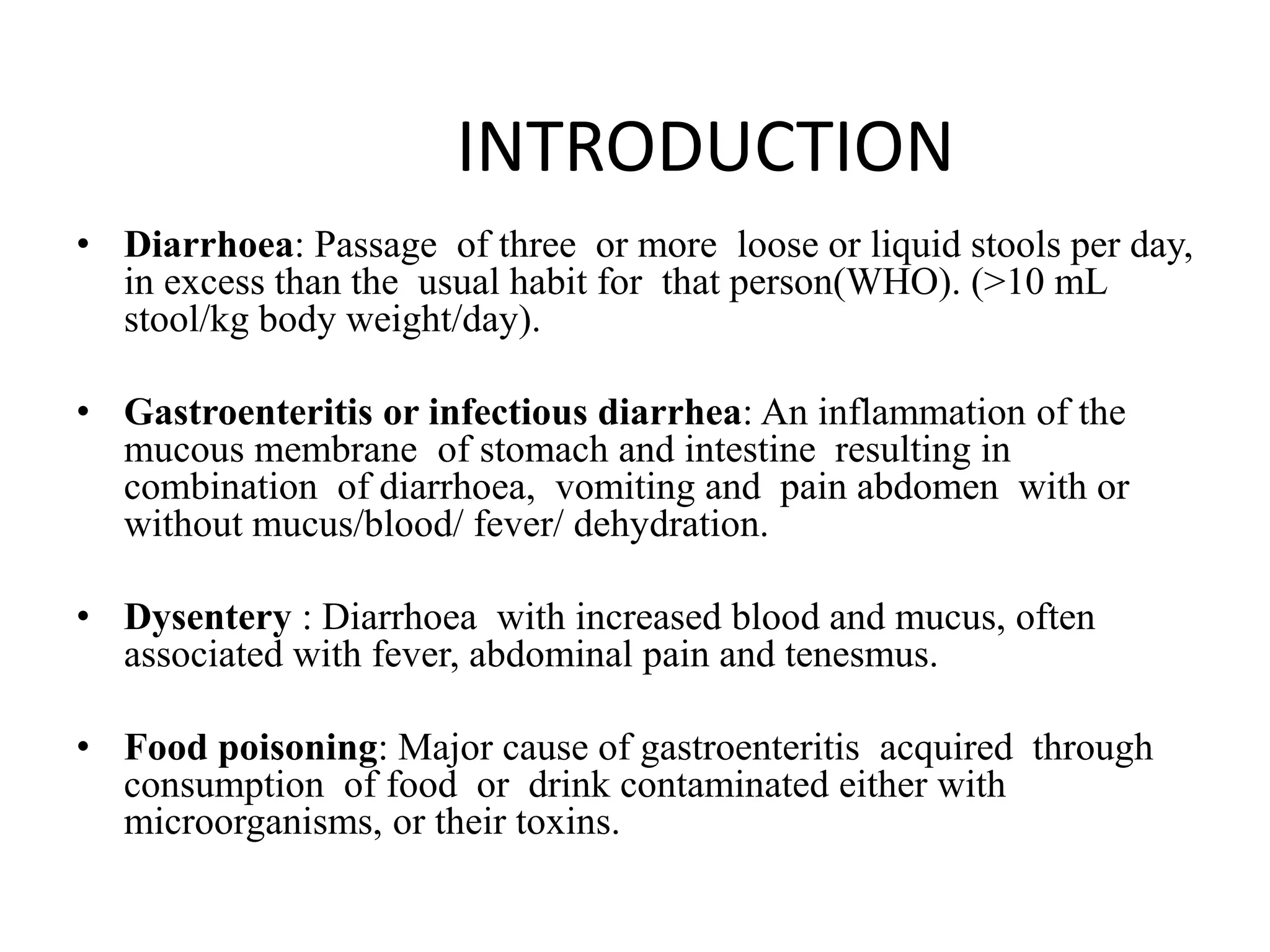
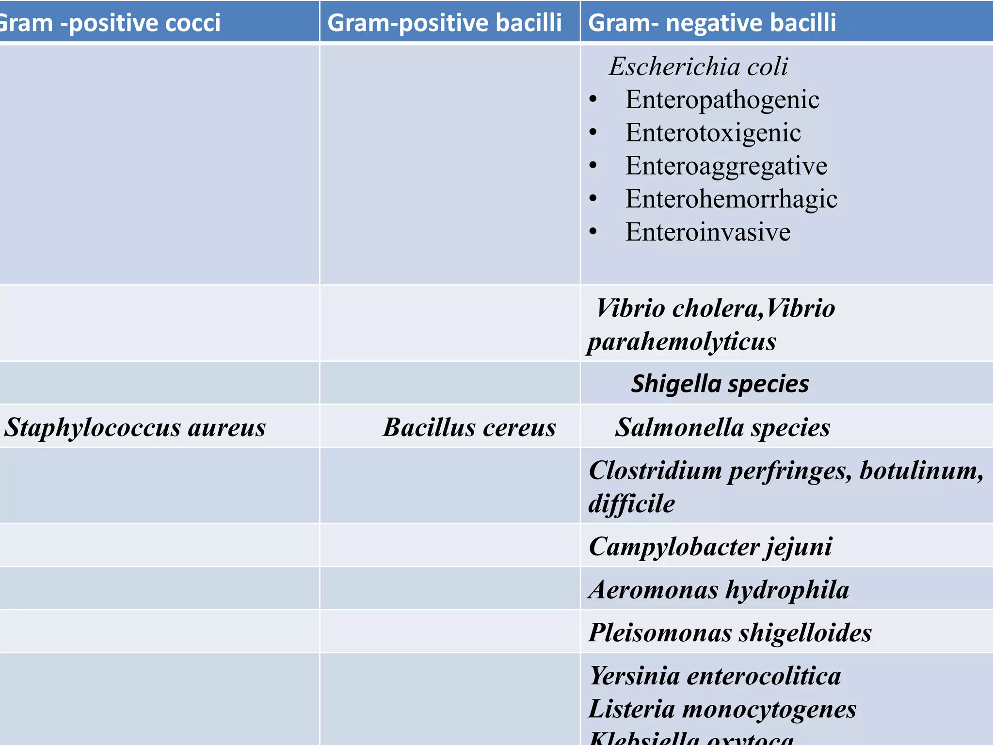
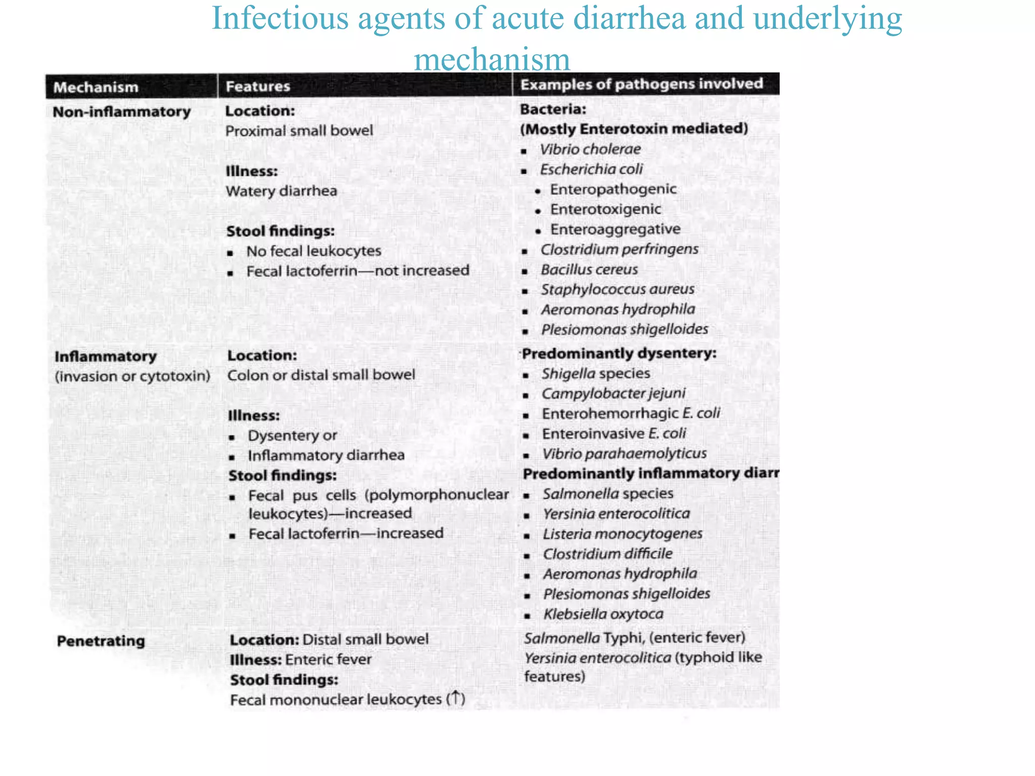
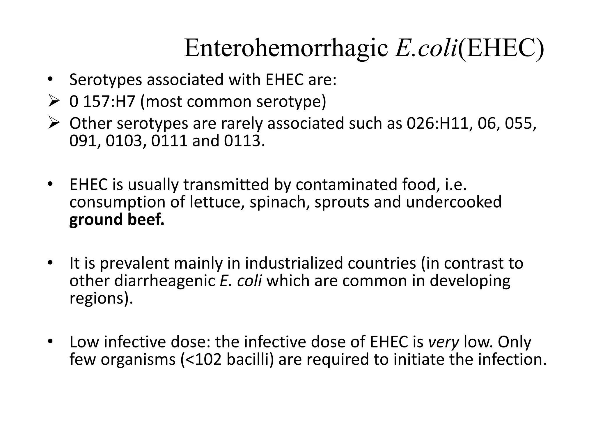
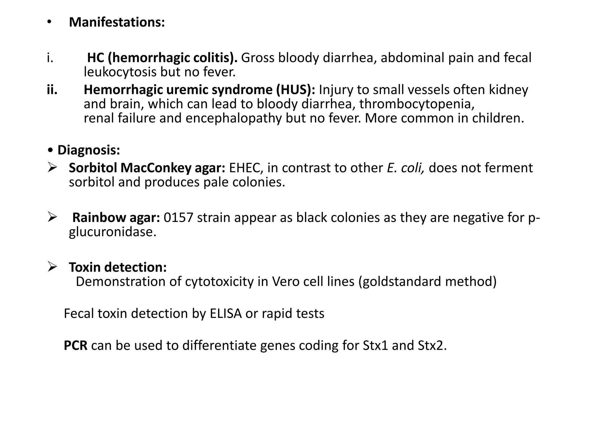
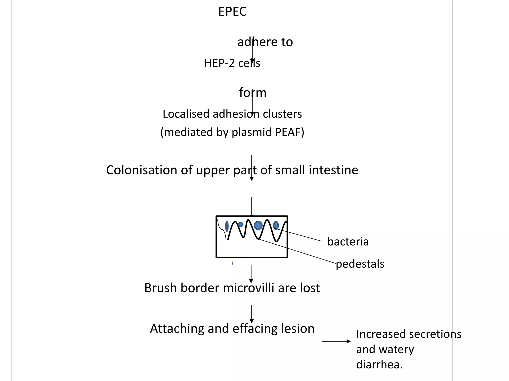
SlideShare a Scribd company logo
1 of 76
Bacterial diarrhea and dysentery
Dr. Dinesh Kr Jain, MD.,
Assistantprofessor,
Department of Microbiology,
SMS Medical college, Jaipur
• Diarrhoea: Passage of three or more loose or liquid stools per day,
in excess than the usual habit for that person(WHO). (>10 mL
stool/kg body weight/day).
• Gastroenteritis or infectious diarrhea: An inflammation of the
mucous membrane of stomach and intestine resulting in
combination of diarrhoea, vomiting and pain abdomen with or
without mucus/blood/ fever/ dehydration.
• Dysentery : Diarrhoea with increased blood and mucus, often
associated with fever, abdominal pain and tenesmus.
• Food poisoning: Major cause of gastroenteritis acquired through
consumption of food or drink contaminated either with
microorganisms, or their toxins.
INTRODUCTION
Bacteria causing diarrhea
Gram -positive cocci Gram-positive bacilli Gram- negative bacilli
Escherichia coli
• Enteropathogenic
• Enterotoxigenic
• Enteroaggregative
• Enterohemorrhagic
• Enteroinvasive
Vibrio cholera,Vibrio
parahemolyticus
Shigella species
Staphylococcus aureus Bacillus cereus Salmonella species
Clostridium perfringes, botulinum,
difficile
Campylobacter jejuni
Aeromonas hydrophila
Pleisomonas shigelloides
Yersinia enterocolitica
Listeria monocytogenes
Infectious agents of acute diarrhea and underlying
mechanism
Cell adherence
• Attachment within or close to mucosal cells.
Enterohemorrhagic E.coli(EHEC)
• Serotypes associated with EHEC are:
 0 157:H7 (most common serotype)
 Other serotypes are rarely associated such as 026:H11, 06, 055,
091, 0103, 0111 and 0113.
• EHEC is usually transmitted by contaminated food, i.e.
consumption of lettuce, spinach, sprouts and undercooked
ground beef.
• It is prevalent mainly in industrialized countries (in contrast to
other diarrheagenic E. coli which are common in developing
regions).
• Low infective dose: the infective dose of EHEC is very low. Only
few organisms (<102 bacilli) are required to initiate the infection.
• Pathogenesis:
• Produces verocytotoxin or Shiga-like toxin
• It is so named because it is cytotoxic to vero
cell lines and it resembles shiga toxin in its
structure and function.
• Detection of VT:
Serologically- Latex agglutination, ELISA
Molecular methods-using specific DNA
probe
Cytotoxicity on Vero and Hela cell lines
• Manifestations:
i. HC (hemorrhagic colitis). Gross bloody diarrhea, abdominal pain and fecal
leukocytosis but no fever.
ii. Hemorrhagic uremic syndrome (HUS): Injury to small vessels often kidney
and brain, which can lead to bloody diarrhea, thrombocytopenia,
renal failure and encephalopathy but no fever. More common in children.
• Diagnosis:
 Sorbitol MacConkey agar: EHEC, in contrast to other E. coli, does not ferment
sorbitol and produces pale colonies.
 Rainbow agar: 0157 strain appear as black colonies as they are negative for p-
glucuronidase.
 Toxin detection:
Demonstration of cytotoxicity in Vero cell lines (goldstandard method)
Fecal toxin detection by ELISA or rapid tests
PCR can be used to differentiate genes coding for Stx1 and Stx2.
Mechanism of action
Enteropathogenic
E.coli(EPEC)
• EPEC frequently causes infantile diarrhea (outbreaks) esp in tropical
countries.
• Occasionally cause sporadic diarrhea in adults.
• Person-to-person spread is seen.
• It is non toxigenic and non invasive.
• Common serogroups- O26, O55, O86, O111, O114, O119, O125, O126,
O127, O128, O142, O158
Pathogenesis:- Plasmid mediated.
 Strains which carry plasmid factor pEAF are called typical EPEC(tEPEC)
 Strains without this plasmid are termed as ‘atypical’ EPEC (aEPEC)
EPEC
adhere to
HEP-2 cells
form
Localised adhesion clusters
(mediated by plasmid PEAF)
Colonisation of upper part of small intestine
Brush border microvilli are lost
Attaching and effacing lesion
pedestals
bacteria
Increased secretions
and watery
diarrhea.
Neurotoxin
• Neurotoxins are toxins that are destructive to nerve
tissue (causing neurotoxicity).
• They are exogenous chemical neurological insults that
can adversely affect function in both developing and
mature nervous tissue
• They inhibit neuron control over ion concentrations
across the cell membrane,or communication between
neurons across a synapse.
• Neurotoxins can also cause glial cell damage, central
nervous system damage such as intellectual
disability persistent memory impairments, epilepsy,
and dementia.
Staphylococcus aureus
• Secretes enterotoxin which acts as neurotoxin.
• It is a preformed toxin.
• Incubation period: 1- 6 hours.
• Causes food poisioning.
• Site of action:
– toxin stimulates the vagus nerve and the vomiting center of the brain.
– also stimulate the intestinal peristaltic activity.
• Symptoms: nausea, vomiting , diarrhea, hypotension, and dehydration (no
fever),resolve within 8- 10 hours.
• Source of infection: food handler(carrier of S. aureus).
• Most common food items involved are milk products, bakery food,
custard, potato salad, or processed meats.
• Heat stable toxin and resistant to gastric juice.
Bacillus cereus
• Normal habitant of soil.
• It produces several toxins implicated in the diarrheal
symptoms, including hemolysin BL (HBL), nonhemolytic
enterotoxin (Nhe), and cytotoxin K (CytK; also referred to
as hemolysin IV).
• The three toxins are believed to act synergistically, with
Nhe responsible for the major symptoms in the diarrheal
presentation of the infection.
• B. cereus group “food poisoning” is associated with the
ingestion of a wide variety of foods including meats,
vegetables, desserts, sauces, and milk.
• A higher incidence is seen after the ingestion of rice dishes.
• After ingestion, patients present with one of two types of
symptoms: diarrhea and abdominal pain within 8 to 16
hours, or nausea and vomiting (emetic food poisoning)
within 1 to 5 hours.
Clostridium botulinum
• Anaerobic bacteria.
• Causes botulism.
• Distributed as saprophyte in soil, animal manure, vegetables and
sea mud.
• Non invasive.
• Transmission: spores ingested or inhaled.
• Pathogenesis: due to production of powerful neurotoxin
‘botulinum toxin’(BT).
• BT:
– 150 kDa zinc dependent protein consisting of 100 kDa heavy chain and a 50
KdA light chain.
– Produced intracellularly and appears outside only after autolysis of
bacterial cell
• 8 serotypes- A, B, C1, C2, D, E, F and G (most severe A)
• Common source: home made canned food(since growth requires
low oxygen content).
• Food borne botulism: results from food contaminated with
preformed botulinum toxin.
• After ingestion of toxin, within 24 hr or less there is progressive
paralysis affecting occulomotor and oropharyngeal muscles
followed by vomiting, diarrhea, abdominal swelling.
• Mechanism of action:After ingestion
of food
BT transported via blood to peripheral cholinergic
nerve terminal
Binds to acetylcholine receptors at NMJ
Blockage of release of Acetylcholine
Flaccid paralysis
Enterotoxin
• An enterotoxin is a protein exotoxin released by
a microorganism that targets the intestines.
• Enterotoxins are frequently cytotoxic and kill cells
by altering the apical membrane permeability of
the mucosal (epithelial) cells of the intestinal
wall.
• They are mostly pore-forming toxins (mostly
chloride pores), secreted by bacteria, that
assemble to form pores in cell membranes which
causes the cells to die.
Vibrio Cholera
• Pathogenesis of Cholera Both V. cholera O1 and 0139 are
capable of producing cholera toxin, thus resulting in cholera.
• Mode of transmission: V. cholerae is transmitted by ingestion
of contaminated water or food.
• Infective dose: Since V. cholerae is extremely acid labile; a high
infective dose of 10 bacilli is required to bypass the gastric
barrier.
• Factors promoting transmission: These include all those
conditions where gastric acidity is reduced, such as
hypochlorhydria, use of antacids, etc.
• Mechanism of action- similar to heat labile enterotoxin of
E.coli.
• Gene for cholera toxin (CTX):- CTXФ
• ToxR gene: It regulates the expression of CT and is itself regulated by
environmental factors, such as heat shock response.
• Chromosomes: V. cholerae has two circular chromosomes, one large and one
small.
 Large chromosome- growth and intestinal survival.
 Small chromosome- environmental survival.
• Other virulence factors include:
 Zona occludens toxin: It disrupts the light junctions between mucosal cells.
 Accessory cholera enterotoxin: Phage packaging and secretion.
 Vero cell toxin: It is analogous to the toxin produced by Shigella dysenteriae 1.
It is important for the survival of the bacilli in aquatic environment.
 Accessory colonization factors: help in adhesion and colonization.
 Siderophore: for iron acquisition .
Clinical Manifestations of Cholera
V. cholerae 01 or O139 infections produce a range of clinical
manifestations such as:
1. Asymptomatic infection (75% of cases)
2. Mild diarrhea or cholera (20% of cases)
3. Sudden onset of explosive and life-threatening diarrhea
(cholera gravis, in 5% of cases).
• Incubation period varies from 24 to 48 hours.
• The usual manifestations include:
 Watery diarrhea
 Rice water stool:
 Vomiting
 No fever
 Muscle cramps
Complications are directly proportional to the fluid loss
resulting in loss of body weight.
Epidemiological Determinants
• Reservoir: Humans.
• Source: either asymptomatic cases or carriers.
• Carriers: healthy people who shed the bacilli in feces.
• Cholera season: high temperatures, heavy rainfall and flooding.
• Risk factors:
– Poor sanitation, poverty, overcrowding, population
mobility.
– Lack of pre-existing immunity
– Persons with 'O' blood group are at greater risk, while
those with type AB
blood group are at least risk.
– Malnutrition
Treatment
• Fluid replacement: to correct hypovolemia and thereafter to be
maintained to replace the ongoing fluid losses.
• In mild to moderate fluid loss: Oral rehydration solution (ORS) should
be given.
• In severe cases: Intravenous fluid replacement with Ringer's lactate (or
normal saline) should be carried out till the consciousness arrives.
Thereafter replaced by ORS
• Antibiotic therapy:
vaccines
• Injectable Killed Vaccines
They are no longer in use, as they provide little
protection, cause adverse effects and fail to induce a
local intestinal mucosal immune response
• Oral Cholera Vaccines (OCV)
Oral cholera vaccines are currently in practice. two types
of oral vaccines are available.
 Killed Whole-cell vaccine:
two preparations are available:
1. Whole-cell (WC) vaccine: It is composed of killed
whole cells of V.cholerae O1 (classical and El Tor, Inaba
and Ogawa).
2. Whole-cell recombinant B subunit cholera
vaccine(WC/rBS) (Dukoral): Composition is same as that of
WC vaccine, in addition it has recombinant cholera toxin B
subunit
• Schedule:
Two doses are given orally, at 7 days gap except for children 2- 5
years (3 doses). Its not licensed for children less than 2 years.
• WHO recommends for using vaccine during epidemics and out
breaks in the community but not during inter epidemic period.
Oral Iive attenuated vaccines (OCV): Several live OCV are under
trial such as:
CVD 103-HgR, Peru-15 and V. cholerae H38 for classical and/ or El
Tor biotypes of V. cholerae 01.
• CVD-112 and Bengal-15 vaccine for V. cholerae 0 139.
• CVD 103-HgR vaccine (Orochol) contains a live attenuated strain
derived from reference strain 569 B (classical, 0 1, Inaba), given as
single dose. Its protection starts after 8 days.
• Use of OCV is recommended to limit the risk
of:
 Occurrence of cholera outbreaks in
displaced populations in endemic areas.
Spread and incidence of cholera during an
outbreak.
NICED: National reference Center for
cholera in India is located at National
lnstitute of Cholera and Enteric Diseases
(NICED), Kolkata.
Vibrio parahemolyticus
• Though V. parahemolyticus was first reported from Japan
(1953), the incidence of infection has greatly increased in
several countries including Japan since 1993. In India, it has
been reported from Kolkata.
ClinicalManifestations
• Food-borne gastroenteritis is the most common
presentation.
• Occurs following raw or uncooked sea food (e.g. oyster)
intake.
• It commonly presents as watery diarrhea or rarely as
dysentery with abdominal cramps
Pathogenesis:
• Virulence factors:
• Polysaccharide capsule which prevents the bacilli from
phagocytosis.
• Hemolysin (thermo -stable)
• Urease enzyme (in few strains): It breaks down urea from food
to release ammonia that buffers gastric acidity.
• It possesses two type III secretion systems in cell wall, which
directly inject toxic bacterial proteins into host cells.
Serotype: V. parahaemolyticus has 13 0 somatic
antigens and more than 60 K capsular antigens. Most
of the infections are caused due to serotypes 03:K6,
04:K68, and 01:K-untypable.
Enterotoxigenic
E.coli(ETEC)• ETEC is the most common cause of traveller's diarrhea causing 25- 75% of cases.
• Occurs in temperate industrialized countries to tropical regions of Asia, Africa, and
Central and South America
• It causes acute watery diarrhoea in infants and adults.
• Responsible for community acquired diarrheal disease in areas of poor sanitation.
• Common serotypes associated are -06, 08, 015, 025, 027, 0153, 0159,etc
• lt is toxigenic, but non invasive .
• Pathogenesis:-by
 Attachment to intestinal mucosa by fimbrial protein called CFA (colonization factor
antigen)
 Toxin production
• Produces: 1)heat labile toxin LT
It resembles cholera toxin in its structure and function, but it is less
potent than the latter.
• Detection of LT:
In vivo test
 Ligated rabbit ileal loop test is positive (fluid accumulation in the
loop) only after 18 hours.
 Adult rabbit skin test is positive
In vitro tests:
Tissue culture tests:
 Steroid production in Y'1 mouse adrenaI cell culture.
 Elongation in Chinese hamster ovary cells
2)-heat stable toxin ST
• Detection of ST:
In vivo tests:
 ligated rabbit ileal loop test ...positive (fluid accumulation) at 6 hours.
 Infant mouse intra gastric test is positive at 4 hours.
In vitro tests:
 Tissue culture tests are negative when compared to both labile toxin
and verocytotoxin
Note: ST and LT can also be detected and differentiated from each other
by
 Serological tests- such as latex agglutination and ELISA.
 Molecular methods- using LT and ST specific DNA probe
Mechanism of
action
Enteroaggregative
E.coli(EAEC)
• It is so named because it adheres to HEp-2 cells in a distinct pattern, layering of the
bacteria aggregated in a stacked-brick fashion.
• Most strains are "O" untypeable but "H” typeable.
• Manifestations: Persistent and acute diarrhea are commonly seen; especially in
developing countries.
• Pathogenesis:
 Intestinal colonization is mediated by aggregative adhesion fimbriae I (regulated by
aggR gene).
 It also produces EAST 1 toxin (entero aggregative heat stable enterotoxin 1).
E. coli 0104: H4 -It is an enteroaggregative strain that has caused major outbreaks in
Germany in 2011. One peculiar feature of this strain is, it produces Shiga- like toxin
and can cause HUS
SALMONELLA
• Elberth-Gaffky bacillus or Eberthella typhi.
• Clinical classification:
– Typhoidal : serotypes S. typhi and S. paratyphi, cause
typhoid fever.
– Non typhoidal : food borne gastroenteritis and
septicemia.
• Antigenic classification: Kauffmann-White Scheme:
– Based on presence somatic(O) and flagellar(H) antigens
– Detected by agglutination
• Molecular classification: based on DNA hybridization:
– Salmonella enterica
– Salmonella bongori
Non typhoidal salmonellosis
• Most common and widely distributed food borne disease.
• Caused by:
• S. typhimurium
• S. enteritidis
• Incidence is higher in rainy season(tropical) and warmer
months(temperate).
• Morbidity and mortality highest among elderly, infants, and
immunocompromised.
• Multiple animal reservoirs.
• Transmission:
– Animal food products: eggs, poultry, undercooked ground meat, dairy
products
MECHANISM OF ACTION
• Clinically varies from self limiting gastroenteritis
to septicemia:
– Nausea, vomiting and diarrhea after 6-48 hrs of
ingestion.
– Abdominal cramping and fever
– Loose, non bloody stool of moderate volume.
• Reference centre for Salmonella:
• National Centre for Salmonella phage typing- Lady Hardinge
Medical College , New Delhi.
• National Salmonella Refrence Centre – Central Research
Institute, Kasauli.
• National Salmonella Refrence Centre for animal origin-
Izatnagar.
Clostridium difficile
• Causes unique colonic disease- pseudomembranous colitis(PMC)
and antibiotic associated diarrhea(AAD).
• Associated with:
– prolonged antibiotic use like cephalosporins (ceftriaxone),
clindamycin, ampicillin and fluoroquinolones (ciprofloxacin)
– prolonged hospital stay.
• Pathogenesis: toxin mediated, produces 2 powerful exotoxins-
– Toxin A (enterotoxin)
– Toxin B (cytotoxin)
MECHANISM OF ACTION
Toxins A and B
Secreted in intestine
Glycosylate the GTP binding protiens that regulate cellular actin
cytoskeleton
Disruption of cytoskeleton and epithelial cell barrier
Results in loss of cell shape, adherence
Diarrhea
Peusdomembrane formatiom
• Clinically:
– Diarrhea, fever, abdominal pain and leukocytosis.
– Pseudomembrane:
• Whitish yellow plaque of ranging from 1-2 mm to large
enough to spread over entire colonic mucosa.
• Composed of necrotic leukocytes, fibrin , mucus and cellular
debris, attaches to underlying mucosa.
Clostridium perfringes
• Commensal in large intestine.
• Saprophyte in soil, dust and air.
• Invasive and toxigenic.
• Virulence factor:
– 4 major toxins-alpha(food poisioning), beta, epsilon and iota,
– 8 minor toxins- gamma, delta, lambda, kappa, theta, eta, mu, and nu,
– Heat labile entertoxin.
• Food poisioning-
– Types A enterotoxin(coded by cpe gene).
– Consumption of improperly cooked contaminated meat.
– Infective dose:108 viable vegetative bacilli.
– Enterotoxin act by forming pore in intestinal mucosal
membrane.
Campylobacter jejuni
• Primarily diarrhea causing accounting for 80-90% cases.
• Mode of transmission:
– By raw or undercooked food products, ingestion of contaminated
poultry , raw unpasteurized milk or untreated water.
– Direct contact with infective animals,
– Travelling to developing countries can cause traveller’s diarrhea
– Oral-anal sexual route.
• Most common in children.
• Incidence peaks during summer and early autumn.
• Pathogenesis :
– Motility of strain
– Capacity to adhere to host tissues
– Toxins: enterotoxin(heat labile) and cytotoxins (cytolethal
distending toxin)
– Protienaceous capsule like structure(S-layer)-prevent
complement mediated killing and opsonisation.
• Clinically :
– Inflammatory diarrhea(several loose stools to groosly bloody
stools),
– Abdominal pain,
– Fever.
Aeromonas hydrophilia
• Gram-negative facultatively anaerobic bacillus resembling
members of the Enterobacteriaceae.
• Motile species have single polar flagellum.
• Ubiquitous in fresh and brackish water.
• Acquired by ingestion of or exposure to contaminated water or
food.
• Associated with gastrointestinal disease:
– Watery diarrhea, vomiting, fever and rarely dysentery and peritonitis.
– Self-limited acute, severe disease in children resembling shigellosis
with blood and leukocytes in the stool.
– 3% carriage rate.
– Mainly in immunocompromised.
CYTOTOXIN
SHIGELLA
• Most important agent of bacillary dysentery.
• CDC classification :
• Japanese microbiologist KIYOSHI SHIGA, isolated first
member S. dysenteriae serotype-1(the shiga bacillus) in
1896 from epidemic dysentery.
• Diarrheal stool contains 106 to 108 shigellae per gram.
Antigens and serotyping of Shigella:
• Somatic O polysacchride antigen-
1. S. dysenteriae (group A)- 15 serotypes, does not
ferment mannitol.
– Serotype 1 (s. shigae): only shiga toxin producing shigella,
catalse negative and indole negative.
– Serotype 2 (s. schmitzi): forms indole and ferments sorbitol
and rhamnose.
– Serotype 3-7( formerly called large sachs group).
2. S. flexneri(group B) -6 serotypes, indole negative
– Based on gas production from sugars 3 biotypes-
manchester, newcastle and boydd 88,
– Based on bacteriophage typing -123 phage types
– Fimbrial antigens present.
Pathogenesis :
• Mode of transmission-
– Ingestion through contaminated fingers(most common), food and water.
– Homosexual transmission
• Minimum infective dose- 10-100 bacilli.
• Resistance to low ph conditions allows shigellae to survive passage
through gastric barrier.
• Toxins:
– Exotoxins : Shigella enterotoxin
– Shiga toxin( S. dysenteriae): cytotoxin, similar to verocytotoxin
of EHEC.
– Endotoxins : induce intestinal inflammation and ulcerations.
Shet1 (S. flexneri2a): structurally
similar to cholera toxin.
Shet2 (S. flexneri): iron uptake
Mechanism of action
Shigellosis(5 phases)
• Incubation period: 1-4 days
• Initial phase:
– Watery diarrhea with fever, malaise, anorexia, vomiting.
• Phase of dysentery: (self limiting)
– Bloody mucopurulent stools,
– Increased tenesmus,
– Abdominal cramps,
– Endoscopically edematous and hemorrhagic mucosa with ulcerations.
• Phase of complications: (less than 5 years of age):
– Intestinal: toxic megacolon, perforations and rectal prolapse.
– Metabolic: hypoglycemia, hyponatremia and dehydration.
– EKIRI SYNDROME / toxic encephalopathy.
• Post infectious phase: only after S. flexneri infection, characterized
by reactive arthritis, ocular inflammation and urethritis(patients
expressing HLA-B27).
• Risk factors:
– Overcrowding,
– Poor hygiene,
– Children less than 5 years.
• Cases caused by S. dysenteriae type 1 are
associated with high mortality.
• Natural host- human.
• Source of infection- cases
INVASION
Enteroinvasive E.coli(EIEC)
• biochemically, genetically and pathogenically closely related to
Shigella .
• Manifestations: ulceration of bowel, dysentery (diarrhea with mucus
and blood, called bacillary dysentery (resembling shigellosis).
• Common serotypes associated with EIEC -028,0112,0114,O124,0136,
0 152,etc.
• Pathogenesis: not toxigenic, but invasive. The
epithelial cell invasion is mediated by a plasmid coded antigen called
virulence marker antigen (VMA)
• Diagnosis:
 Detection of VMA by ELISA
 HeLa cell invasion assay
 Sereny test (inoculation of bacterial suspension into guinea pig eyes produces
conjunctivitis)
Plesiomonas shigelloides
• Antigenically related to S.sonnie.
• Closely related to Proteus & now classified as Enterobacteriaceae
despite differences:
 Oxidase positive
 Multiple polar flagella.
 Saprophyte in water and Soil.
• Causes Self-limited gastroenteritis, secretory colitis .
• Risk factors:
– Consumption of untreated water and fresh-water sources.
– Immunocompromised persons.
• Putative virulence factors include: endotoxin; hemolysins;
eneterotoxin; proteases; siderophores; adhesins.
• Pathogenicity:
– Tissue adherence mediated by adhesions such as S- layer and
fimbriae.
– Capsular polysaccharide ( prevents phagocytosis).
– Exotoxins such as aerolysin, phospholipases, hemolysins,
enterotoxin and cytotoxin similar to Shiga toxin.
– Endotoxin or LPS.
Yersinia enterocolitica
• Causes yersinosis.
• Due to consumption of contaminated food such as raw pork,
milk etc.
• More common in children and colder climates.
• Characterized biochemically into 6 biotypes and antigenically
based on somatic O Antigen into 60 biotypes.
• Infection due to O:3 and O:9.
• Virulence factors:
– Invasion protein ( inv)- binds to B-1 integrins on M cells of GI mucosa
and helps in invasion,
– Ail protein ( attachment and invasion),
– Yersinia adhesin A ( Yad A),
– Myf antigen( fimbrial),
– Heat stable toxin,
– Ph6 antigen.
• Clinically:
– Self limited gastroenteritis
– Intestinal complications in older children ( terminal
ileitis and mesenteric adenitis).
– May mimic pseudoappendicitis
Listeria monocytogenes
• Food borne pathogen.
• Ubiquitous saprophyte.
• Human infection due to serotypes 1/2a, 1/2b and 4.
• Mode of transmission: contaminated food followed by vertical
transmission.
• Common among extremes of age, pregnant women and
immunocompromised.
• Sources: stored food especially ages soft cheeses, packaged meats,
milk and cold salads.
• Pathogenesis:
– Entry into intestinal epithelium by host surface protein
internalins.
– Intracellular survival due to inhibition and lysis of phagosome by
forming pores ( listeriolysin O).
– Direct cell to cell spread: with surface protein Act A, that
mediates the nucleation of host actin filaments which in turn
helps in bacterium to reach cell membrane and migrate further(
listeriopods).
• Clinically causes gastroenteritis.
Klebsiella Oxytoca
• Causes Antibiotic associated diarrhea.
• Antibiotic- penicillin like antibiotic
• Virulence factor:
– Outer membrane composed of lipopolysaccharides(LPS) with O
antigen(endotoxins),
– Capsules,
– Resistance to multiple antimicrobial agents.
• Clinically bloody diarrhea with severe abdominal cramps
FOOD POISIONING
Laboratory diagnosis
• Specimen and collection: Feces-
– passed into clean bedpan,
– Unmixed with urine or disinfectant
– Container: 25 ml screwcapped, wide-mouthed glass or
plastic bottle, preferably with a spoon projecting from
underside of the cap.
– Collect 1-2 ml of stool on spoon into bottle.
– Transmit quickly.
– If delay unavoidable, collect in a container holding 6 ml
buffered glycerol saline transport medium.
– Note: rectal swab is unsatisfactory unless it is heavily
charged and visibly stained with feces collected from
rectum.
Universal container for stool
• Microscopy :
– With naked eye: consistency, whether formed or fluid,
presence of mucus or pus and blood,
– Hanging drop preparation: darting motility of vibrio,
– Gram stain:
• Comma shaped bacilli: Vibrio cholerae
• Culture: fecal suspension is prepared in 1 in 10 dilution in 2-3 ml of
phosphate-buffered saline(ph 7.3) or 0.1% peptone water.
– Shigella :
• Enrichment broth- Selenite F Broth, Tetrathionate broth and Gram
Negative broth, after 24 hrs subculture onto selective media.
• Selective media-
o Mildly selective: Macconkey agar
o Highly selective(increased bile salts as inhibitory): Deoxycholate
Citrate Agar, Xylose Lysine Deoxycholate, Salmonella Shigella Agar
and Hektoen Enteric Agar.
– Salmonella:
• Highly selective: Deoxycholate Citrate Agar, Xylose Lysine
Deoxycholate, Salmonella Shigella Agar and Wilson Blairs’s bismuth
sulphite medium( jet black colonies).
– Campylobacter:
• Selective media: skirrows’s selective medium, Butzler’s selective
medium, Campy BAP selective media( contains lysed blood agar,
vancomycin, polymyxin B, trimethoprim, cephalothin and
amphotericin B)
• Vibrio cholerae:
Transport media: VR medium, Cary·Blair medium,
Enrichment broth: APW; Monsur's taurocholate tellurite peptone
water,
Selective media: Bile salt agar, Monsur's GTTT agar, TCBS agar
(yellow colonies),
MacConkey agar
• Clostridium difficile: (whenever there is severe, blood stained
diarrhea and toxemia in patient who has been receiving antibiotics)
– Selective media: cefoxitin cycloserine fructose agar(CCFA) or
cefoxitin cycloserine egg yolk agar(CCYA).
• Aeromonas hydrophila:
– Selective media: sheep blood agar with 15 mg ampicillin/litre,
after incubation flood the plate with 1% sodium dimethyl-p-
phenylenediamine monohydrochloride(oxidase reagent) gives
purple black color with narrow zone of hemolysis.
• Food poisoining bacteria: for outbreak examine for:
– Clostridium perfringes:
• 106 or more C. perfringes per gram reported as significant.
• Media : Robertson cooked meat broth,Egg yolk agar.
– Staphylococcal aureus:
• in an outbreak identity of phage type among the isolates from
several patients and food stuffs is fair evidence.
• Reverse passive latex agglutination for detection of staphylococcal
enterotoxin.
– Bacillius cereus:
• Selective media: MYPA (mannitol, egg yolk, polymyxin, phenol red
and agar), PEMBA( Polymyxin B, ehh yolk, mannitol, bromothymol,
blue agar).
– Vibrio parahemolyticus:
• Wagatsuma agar- beta hemolysis
• TCBS- green colonies,
• Blood agar- swarming seen
• Appropriate biochemical tests
• Antibmicrobial susceptibility test: various drugs for
sensitivity testing are- amikacin, ciprofloxacin,
cotrimoxazole, chloramphenicol, ceftriaxone, imipenam,
tetracycline, furazolidine.
• Tissue culture: for E.coli
• Molecular methods: PCR assays.
• Toxin detection: ELISA and PCR
THANKYOU

More Related Content

What's hot (20)

Vibrio cholerae PPT for students
Vibrio cholerae PPT for studentsVibrio cholerae PPT for students
Vibrio cholerae PPT for students
 
Approach to acute diarrhoea
Approach to acute diarrhoea Approach to acute diarrhoea
Approach to acute diarrhoea
 
Meningococci
MeningococciMeningococci
Meningococci
 
H. pylori
H. pyloriH. pylori
H. pylori
 
16. vibrio cholera
16. vibrio cholera16. vibrio cholera
16. vibrio cholera
 
Antibiotic associated diarrhea
Antibiotic associated diarrheaAntibiotic associated diarrhea
Antibiotic associated diarrhea
 
Measles
MeaslesMeasles
Measles
 
Leishmaniasis
LeishmaniasisLeishmaniasis
Leishmaniasis
 
Vibrio cholera
Vibrio choleraVibrio cholera
Vibrio cholera
 
Lecture enteroviruses
Lecture enterovirusesLecture enteroviruses
Lecture enteroviruses
 
Differences between amoebic and bacillary dysentery
Differences between amoebic and bacillary dysenteryDifferences between amoebic and bacillary dysentery
Differences between amoebic and bacillary dysentery
 
Laboratory diagnosis of Tuberculosis gs
Laboratory diagnosis of Tuberculosis gs Laboratory diagnosis of Tuberculosis gs
Laboratory diagnosis of Tuberculosis gs
 
Genus Escherichia coli
Genus Escherichia coliGenus Escherichia coli
Genus Escherichia coli
 
Diarrhea clinical diagnosis
Diarrhea clinical diagnosisDiarrhea clinical diagnosis
Diarrhea clinical diagnosis
 
Hepatitis B virus
Hepatitis B virusHepatitis B virus
Hepatitis B virus
 
Gastrointestinal infections - bacteriology
Gastrointestinal infections - bacteriologyGastrointestinal infections - bacteriology
Gastrointestinal infections - bacteriology
 
Salmonella infections
Salmonella infectionsSalmonella infections
Salmonella infections
 
Shigella
ShigellaShigella
Shigella
 
E.coli
E.coliE.coli
E.coli
 
14. salmonella typhi
14. salmonella typhi14. salmonella typhi
14. salmonella typhi
 

Similar to Bacterial diarrhea and dysentery

Medi 4303-i-bacterial infections of the gastrointestinal tract-i--diarrhoea
Medi 4303-i-bacterial infections of the gastrointestinal tract-i--diarrhoeaMedi 4303-i-bacterial infections of the gastrointestinal tract-i--diarrhoea
Medi 4303-i-bacterial infections of the gastrointestinal tract-i--diarrhoeaMeher Rizvi
 
E.coli and others dr .ihsan alsaimary
E.coli and others dr .ihsan alsaimaryE.coli and others dr .ihsan alsaimary
E.coli and others dr .ihsan alsaimarydr.Ihsan alsaimary
 
Pharmacotherapy of Gastroenteritis infection.pptx
Pharmacotherapy of Gastroenteritis infection.pptxPharmacotherapy of Gastroenteritis infection.pptx
Pharmacotherapy of Gastroenteritis infection.pptxdrsriram2001
 
Gastroenteritis
Gastroenteritis Gastroenteritis
Gastroenteritis DrSafwan1
 
Acute diarrhea and Gastroenteritis in Children.pptx
Acute diarrhea and Gastroenteritis in Children.pptxAcute diarrhea and Gastroenteritis in Children.pptx
Acute diarrhea and Gastroenteritis in Children.pptxJwan AlSofi
 
Acute gastroenteritis
Acute gastroenteritis  Acute gastroenteritis
Acute gastroenteritis Pediatrics
 
PEPTIC ULCER DISEASE-1.pptx
PEPTIC ULCER DISEASE-1.pptxPEPTIC ULCER DISEASE-1.pptx
PEPTIC ULCER DISEASE-1.pptxWassahIsaac
 
Diarrheal diseases.pptx
Diarrheal diseases.pptxDiarrheal diseases.pptx
Diarrheal diseases.pptxabdiazizhamud1
 
Gram negative bacilli (Enterobacteriaceae)
Gram negative bacilli (Enterobacteriaceae)Gram negative bacilli (Enterobacteriaceae)
Gram negative bacilli (Enterobacteriaceae)Sijo A
 
Acute and chronic diarrhea
Acute and chronic diarrheaAcute and chronic diarrhea
Acute and chronic diarrheadrfarhatbashir
 

Similar to Bacterial diarrhea and dysentery (20)

Medi 4303-i-bacterial infections of the gastrointestinal tract-i--diarrhoea
Medi 4303-i-bacterial infections of the gastrointestinal tract-i--diarrhoeaMedi 4303-i-bacterial infections of the gastrointestinal tract-i--diarrhoea
Medi 4303-i-bacterial infections of the gastrointestinal tract-i--diarrhoea
 
GI tract infection
GI tract infectionGI tract infection
GI tract infection
 
2 food.ppt
2 food.ppt2 food.ppt
2 food.ppt
 
E.coli and others dr .ihsan alsaimary
E.coli and others dr .ihsan alsaimaryE.coli and others dr .ihsan alsaimary
E.coli and others dr .ihsan alsaimary
 
Pharmacotherapy of Gastroenteritis infection.pptx
Pharmacotherapy of Gastroenteritis infection.pptxPharmacotherapy of Gastroenteritis infection.pptx
Pharmacotherapy of Gastroenteritis infection.pptx
 
Gastroenteritis
GastroenteritisGastroenteritis
Gastroenteritis
 
Enterobactaraecae.pptx
Enterobactaraecae.pptxEnterobactaraecae.pptx
Enterobactaraecae.pptx
 
Gastroenteritis
Gastroenteritis Gastroenteritis
Gastroenteritis
 
E. coli
E. coli  E. coli
E. coli
 
Acute diarrhea and Gastroenteritis in Children.pptx
Acute diarrhea and Gastroenteritis in Children.pptxAcute diarrhea and Gastroenteritis in Children.pptx
Acute diarrhea and Gastroenteritis in Children.pptx
 
Acute gastroenteritis
Acute gastroenteritis  Acute gastroenteritis
Acute gastroenteritis
 
PEPTIC ULCER DISEASE-1.pptx
PEPTIC ULCER DISEASE-1.pptxPEPTIC ULCER DISEASE-1.pptx
PEPTIC ULCER DISEASE-1.pptx
 
Diarrheal diseases.pptx
Diarrheal diseases.pptxDiarrheal diseases.pptx
Diarrheal diseases.pptx
 
Gram negative bacilli (Enterobacteriaceae)
Gram negative bacilli (Enterobacteriaceae)Gram negative bacilli (Enterobacteriaceae)
Gram negative bacilli (Enterobacteriaceae)
 
Acute and chronic diarrhea
Acute and chronic diarrheaAcute and chronic diarrhea
Acute and chronic diarrhea
 
Micro notes gi_13_student
Micro notes gi_13_studentMicro notes gi_13_student
Micro notes gi_13_student
 
Gastroenteritis
GastroenteritisGastroenteritis
Gastroenteritis
 
Diarrhoea by Dr Peter Soltau 2014
Diarrhoea by Dr Peter Soltau 2014Diarrhoea by Dr Peter Soltau 2014
Diarrhoea by Dr Peter Soltau 2014
 
6-cholera .pdf
6-cholera .pdf6-cholera .pdf
6-cholera .pdf
 
6-cholera .pdf
6-cholera .pdf6-cholera .pdf
6-cholera .pdf
 

More from Dr.Dinesh Jain

MOLECULAR MICROBIOLOGY.pptx
MOLECULAR MICROBIOLOGY.pptxMOLECULAR MICROBIOLOGY.pptx
MOLECULAR MICROBIOLOGY.pptxDr.Dinesh Jain
 
Antimicrobial sensitivity testing
Antimicrobial sensitivity testing  Antimicrobial sensitivity testing
Antimicrobial sensitivity testing Dr.Dinesh Jain
 
Antigen processing-and-presentation-09
Antigen processing-and-presentation-09Antigen processing-and-presentation-09
Antigen processing-and-presentation-09Dr.Dinesh Jain
 
Abnormal immunoglobulins and immunoglobulin specificities (1)
Abnormal immunoglobulins and immunoglobulin specificities (1)Abnormal immunoglobulins and immunoglobulin specificities (1)
Abnormal immunoglobulins and immunoglobulin specificities (1)Dr.Dinesh Jain
 
Various types of microscopes and microscopy
Various types of microscopes and microscopy   Various types of microscopes and microscopy
Various types of microscopes and microscopy Dr.Dinesh Jain
 
Overview of medical mycology
Overview of medical mycology Overview of medical mycology
Overview of medical mycology Dr.Dinesh Jain
 
Laboratory diagnosis of fungal infections
Laboratory diagnosis of fungal infectionsLaboratory diagnosis of fungal infections
Laboratory diagnosis of fungal infectionsDr.Dinesh Jain
 
Viral zoonotic disease
Viral zoonotic diseaseViral zoonotic disease
Viral zoonotic diseaseDr.Dinesh Jain
 
samplecollection and transport of sample
samplecollection and transport of samplesamplecollection and transport of sample
samplecollection and transport of sampleDr.Dinesh Jain
 
Phase and antigenic variations in bacteria
Phase and antigenic variations in bacteriaPhase and antigenic variations in bacteria
Phase and antigenic variations in bacteriaDr.Dinesh Jain
 
Methods of chemical disinfection
Methods of chemical disinfectionMethods of chemical disinfection
Methods of chemical disinfectionDr.Dinesh Jain
 

More from Dr.Dinesh Jain (20)

MOLECULAR MICROBIOLOGY.pptx
MOLECULAR MICROBIOLOGY.pptxMOLECULAR MICROBIOLOGY.pptx
MOLECULAR MICROBIOLOGY.pptx
 
PCR.pptx
PCR.pptxPCR.pptx
PCR.pptx
 
Antimicrobial sensitivity testing
Antimicrobial sensitivity testing  Antimicrobial sensitivity testing
Antimicrobial sensitivity testing
 
Antigen processing-and-presentation-09
Antigen processing-and-presentation-09Antigen processing-and-presentation-09
Antigen processing-and-presentation-09
 
Anaerobiosis
AnaerobiosisAnaerobiosis
Anaerobiosis
 
Abnormal immunoglobulins and immunoglobulin specificities (1)
Abnormal immunoglobulins and immunoglobulin specificities (1)Abnormal immunoglobulins and immunoglobulin specificities (1)
Abnormal immunoglobulins and immunoglobulin specificities (1)
 
Mycotoxins
MycotoxinsMycotoxins
Mycotoxins
 
Various types of microscopes and microscopy
Various types of microscopes and microscopy   Various types of microscopes and microscopy
Various types of microscopes and microscopy
 
Systemic mycosis
Systemic mycosisSystemic mycosis
Systemic mycosis
 
Superficial mycosis
Superficial mycosisSuperficial mycosis
Superficial mycosis
 
Subcutaneous mycoses
Subcutaneous mycosesSubcutaneous mycoses
Subcutaneous mycoses
 
Overview of medical mycology
Overview of medical mycology Overview of medical mycology
Overview of medical mycology
 
Opportunisticmycoses
OpportunisticmycosesOpportunisticmycoses
Opportunisticmycoses
 
Laboratory diagnosis of fungal infections
Laboratory diagnosis of fungal infectionsLaboratory diagnosis of fungal infections
Laboratory diagnosis of fungal infections
 
Introduction mycology
Introduction mycologyIntroduction mycology
Introduction mycology
 
Viral zoonotic disease
Viral zoonotic diseaseViral zoonotic disease
Viral zoonotic disease
 
Sterilization by heat
Sterilization by heatSterilization by heat
Sterilization by heat
 
samplecollection and transport of sample
samplecollection and transport of samplesamplecollection and transport of sample
samplecollection and transport of sample
 
Phase and antigenic variations in bacteria
Phase and antigenic variations in bacteriaPhase and antigenic variations in bacteria
Phase and antigenic variations in bacteria
 
Methods of chemical disinfection
Methods of chemical disinfectionMethods of chemical disinfection
Methods of chemical disinfection
 

Recently uploaded

Call Girls Electronic City Just Call 7001305949 Top Class Call Girl Service A...
Call Girls Electronic City Just Call 7001305949 Top Class Call Girl Service A...Call Girls Electronic City Just Call 7001305949 Top Class Call Girl Service A...
Call Girls Electronic City Just Call 7001305949 Top Class Call Girl Service A...narwatsonia7
 
Call Girls Frazer Town Just Call 7001305949 Top Class Call Girl Service Avail...
Call Girls Frazer Town Just Call 7001305949 Top Class Call Girl Service Avail...Call Girls Frazer Town Just Call 7001305949 Top Class Call Girl Service Avail...
Call Girls Frazer Town Just Call 7001305949 Top Class Call Girl Service Avail...narwatsonia7
 
Call Girl Bangalore Nandini 7001305949 Independent Escort Service Bangalore
Call Girl Bangalore Nandini 7001305949 Independent Escort Service BangaloreCall Girl Bangalore Nandini 7001305949 Independent Escort Service Bangalore
Call Girl Bangalore Nandini 7001305949 Independent Escort Service Bangalorenarwatsonia7
 
Vip Call Girls Anna Salai Chennai 👉 8250192130 ❣️💯 Top Class Girls Available
Vip Call Girls Anna Salai Chennai 👉 8250192130 ❣️💯 Top Class Girls AvailableVip Call Girls Anna Salai Chennai 👉 8250192130 ❣️💯 Top Class Girls Available
Vip Call Girls Anna Salai Chennai 👉 8250192130 ❣️💯 Top Class Girls AvailableNehru place Escorts
 
Call Girls Kanakapura Road Just Call 7001305949 Top Class Call Girl Service A...
Call Girls Kanakapura Road Just Call 7001305949 Top Class Call Girl Service A...Call Girls Kanakapura Road Just Call 7001305949 Top Class Call Girl Service A...
Call Girls Kanakapura Road Just Call 7001305949 Top Class Call Girl Service A...narwatsonia7
 
Call Girls Service Nandiambakkam | 7001305949 At Low Cost Cash Payment Booking
Call Girls Service Nandiambakkam | 7001305949 At Low Cost Cash Payment BookingCall Girls Service Nandiambakkam | 7001305949 At Low Cost Cash Payment Booking
Call Girls Service Nandiambakkam | 7001305949 At Low Cost Cash Payment BookingNehru place Escorts
 
Hemostasis Physiology and Clinical correlations by Dr Faiza.pdf
Hemostasis Physiology and Clinical correlations by Dr Faiza.pdfHemostasis Physiology and Clinical correlations by Dr Faiza.pdf
Hemostasis Physiology and Clinical correlations by Dr Faiza.pdfMedicoseAcademics
 
Aspirin presentation slides by Dr. Rewas Ali
Aspirin presentation slides by Dr. Rewas AliAspirin presentation slides by Dr. Rewas Ali
Aspirin presentation slides by Dr. Rewas AliRewAs ALI
 
Low Rate Call Girls Pune Esha 9907093804 Short 1500 Night 6000 Best call girl...
Low Rate Call Girls Pune Esha 9907093804 Short 1500 Night 6000 Best call girl...Low Rate Call Girls Pune Esha 9907093804 Short 1500 Night 6000 Best call girl...
Low Rate Call Girls Pune Esha 9907093804 Short 1500 Night 6000 Best call girl...Miss joya
 
Call Girls Service Chennai Jiya 7001305949 Independent Escort Service Chennai
Call Girls Service Chennai Jiya 7001305949 Independent Escort Service ChennaiCall Girls Service Chennai Jiya 7001305949 Independent Escort Service Chennai
Call Girls Service Chennai Jiya 7001305949 Independent Escort Service ChennaiNehru place Escorts
 
Artifacts in Nuclear Medicine with Identifying and resolving artifacts.
Artifacts in Nuclear Medicine with Identifying and resolving artifacts.Artifacts in Nuclear Medicine with Identifying and resolving artifacts.
Artifacts in Nuclear Medicine with Identifying and resolving artifacts.MiadAlsulami
 
Call Girls Thane Just Call 9910780858 Get High Class Call Girls Service
Call Girls Thane Just Call 9910780858 Get High Class Call Girls ServiceCall Girls Thane Just Call 9910780858 Get High Class Call Girls Service
Call Girls Thane Just Call 9910780858 Get High Class Call Girls Servicesonalikaur4
 
Call Girls Budhwar Peth 7001305949 All Area Service COD available Any Time
Call Girls Budhwar Peth 7001305949 All Area Service COD available Any TimeCall Girls Budhwar Peth 7001305949 All Area Service COD available Any Time
Call Girls Budhwar Peth 7001305949 All Area Service COD available Any Timevijaych2041
 
VIP Call Girls Mumbai Arpita 9910780858 Independent Escort Service Mumbai
VIP Call Girls Mumbai Arpita 9910780858 Independent Escort Service MumbaiVIP Call Girls Mumbai Arpita 9910780858 Independent Escort Service Mumbai
VIP Call Girls Mumbai Arpita 9910780858 Independent Escort Service Mumbaisonalikaur4
 
Call Girls Whitefield Just Call 7001305949 Top Class Call Girl Service Available
Call Girls Whitefield Just Call 7001305949 Top Class Call Girl Service AvailableCall Girls Whitefield Just Call 7001305949 Top Class Call Girl Service Available
Call Girls Whitefield Just Call 7001305949 Top Class Call Girl Service Availablenarwatsonia7
 
Call Girl Surat Madhuri 7001305949 Independent Escort Service Surat
Call Girl Surat Madhuri 7001305949 Independent Escort Service SuratCall Girl Surat Madhuri 7001305949 Independent Escort Service Surat
Call Girl Surat Madhuri 7001305949 Independent Escort Service Suratnarwatsonia7
 
Call Girl Lucknow Mallika 7001305949 Independent Escort Service Lucknow
Call Girl Lucknow Mallika 7001305949 Independent Escort Service LucknowCall Girl Lucknow Mallika 7001305949 Independent Escort Service Lucknow
Call Girl Lucknow Mallika 7001305949 Independent Escort Service Lucknownarwatsonia7
 
Call Girls Service In Shyam Nagar Whatsapp 8445551418 Independent Escort Service
Call Girls Service In Shyam Nagar Whatsapp 8445551418 Independent Escort ServiceCall Girls Service In Shyam Nagar Whatsapp 8445551418 Independent Escort Service
Call Girls Service In Shyam Nagar Whatsapp 8445551418 Independent Escort Serviceparulsinha
 
Call Girls Jp Nagar Just Call 7001305949 Top Class Call Girl Service Available
Call Girls Jp Nagar Just Call 7001305949 Top Class Call Girl Service AvailableCall Girls Jp Nagar Just Call 7001305949 Top Class Call Girl Service Available
Call Girls Jp Nagar Just Call 7001305949 Top Class Call Girl Service Availablenarwatsonia7
 

Recently uploaded (20)

Call Girls Electronic City Just Call 7001305949 Top Class Call Girl Service A...
Call Girls Electronic City Just Call 7001305949 Top Class Call Girl Service A...Call Girls Electronic City Just Call 7001305949 Top Class Call Girl Service A...
Call Girls Electronic City Just Call 7001305949 Top Class Call Girl Service A...
 
Call Girls Frazer Town Just Call 7001305949 Top Class Call Girl Service Avail...
Call Girls Frazer Town Just Call 7001305949 Top Class Call Girl Service Avail...Call Girls Frazer Town Just Call 7001305949 Top Class Call Girl Service Avail...
Call Girls Frazer Town Just Call 7001305949 Top Class Call Girl Service Avail...
 
Call Girl Bangalore Nandini 7001305949 Independent Escort Service Bangalore
Call Girl Bangalore Nandini 7001305949 Independent Escort Service BangaloreCall Girl Bangalore Nandini 7001305949 Independent Escort Service Bangalore
Call Girl Bangalore Nandini 7001305949 Independent Escort Service Bangalore
 
Vip Call Girls Anna Salai Chennai 👉 8250192130 ❣️💯 Top Class Girls Available
Vip Call Girls Anna Salai Chennai 👉 8250192130 ❣️💯 Top Class Girls AvailableVip Call Girls Anna Salai Chennai 👉 8250192130 ❣️💯 Top Class Girls Available
Vip Call Girls Anna Salai Chennai 👉 8250192130 ❣️💯 Top Class Girls Available
 
Call Girls Kanakapura Road Just Call 7001305949 Top Class Call Girl Service A...
Call Girls Kanakapura Road Just Call 7001305949 Top Class Call Girl Service A...Call Girls Kanakapura Road Just Call 7001305949 Top Class Call Girl Service A...
Call Girls Kanakapura Road Just Call 7001305949 Top Class Call Girl Service A...
 
Call Girls Service Nandiambakkam | 7001305949 At Low Cost Cash Payment Booking
Call Girls Service Nandiambakkam | 7001305949 At Low Cost Cash Payment BookingCall Girls Service Nandiambakkam | 7001305949 At Low Cost Cash Payment Booking
Call Girls Service Nandiambakkam | 7001305949 At Low Cost Cash Payment Booking
 
Hemostasis Physiology and Clinical correlations by Dr Faiza.pdf
Hemostasis Physiology and Clinical correlations by Dr Faiza.pdfHemostasis Physiology and Clinical correlations by Dr Faiza.pdf
Hemostasis Physiology and Clinical correlations by Dr Faiza.pdf
 
Aspirin presentation slides by Dr. Rewas Ali
Aspirin presentation slides by Dr. Rewas AliAspirin presentation slides by Dr. Rewas Ali
Aspirin presentation slides by Dr. Rewas Ali
 
Low Rate Call Girls Pune Esha 9907093804 Short 1500 Night 6000 Best call girl...
Low Rate Call Girls Pune Esha 9907093804 Short 1500 Night 6000 Best call girl...Low Rate Call Girls Pune Esha 9907093804 Short 1500 Night 6000 Best call girl...
Low Rate Call Girls Pune Esha 9907093804 Short 1500 Night 6000 Best call girl...
 
Call Girls Service Chennai Jiya 7001305949 Independent Escort Service Chennai
Call Girls Service Chennai Jiya 7001305949 Independent Escort Service ChennaiCall Girls Service Chennai Jiya 7001305949 Independent Escort Service Chennai
Call Girls Service Chennai Jiya 7001305949 Independent Escort Service Chennai
 
Artifacts in Nuclear Medicine with Identifying and resolving artifacts.
Artifacts in Nuclear Medicine with Identifying and resolving artifacts.Artifacts in Nuclear Medicine with Identifying and resolving artifacts.
Artifacts in Nuclear Medicine with Identifying and resolving artifacts.
 
Call Girls Thane Just Call 9910780858 Get High Class Call Girls Service
Call Girls Thane Just Call 9910780858 Get High Class Call Girls ServiceCall Girls Thane Just Call 9910780858 Get High Class Call Girls Service
Call Girls Thane Just Call 9910780858 Get High Class Call Girls Service
 
Call Girls Budhwar Peth 7001305949 All Area Service COD available Any Time
Call Girls Budhwar Peth 7001305949 All Area Service COD available Any TimeCall Girls Budhwar Peth 7001305949 All Area Service COD available Any Time
Call Girls Budhwar Peth 7001305949 All Area Service COD available Any Time
 
VIP Call Girls Mumbai Arpita 9910780858 Independent Escort Service Mumbai
VIP Call Girls Mumbai Arpita 9910780858 Independent Escort Service MumbaiVIP Call Girls Mumbai Arpita 9910780858 Independent Escort Service Mumbai
VIP Call Girls Mumbai Arpita 9910780858 Independent Escort Service Mumbai
 
Call Girls Whitefield Just Call 7001305949 Top Class Call Girl Service Available
Call Girls Whitefield Just Call 7001305949 Top Class Call Girl Service AvailableCall Girls Whitefield Just Call 7001305949 Top Class Call Girl Service Available
Call Girls Whitefield Just Call 7001305949 Top Class Call Girl Service Available
 
Call Girl Surat Madhuri 7001305949 Independent Escort Service Surat
Call Girl Surat Madhuri 7001305949 Independent Escort Service SuratCall Girl Surat Madhuri 7001305949 Independent Escort Service Surat
Call Girl Surat Madhuri 7001305949 Independent Escort Service Surat
 
Escort Service Call Girls In Sarita Vihar,, 99530°56974 Delhi NCR
Escort Service Call Girls In Sarita Vihar,, 99530°56974 Delhi NCREscort Service Call Girls In Sarita Vihar,, 99530°56974 Delhi NCR
Escort Service Call Girls In Sarita Vihar,, 99530°56974 Delhi NCR
 
Call Girl Lucknow Mallika 7001305949 Independent Escort Service Lucknow
Call Girl Lucknow Mallika 7001305949 Independent Escort Service LucknowCall Girl Lucknow Mallika 7001305949 Independent Escort Service Lucknow
Call Girl Lucknow Mallika 7001305949 Independent Escort Service Lucknow
 
Call Girls Service In Shyam Nagar Whatsapp 8445551418 Independent Escort Service
Call Girls Service In Shyam Nagar Whatsapp 8445551418 Independent Escort ServiceCall Girls Service In Shyam Nagar Whatsapp 8445551418 Independent Escort Service
Call Girls Service In Shyam Nagar Whatsapp 8445551418 Independent Escort Service
 
Call Girls Jp Nagar Just Call 7001305949 Top Class Call Girl Service Available
Call Girls Jp Nagar Just Call 7001305949 Top Class Call Girl Service AvailableCall Girls Jp Nagar Just Call 7001305949 Top Class Call Girl Service Available
Call Girls Jp Nagar Just Call 7001305949 Top Class Call Girl Service Available
 

Bacterial diarrhea and dysentery

  • 1. Bacterial diarrhea and dysentery Dr. Dinesh Kr Jain, MD., Assistantprofessor, Department of Microbiology, SMS Medical college, Jaipur
  • 2. • Diarrhoea: Passage of three or more loose or liquid stools per day, in excess than the usual habit for that person(WHO). (>10 mL stool/kg body weight/day). • Gastroenteritis or infectious diarrhea: An inflammation of the mucous membrane of stomach and intestine resulting in combination of diarrhoea, vomiting and pain abdomen with or without mucus/blood/ fever/ dehydration. • Dysentery : Diarrhoea with increased blood and mucus, often associated with fever, abdominal pain and tenesmus. • Food poisoning: Major cause of gastroenteritis acquired through consumption of food or drink contaminated either with microorganisms, or their toxins. INTRODUCTION
  • 4. Gram -positive cocci Gram-positive bacilli Gram- negative bacilli Escherichia coli • Enteropathogenic • Enterotoxigenic • Enteroaggregative • Enterohemorrhagic • Enteroinvasive Vibrio cholera,Vibrio parahemolyticus Shigella species Staphylococcus aureus Bacillus cereus Salmonella species Clostridium perfringes, botulinum, difficile Campylobacter jejuni Aeromonas hydrophila Pleisomonas shigelloides Yersinia enterocolitica Listeria monocytogenes
  • 5. Infectious agents of acute diarrhea and underlying mechanism
  • 6.
  • 7. Cell adherence • Attachment within or close to mucosal cells.
  • 8. Enterohemorrhagic E.coli(EHEC) • Serotypes associated with EHEC are:  0 157:H7 (most common serotype)  Other serotypes are rarely associated such as 026:H11, 06, 055, 091, 0103, 0111 and 0113. • EHEC is usually transmitted by contaminated food, i.e. consumption of lettuce, spinach, sprouts and undercooked ground beef. • It is prevalent mainly in industrialized countries (in contrast to other diarrheagenic E. coli which are common in developing regions). • Low infective dose: the infective dose of EHEC is very low. Only few organisms (<102 bacilli) are required to initiate the infection.
  • 9. • Pathogenesis: • Produces verocytotoxin or Shiga-like toxin • It is so named because it is cytotoxic to vero cell lines and it resembles shiga toxin in its structure and function. • Detection of VT: Serologically- Latex agglutination, ELISA Molecular methods-using specific DNA probe Cytotoxicity on Vero and Hela cell lines
  • 10. • Manifestations: i. HC (hemorrhagic colitis). Gross bloody diarrhea, abdominal pain and fecal leukocytosis but no fever. ii. Hemorrhagic uremic syndrome (HUS): Injury to small vessels often kidney and brain, which can lead to bloody diarrhea, thrombocytopenia, renal failure and encephalopathy but no fever. More common in children. • Diagnosis:  Sorbitol MacConkey agar: EHEC, in contrast to other E. coli, does not ferment sorbitol and produces pale colonies.  Rainbow agar: 0157 strain appear as black colonies as they are negative for p- glucuronidase.  Toxin detection: Demonstration of cytotoxicity in Vero cell lines (goldstandard method) Fecal toxin detection by ELISA or rapid tests PCR can be used to differentiate genes coding for Stx1 and Stx2.
  • 12. Enteropathogenic E.coli(EPEC) • EPEC frequently causes infantile diarrhea (outbreaks) esp in tropical countries. • Occasionally cause sporadic diarrhea in adults. • Person-to-person spread is seen. • It is non toxigenic and non invasive. • Common serogroups- O26, O55, O86, O111, O114, O119, O125, O126, O127, O128, O142, O158 Pathogenesis:- Plasmid mediated.  Strains which carry plasmid factor pEAF are called typical EPEC(tEPEC)  Strains without this plasmid are termed as ‘atypical’ EPEC (aEPEC)
  • 13. EPEC adhere to HEP-2 cells form Localised adhesion clusters (mediated by plasmid PEAF) Colonisation of upper part of small intestine Brush border microvilli are lost Attaching and effacing lesion pedestals bacteria Increased secretions and watery diarrhea.
  • 14. Neurotoxin • Neurotoxins are toxins that are destructive to nerve tissue (causing neurotoxicity). • They are exogenous chemical neurological insults that can adversely affect function in both developing and mature nervous tissue • They inhibit neuron control over ion concentrations across the cell membrane,or communication between neurons across a synapse. • Neurotoxins can also cause glial cell damage, central nervous system damage such as intellectual disability persistent memory impairments, epilepsy, and dementia.
  • 15. Staphylococcus aureus • Secretes enterotoxin which acts as neurotoxin. • It is a preformed toxin. • Incubation period: 1- 6 hours. • Causes food poisioning. • Site of action: – toxin stimulates the vagus nerve and the vomiting center of the brain. – also stimulate the intestinal peristaltic activity. • Symptoms: nausea, vomiting , diarrhea, hypotension, and dehydration (no fever),resolve within 8- 10 hours. • Source of infection: food handler(carrier of S. aureus). • Most common food items involved are milk products, bakery food, custard, potato salad, or processed meats. • Heat stable toxin and resistant to gastric juice.
  • 16. Bacillus cereus • Normal habitant of soil. • It produces several toxins implicated in the diarrheal symptoms, including hemolysin BL (HBL), nonhemolytic enterotoxin (Nhe), and cytotoxin K (CytK; also referred to as hemolysin IV). • The three toxins are believed to act synergistically, with Nhe responsible for the major symptoms in the diarrheal presentation of the infection. • B. cereus group “food poisoning” is associated with the ingestion of a wide variety of foods including meats, vegetables, desserts, sauces, and milk. • A higher incidence is seen after the ingestion of rice dishes. • After ingestion, patients present with one of two types of symptoms: diarrhea and abdominal pain within 8 to 16 hours, or nausea and vomiting (emetic food poisoning) within 1 to 5 hours.
  • 17. Clostridium botulinum • Anaerobic bacteria. • Causes botulism. • Distributed as saprophyte in soil, animal manure, vegetables and sea mud. • Non invasive. • Transmission: spores ingested or inhaled. • Pathogenesis: due to production of powerful neurotoxin ‘botulinum toxin’(BT). • BT: – 150 kDa zinc dependent protein consisting of 100 kDa heavy chain and a 50 KdA light chain. – Produced intracellularly and appears outside only after autolysis of bacterial cell • 8 serotypes- A, B, C1, C2, D, E, F and G (most severe A)
  • 18. • Common source: home made canned food(since growth requires low oxygen content). • Food borne botulism: results from food contaminated with preformed botulinum toxin. • After ingestion of toxin, within 24 hr or less there is progressive paralysis affecting occulomotor and oropharyngeal muscles followed by vomiting, diarrhea, abdominal swelling. • Mechanism of action:After ingestion of food BT transported via blood to peripheral cholinergic nerve terminal Binds to acetylcholine receptors at NMJ Blockage of release of Acetylcholine Flaccid paralysis
  • 19. Enterotoxin • An enterotoxin is a protein exotoxin released by a microorganism that targets the intestines. • Enterotoxins are frequently cytotoxic and kill cells by altering the apical membrane permeability of the mucosal (epithelial) cells of the intestinal wall. • They are mostly pore-forming toxins (mostly chloride pores), secreted by bacteria, that assemble to form pores in cell membranes which causes the cells to die.
  • 20. Vibrio Cholera • Pathogenesis of Cholera Both V. cholera O1 and 0139 are capable of producing cholera toxin, thus resulting in cholera. • Mode of transmission: V. cholerae is transmitted by ingestion of contaminated water or food. • Infective dose: Since V. cholerae is extremely acid labile; a high infective dose of 10 bacilli is required to bypass the gastric barrier. • Factors promoting transmission: These include all those conditions where gastric acidity is reduced, such as hypochlorhydria, use of antacids, etc. • Mechanism of action- similar to heat labile enterotoxin of E.coli.
  • 21.
  • 22. • Gene for cholera toxin (CTX):- CTXФ • ToxR gene: It regulates the expression of CT and is itself regulated by environmental factors, such as heat shock response. • Chromosomes: V. cholerae has two circular chromosomes, one large and one small.  Large chromosome- growth and intestinal survival.  Small chromosome- environmental survival. • Other virulence factors include:  Zona occludens toxin: It disrupts the light junctions between mucosal cells.  Accessory cholera enterotoxin: Phage packaging and secretion.  Vero cell toxin: It is analogous to the toxin produced by Shigella dysenteriae 1. It is important for the survival of the bacilli in aquatic environment.  Accessory colonization factors: help in adhesion and colonization.  Siderophore: for iron acquisition .
  • 23. Clinical Manifestations of Cholera V. cholerae 01 or O139 infections produce a range of clinical manifestations such as: 1. Asymptomatic infection (75% of cases) 2. Mild diarrhea or cholera (20% of cases) 3. Sudden onset of explosive and life-threatening diarrhea (cholera gravis, in 5% of cases). • Incubation period varies from 24 to 48 hours. • The usual manifestations include:  Watery diarrhea  Rice water stool:  Vomiting  No fever  Muscle cramps
  • 24. Complications are directly proportional to the fluid loss resulting in loss of body weight.
  • 25. Epidemiological Determinants • Reservoir: Humans. • Source: either asymptomatic cases or carriers. • Carriers: healthy people who shed the bacilli in feces. • Cholera season: high temperatures, heavy rainfall and flooding. • Risk factors: – Poor sanitation, poverty, overcrowding, population mobility. – Lack of pre-existing immunity – Persons with 'O' blood group are at greater risk, while those with type AB blood group are at least risk. – Malnutrition
  • 26. Treatment • Fluid replacement: to correct hypovolemia and thereafter to be maintained to replace the ongoing fluid losses. • In mild to moderate fluid loss: Oral rehydration solution (ORS) should be given. • In severe cases: Intravenous fluid replacement with Ringer's lactate (or normal saline) should be carried out till the consciousness arrives. Thereafter replaced by ORS • Antibiotic therapy:
  • 27. vaccines • Injectable Killed Vaccines They are no longer in use, as they provide little protection, cause adverse effects and fail to induce a local intestinal mucosal immune response • Oral Cholera Vaccines (OCV) Oral cholera vaccines are currently in practice. two types of oral vaccines are available.  Killed Whole-cell vaccine: two preparations are available: 1. Whole-cell (WC) vaccine: It is composed of killed whole cells of V.cholerae O1 (classical and El Tor, Inaba and Ogawa). 2. Whole-cell recombinant B subunit cholera vaccine(WC/rBS) (Dukoral): Composition is same as that of WC vaccine, in addition it has recombinant cholera toxin B subunit
  • 28. • Schedule: Two doses are given orally, at 7 days gap except for children 2- 5 years (3 doses). Its not licensed for children less than 2 years. • WHO recommends for using vaccine during epidemics and out breaks in the community but not during inter epidemic period. Oral Iive attenuated vaccines (OCV): Several live OCV are under trial such as: CVD 103-HgR, Peru-15 and V. cholerae H38 for classical and/ or El Tor biotypes of V. cholerae 01. • CVD-112 and Bengal-15 vaccine for V. cholerae 0 139. • CVD 103-HgR vaccine (Orochol) contains a live attenuated strain derived from reference strain 569 B (classical, 0 1, Inaba), given as single dose. Its protection starts after 8 days.
  • 29. • Use of OCV is recommended to limit the risk of:  Occurrence of cholera outbreaks in displaced populations in endemic areas. Spread and incidence of cholera during an outbreak. NICED: National reference Center for cholera in India is located at National lnstitute of Cholera and Enteric Diseases (NICED), Kolkata.
  • 30.
  • 31. Vibrio parahemolyticus • Though V. parahemolyticus was first reported from Japan (1953), the incidence of infection has greatly increased in several countries including Japan since 1993. In India, it has been reported from Kolkata. ClinicalManifestations • Food-borne gastroenteritis is the most common presentation. • Occurs following raw or uncooked sea food (e.g. oyster) intake. • It commonly presents as watery diarrhea or rarely as dysentery with abdominal cramps
  • 32. Pathogenesis: • Virulence factors: • Polysaccharide capsule which prevents the bacilli from phagocytosis. • Hemolysin (thermo -stable) • Urease enzyme (in few strains): It breaks down urea from food to release ammonia that buffers gastric acidity. • It possesses two type III secretion systems in cell wall, which directly inject toxic bacterial proteins into host cells. Serotype: V. parahaemolyticus has 13 0 somatic antigens and more than 60 K capsular antigens. Most of the infections are caused due to serotypes 03:K6, 04:K68, and 01:K-untypable.
  • 33. Enterotoxigenic E.coli(ETEC)• ETEC is the most common cause of traveller's diarrhea causing 25- 75% of cases. • Occurs in temperate industrialized countries to tropical regions of Asia, Africa, and Central and South America • It causes acute watery diarrhoea in infants and adults. • Responsible for community acquired diarrheal disease in areas of poor sanitation. • Common serotypes associated are -06, 08, 015, 025, 027, 0153, 0159,etc • lt is toxigenic, but non invasive . • Pathogenesis:-by  Attachment to intestinal mucosa by fimbrial protein called CFA (colonization factor antigen)  Toxin production
  • 34. • Produces: 1)heat labile toxin LT It resembles cholera toxin in its structure and function, but it is less potent than the latter. • Detection of LT: In vivo test  Ligated rabbit ileal loop test is positive (fluid accumulation in the loop) only after 18 hours.  Adult rabbit skin test is positive In vitro tests: Tissue culture tests:  Steroid production in Y'1 mouse adrenaI cell culture.  Elongation in Chinese hamster ovary cells
  • 35. 2)-heat stable toxin ST • Detection of ST: In vivo tests:  ligated rabbit ileal loop test ...positive (fluid accumulation) at 6 hours.  Infant mouse intra gastric test is positive at 4 hours. In vitro tests:  Tissue culture tests are negative when compared to both labile toxin and verocytotoxin Note: ST and LT can also be detected and differentiated from each other by  Serological tests- such as latex agglutination and ELISA.  Molecular methods- using LT and ST specific DNA probe
  • 37. Enteroaggregative E.coli(EAEC) • It is so named because it adheres to HEp-2 cells in a distinct pattern, layering of the bacteria aggregated in a stacked-brick fashion. • Most strains are "O" untypeable but "H” typeable. • Manifestations: Persistent and acute diarrhea are commonly seen; especially in developing countries. • Pathogenesis:  Intestinal colonization is mediated by aggregative adhesion fimbriae I (regulated by aggR gene).  It also produces EAST 1 toxin (entero aggregative heat stable enterotoxin 1). E. coli 0104: H4 -It is an enteroaggregative strain that has caused major outbreaks in Germany in 2011. One peculiar feature of this strain is, it produces Shiga- like toxin and can cause HUS
  • 38. SALMONELLA • Elberth-Gaffky bacillus or Eberthella typhi. • Clinical classification: – Typhoidal : serotypes S. typhi and S. paratyphi, cause typhoid fever. – Non typhoidal : food borne gastroenteritis and septicemia. • Antigenic classification: Kauffmann-White Scheme: – Based on presence somatic(O) and flagellar(H) antigens – Detected by agglutination • Molecular classification: based on DNA hybridization: – Salmonella enterica – Salmonella bongori
  • 39. Non typhoidal salmonellosis • Most common and widely distributed food borne disease. • Caused by: • S. typhimurium • S. enteritidis • Incidence is higher in rainy season(tropical) and warmer months(temperate). • Morbidity and mortality highest among elderly, infants, and immunocompromised. • Multiple animal reservoirs. • Transmission: – Animal food products: eggs, poultry, undercooked ground meat, dairy products
  • 41. • Clinically varies from self limiting gastroenteritis to septicemia: – Nausea, vomiting and diarrhea after 6-48 hrs of ingestion. – Abdominal cramping and fever – Loose, non bloody stool of moderate volume. • Reference centre for Salmonella: • National Centre for Salmonella phage typing- Lady Hardinge Medical College , New Delhi. • National Salmonella Refrence Centre – Central Research Institute, Kasauli. • National Salmonella Refrence Centre for animal origin- Izatnagar.
  • 42. Clostridium difficile • Causes unique colonic disease- pseudomembranous colitis(PMC) and antibiotic associated diarrhea(AAD). • Associated with: – prolonged antibiotic use like cephalosporins (ceftriaxone), clindamycin, ampicillin and fluoroquinolones (ciprofloxacin) – prolonged hospital stay. • Pathogenesis: toxin mediated, produces 2 powerful exotoxins- – Toxin A (enterotoxin) – Toxin B (cytotoxin)
  • 43. MECHANISM OF ACTION Toxins A and B Secreted in intestine Glycosylate the GTP binding protiens that regulate cellular actin cytoskeleton Disruption of cytoskeleton and epithelial cell barrier Results in loss of cell shape, adherence Diarrhea Peusdomembrane formatiom
  • 44. • Clinically: – Diarrhea, fever, abdominal pain and leukocytosis. – Pseudomembrane: • Whitish yellow plaque of ranging from 1-2 mm to large enough to spread over entire colonic mucosa. • Composed of necrotic leukocytes, fibrin , mucus and cellular debris, attaches to underlying mucosa.
  • 45. Clostridium perfringes • Commensal in large intestine. • Saprophyte in soil, dust and air. • Invasive and toxigenic. • Virulence factor: – 4 major toxins-alpha(food poisioning), beta, epsilon and iota, – 8 minor toxins- gamma, delta, lambda, kappa, theta, eta, mu, and nu, – Heat labile entertoxin. • Food poisioning- – Types A enterotoxin(coded by cpe gene). – Consumption of improperly cooked contaminated meat. – Infective dose:108 viable vegetative bacilli. – Enterotoxin act by forming pore in intestinal mucosal membrane.
  • 46. Campylobacter jejuni • Primarily diarrhea causing accounting for 80-90% cases. • Mode of transmission: – By raw or undercooked food products, ingestion of contaminated poultry , raw unpasteurized milk or untreated water. – Direct contact with infective animals, – Travelling to developing countries can cause traveller’s diarrhea – Oral-anal sexual route. • Most common in children. • Incidence peaks during summer and early autumn.
  • 47. • Pathogenesis : – Motility of strain – Capacity to adhere to host tissues – Toxins: enterotoxin(heat labile) and cytotoxins (cytolethal distending toxin) – Protienaceous capsule like structure(S-layer)-prevent complement mediated killing and opsonisation. • Clinically : – Inflammatory diarrhea(several loose stools to groosly bloody stools), – Abdominal pain, – Fever.
  • 48. Aeromonas hydrophilia • Gram-negative facultatively anaerobic bacillus resembling members of the Enterobacteriaceae. • Motile species have single polar flagellum. • Ubiquitous in fresh and brackish water. • Acquired by ingestion of or exposure to contaminated water or food. • Associated with gastrointestinal disease: – Watery diarrhea, vomiting, fever and rarely dysentery and peritonitis. – Self-limited acute, severe disease in children resembling shigellosis with blood and leukocytes in the stool. – 3% carriage rate. – Mainly in immunocompromised.
  • 50. SHIGELLA • Most important agent of bacillary dysentery. • CDC classification : • Japanese microbiologist KIYOSHI SHIGA, isolated first member S. dysenteriae serotype-1(the shiga bacillus) in 1896 from epidemic dysentery. • Diarrheal stool contains 106 to 108 shigellae per gram.
  • 51. Antigens and serotyping of Shigella: • Somatic O polysacchride antigen- 1. S. dysenteriae (group A)- 15 serotypes, does not ferment mannitol. – Serotype 1 (s. shigae): only shiga toxin producing shigella, catalse negative and indole negative. – Serotype 2 (s. schmitzi): forms indole and ferments sorbitol and rhamnose. – Serotype 3-7( formerly called large sachs group). 2. S. flexneri(group B) -6 serotypes, indole negative – Based on gas production from sugars 3 biotypes- manchester, newcastle and boydd 88, – Based on bacteriophage typing -123 phage types – Fimbrial antigens present.
  • 52. Pathogenesis : • Mode of transmission- – Ingestion through contaminated fingers(most common), food and water. – Homosexual transmission • Minimum infective dose- 10-100 bacilli. • Resistance to low ph conditions allows shigellae to survive passage through gastric barrier. • Toxins: – Exotoxins : Shigella enterotoxin – Shiga toxin( S. dysenteriae): cytotoxin, similar to verocytotoxin of EHEC. – Endotoxins : induce intestinal inflammation and ulcerations. Shet1 (S. flexneri2a): structurally similar to cholera toxin. Shet2 (S. flexneri): iron uptake
  • 54. Shigellosis(5 phases) • Incubation period: 1-4 days • Initial phase: – Watery diarrhea with fever, malaise, anorexia, vomiting. • Phase of dysentery: (self limiting) – Bloody mucopurulent stools, – Increased tenesmus, – Abdominal cramps, – Endoscopically edematous and hemorrhagic mucosa with ulcerations. • Phase of complications: (less than 5 years of age): – Intestinal: toxic megacolon, perforations and rectal prolapse. – Metabolic: hypoglycemia, hyponatremia and dehydration. – EKIRI SYNDROME / toxic encephalopathy. • Post infectious phase: only after S. flexneri infection, characterized by reactive arthritis, ocular inflammation and urethritis(patients expressing HLA-B27).
  • 55. • Risk factors: – Overcrowding, – Poor hygiene, – Children less than 5 years. • Cases caused by S. dysenteriae type 1 are associated with high mortality. • Natural host- human. • Source of infection- cases
  • 57. Enteroinvasive E.coli(EIEC) • biochemically, genetically and pathogenically closely related to Shigella . • Manifestations: ulceration of bowel, dysentery (diarrhea with mucus and blood, called bacillary dysentery (resembling shigellosis). • Common serotypes associated with EIEC -028,0112,0114,O124,0136, 0 152,etc. • Pathogenesis: not toxigenic, but invasive. The epithelial cell invasion is mediated by a plasmid coded antigen called virulence marker antigen (VMA) • Diagnosis:  Detection of VMA by ELISA  HeLa cell invasion assay  Sereny test (inoculation of bacterial suspension into guinea pig eyes produces conjunctivitis)
  • 58. Plesiomonas shigelloides • Antigenically related to S.sonnie. • Closely related to Proteus & now classified as Enterobacteriaceae despite differences:  Oxidase positive  Multiple polar flagella.  Saprophyte in water and Soil. • Causes Self-limited gastroenteritis, secretory colitis . • Risk factors: – Consumption of untreated water and fresh-water sources. – Immunocompromised persons.
  • 59. • Putative virulence factors include: endotoxin; hemolysins; eneterotoxin; proteases; siderophores; adhesins. • Pathogenicity: – Tissue adherence mediated by adhesions such as S- layer and fimbriae. – Capsular polysaccharide ( prevents phagocytosis). – Exotoxins such as aerolysin, phospholipases, hemolysins, enterotoxin and cytotoxin similar to Shiga toxin. – Endotoxin or LPS.
  • 60. Yersinia enterocolitica • Causes yersinosis. • Due to consumption of contaminated food such as raw pork, milk etc. • More common in children and colder climates. • Characterized biochemically into 6 biotypes and antigenically based on somatic O Antigen into 60 biotypes. • Infection due to O:3 and O:9. • Virulence factors: – Invasion protein ( inv)- binds to B-1 integrins on M cells of GI mucosa and helps in invasion, – Ail protein ( attachment and invasion), – Yersinia adhesin A ( Yad A), – Myf antigen( fimbrial), – Heat stable toxin, – Ph6 antigen.
  • 61. • Clinically: – Self limited gastroenteritis – Intestinal complications in older children ( terminal ileitis and mesenteric adenitis). – May mimic pseudoappendicitis
  • 62. Listeria monocytogenes • Food borne pathogen. • Ubiquitous saprophyte. • Human infection due to serotypes 1/2a, 1/2b and 4. • Mode of transmission: contaminated food followed by vertical transmission. • Common among extremes of age, pregnant women and immunocompromised. • Sources: stored food especially ages soft cheeses, packaged meats, milk and cold salads.
  • 63. • Pathogenesis: – Entry into intestinal epithelium by host surface protein internalins. – Intracellular survival due to inhibition and lysis of phagosome by forming pores ( listeriolysin O). – Direct cell to cell spread: with surface protein Act A, that mediates the nucleation of host actin filaments which in turn helps in bacterium to reach cell membrane and migrate further( listeriopods). • Clinically causes gastroenteritis.
  • 64. Klebsiella Oxytoca • Causes Antibiotic associated diarrhea. • Antibiotic- penicillin like antibiotic • Virulence factor: – Outer membrane composed of lipopolysaccharides(LPS) with O antigen(endotoxins), – Capsules, – Resistance to multiple antimicrobial agents. • Clinically bloody diarrhea with severe abdominal cramps
  • 66.
  • 67. Laboratory diagnosis • Specimen and collection: Feces- – passed into clean bedpan, – Unmixed with urine or disinfectant – Container: 25 ml screwcapped, wide-mouthed glass or plastic bottle, preferably with a spoon projecting from underside of the cap. – Collect 1-2 ml of stool on spoon into bottle. – Transmit quickly. – If delay unavoidable, collect in a container holding 6 ml buffered glycerol saline transport medium. – Note: rectal swab is unsatisfactory unless it is heavily charged and visibly stained with feces collected from rectum.
  • 69. • Microscopy : – With naked eye: consistency, whether formed or fluid, presence of mucus or pus and blood, – Hanging drop preparation: darting motility of vibrio, – Gram stain: • Comma shaped bacilli: Vibrio cholerae
  • 70. • Culture: fecal suspension is prepared in 1 in 10 dilution in 2-3 ml of phosphate-buffered saline(ph 7.3) or 0.1% peptone water. – Shigella : • Enrichment broth- Selenite F Broth, Tetrathionate broth and Gram Negative broth, after 24 hrs subculture onto selective media. • Selective media- o Mildly selective: Macconkey agar o Highly selective(increased bile salts as inhibitory): Deoxycholate Citrate Agar, Xylose Lysine Deoxycholate, Salmonella Shigella Agar and Hektoen Enteric Agar. – Salmonella: • Highly selective: Deoxycholate Citrate Agar, Xylose Lysine Deoxycholate, Salmonella Shigella Agar and Wilson Blairs’s bismuth sulphite medium( jet black colonies). – Campylobacter: • Selective media: skirrows’s selective medium, Butzler’s selective medium, Campy BAP selective media( contains lysed blood agar, vancomycin, polymyxin B, trimethoprim, cephalothin and amphotericin B)
  • 71. • Vibrio cholerae: Transport media: VR medium, Cary·Blair medium, Enrichment broth: APW; Monsur's taurocholate tellurite peptone water, Selective media: Bile salt agar, Monsur's GTTT agar, TCBS agar (yellow colonies), MacConkey agar
  • 72. • Clostridium difficile: (whenever there is severe, blood stained diarrhea and toxemia in patient who has been receiving antibiotics) – Selective media: cefoxitin cycloserine fructose agar(CCFA) or cefoxitin cycloserine egg yolk agar(CCYA). • Aeromonas hydrophila: – Selective media: sheep blood agar with 15 mg ampicillin/litre, after incubation flood the plate with 1% sodium dimethyl-p- phenylenediamine monohydrochloride(oxidase reagent) gives purple black color with narrow zone of hemolysis.
  • 73. • Food poisoining bacteria: for outbreak examine for: – Clostridium perfringes: • 106 or more C. perfringes per gram reported as significant. • Media : Robertson cooked meat broth,Egg yolk agar. – Staphylococcal aureus: • in an outbreak identity of phage type among the isolates from several patients and food stuffs is fair evidence. • Reverse passive latex agglutination for detection of staphylococcal enterotoxin. – Bacillius cereus: • Selective media: MYPA (mannitol, egg yolk, polymyxin, phenol red and agar), PEMBA( Polymyxin B, ehh yolk, mannitol, bromothymol, blue agar). – Vibrio parahemolyticus: • Wagatsuma agar- beta hemolysis • TCBS- green colonies, • Blood agar- swarming seen
  • 74. • Appropriate biochemical tests • Antibmicrobial susceptibility test: various drugs for sensitivity testing are- amikacin, ciprofloxacin, cotrimoxazole, chloramphenicol, ceftriaxone, imipenam, tetracycline, furazolidine. • Tissue culture: for E.coli • Molecular methods: PCR assays. • Toxin detection: ELISA and PCR
  • 75.