Downloaded 453 times

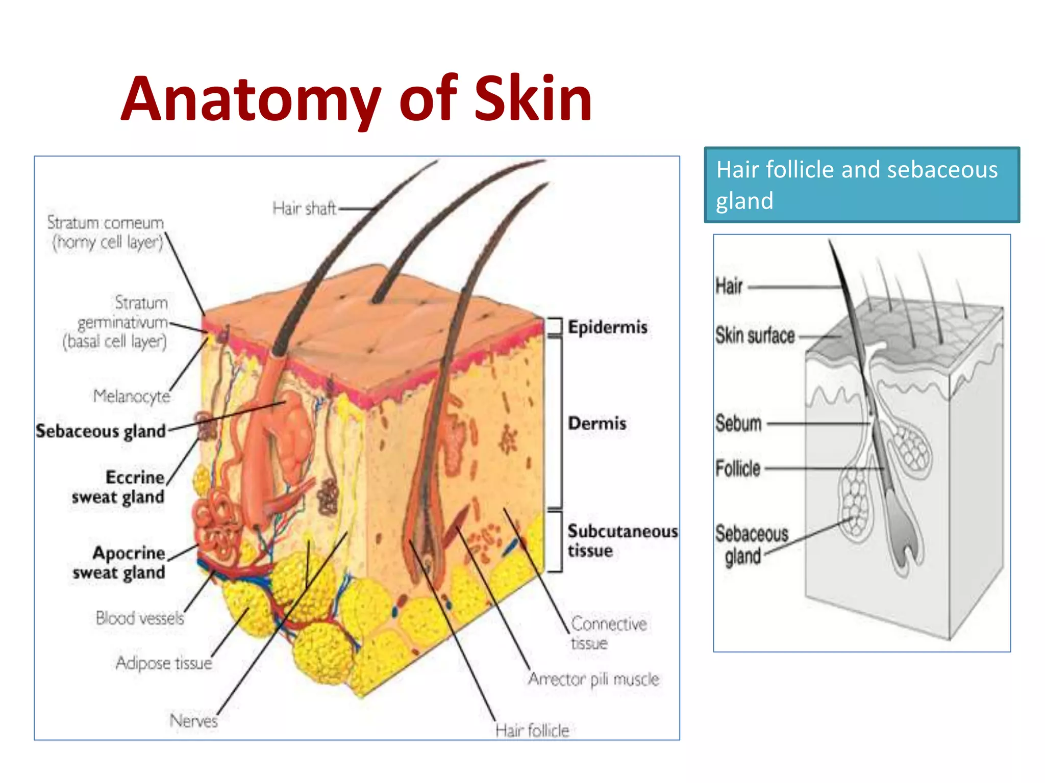

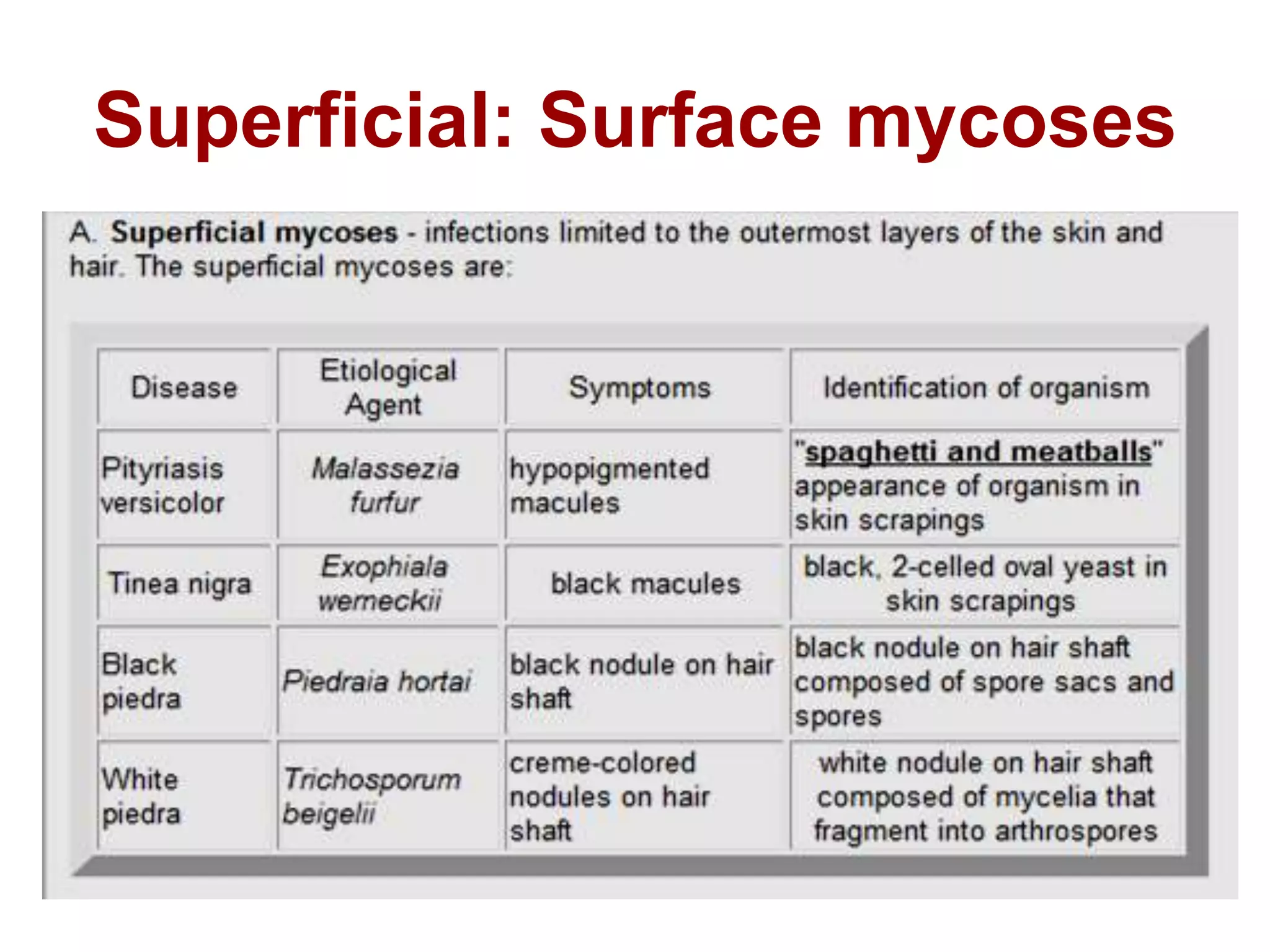

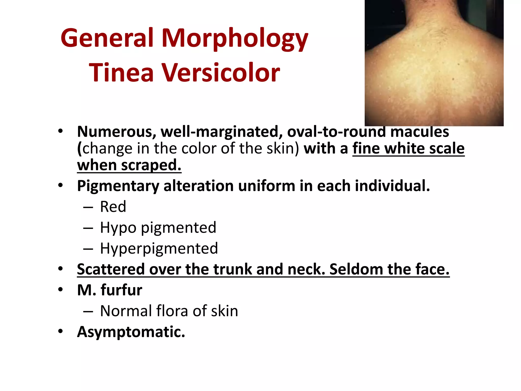


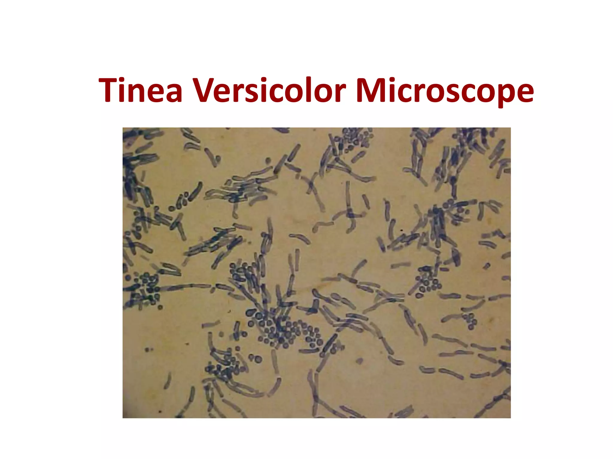


Pityriasis versicolor is a common superficial fungal infection of the skin caused by Malassezia furfur. It produces hypopigmented, hyperpigmented, or scaly macules on the trunk and neck. Diagnosis involves scraping skin lesions and examining under a microscope using potassium hydroxide, which reveals short hyphal fragments and clusters of yeast. Wood's lamp examination shows pale yellow-white fluorescence of lesions. M. furfur is normally present on human skin but overgrowth causes pityriasis versicolor. The fungus can be cultured using Sabouraud dextrose agar covered with oil, as M. furfur requires lipids for growth.


![Fungal infections of skin [compatibility mode]](https://cdn.slidesharecdn.com/ss_thumbnails/fungalinfectionsofskincompatibilitymode-130321223403-phpapp01-thumbnail.jpg?width=640&height=640&fit=bounds)


