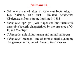
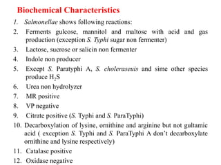
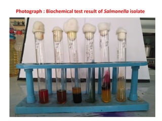
This document discusses the laboratory diagnosis of Salmonella species. It begins by describing Salmonella bacteria and the diseases they can cause in humans, including typhoid fever, paratyphoid fever, and gastroenteritis. It then discusses the habitats of different Salmonella serotypes and outlines several methods for laboratory diagnosis, including culture-based isolation and identification using biochemical tests and serological or molecular techniques. The document provides details on the morphology, cultural characteristics, enrichment and selective media used for Salmonella as well as their typical biochemical reactions that are used for identification.
pseudomonas aeruginosa is one of the leading cause of hospital-associated infection. mainly Pseudomonas is a multi drug resistant bacteria.
they are oxidase positive, non fermenters, strictly aerobic bacteria.
they are pigment producing, pigment can be appreciated on nutrient agar.
The genus Shigella exclusively infects human intestine.
Shigella dysenteriae is the causative agent of bacillary dysentery or shigellosis in humans.
It is a diarrheal illness which is characterized by frequent passage of blood stained mucopurulent stools.
The four important species of the genus Shigella are:
Shigella dysenteriae
Shigella flexneri
Shigella sonnei
Shigella boydii.
Microbiology of E coli giving basic of Escherichia coli, its morphology, cultural and biochemical characteristics, Antigenic character, pathogenesis, laboratory diagnosis, prevention and control
Cholera is a serious bacterial disease that usually
causes severe diarrhea and dehydration. The disease is typically spread through contaminated water.
Modern sewage and water treatment have effectively eliminated cholera in most countries. It’s still a problem in countries like Asia, America and Africa. Mostly in India.
Countries affected by war, poverty, and natural disasters have the greatest risk for a cholera outbreak.
Taxonomy:
class : Gamma Proteobacteria
Order: Vibrionales
Family: Vibrionaceae
Genus: Vibrio
Species: v.cholerae, v.parahaemolyticus,
v. vulnificus, v. alginolyticus
MORPHOLOGY:
Gram negative, actively motile, short, rigid curved bacilli
Resembling letter “V”
about 34 genus
most common in water
1.5µ X 0.2 -0.4 µ in size
polar flagellum , strongly aerobic
Smear – fish in stream appearance
PATHOGENESIS:
Source: Ingestion of contaminated water, food,
fruits and vegetables etc.,
Incubation periods: 1-5 days
Symptoms: Watery diarrhoea, vomiting, thirst, dehydration, muscle cramps
Complications: muscular pain, renal failure, pulmonary edema, cardiac arrhythrnias
DIAGNOSIS:
Specimen: stool sample, water sample(envt)
Microscopy: a) Hanging drop : +ve
b) Gram stain :-ve
Culture: Mac conkey Agar :colourless to light pink
TCBS : yellow colonies
Serology: serological tests are no diagnostic value
TREATMENT:
Adequate replacement of fluids and electrolytes.
Oral tetracycline reduces the period of vibrio excreation.
PREVENTION:
Drink and use bottled water
Frequent washing
Sanitary environment
Defecate in water
Cook food thoroughly
As the channel name suggests, our channel will be a perfect lounge for the malayali medicos..we wil be covering videos which will be like lecture classes related to the subjects biochemistry and microbiology in which we are specialised.. It will be a better learning experience for the students especially for those who are not able to understand and follow the normal classes in college..we assure the students that you will get a basic idea regarding the topic and extra reading can be done from the reference textbooks..
Qalification
AHLAD T O
MSc MLT (Biochemistry)
Assistant Professor
Baby memorial college of allied Health science
Kozhikode
Maneesha M Joseph
MSc MLT (Microbiology)
Assistant Professor
Baby memorial college of allied Health science
Kozhikode
Our Partner Channel
Health & Voyage channel link - https://youtu.be/nzKqRVjlwc0
#Proteus microbiology
#Medical
#Microbiology
#Biochemistry
#Mallu Medicos Lounge
##MalluMedicosLounge
#MLT
#Channel introduction
#HealthAndVoyage
#New Youtube Channel introduction
#Gram-negative
#Enterobactericea
#Weil Felix Test
#PROTEUS - causes, symptoms, diagnosis, treatment, pathology
Aero tolerant organisms cannot use oxygen for growth but are tolerate its presence. use fermentation to produce ATP.
Characteristics:
They don’t possess cytochromes.
The cells are arranged in pairs, chain or tetrads.
They have only fermentative type of metabolism and don’t respire
They can grow anaerobically or aerobically.
Genera
Streptococcus
Leuconostoc
Pediococcus
Non spore forming gram positive rods of regular shape:
Non spore forming irregular shape
Salmonella & Vibrio Cholerae an overview.pdfAlanShwan2
medical Bacteriology
Salmonella & Vibrio
Salmonella infection (salmonellosis) is a common bacterial disease that affects the intestinal tract. Salmonella bacteria typically live in animal and human intestines and are shed through stool (feces). Humans become infected most frequently through contaminated water or food.
Some people with salmonella infection have no symptoms. Most people develop diarrhea, fever and stomach (abdominal) cramps within 8 to 72 hours after exposure. Most healthy people recover within a few days to a week without specific treatment.
Cholera is a bacterial disease usually spread through contaminated water. Cholera causes severe diarrhea and dehydration. Left untreated, cholera can be fatal within hours, even in previously healthy people.
Modern sewage and water treatment have virtually eliminated cholera in industrialized countries. But cholera still exists in Africa, Southeast Asia and Haiti. The risk of a cholera epidemic is highest when poverty, war or natural disasters force people to live in crowded conditions without adequate sanitation.
Gram-positive cocci include Staphylococcus (catalase-positive), which grows clusters, and Streptococcus (catalase-negative), which grows in chains. The staphylococci further subdivide into coagulase-positive (S. aureus) and coagulase-negative (S. epidermidis and S. saprophyticus) species. Streptococcus bacteria subdivide into Strep. pyogenes (Group A), Strep. agalactiae (Group B), enterococci (Group D), Strep viridans, and Strep pneumonia.
Gram-positive bacilli (rods) subdivide according to their ability to produce spores. Bacillus and Clostridia are spore-forming rods while Listeria and Corynebacterium are not. Spore-forming rods that produce spores can survive in environments for many years. Also, the branching filament rods encompass Nocardia and actinomyces.
Gram-positive organisms have a thicker peptidoglycan cell wall compared with gram-negative bacteria. It is a 20 to 80 nm thick polymer while the peptidoglycan layer of the gram-negative cell wall is 2 to 3 nm thick and covered with an outer lipid bilayer membrane.
Bloodstream infection mortality rates have increased by 78% in just two decades[1]. Gram-positive organisms have highly variable growth and resistance patterns. The SCOPE project (Surveillance and Control of Pathogens of Epidemiologic Importance) found that gram-positive organisms in those with an underlying malignancy accounted for 62% of all bloodstream infections in 1995 and 76% in 2000 while gram-negative organisms accounted for 22% and 14% of infections for these years.[2]
This pdf is about the Schizophrenia.
For more details visit on YouTube; @SELF-EXPLANATORY;
https://www.youtube.com/channel/UCAiarMZDNhe1A3Rnpr_WkzA/videos
Thanks...!
The increased availability of biomedical data, particularly in the public domain, offers the opportunity to better understand human health and to develop effective therapeutics for a wide range of unmet medical needs. However, data scientists remain stymied by the fact that data remain hard to find and to productively reuse because data and their metadata i) are wholly inaccessible, ii) are in non-standard or incompatible representations, iii) do not conform to community standards, and iv) have unclear or highly restricted terms and conditions that preclude legitimate reuse. These limitations require a rethink on data can be made machine and AI-ready - the key motivation behind the FAIR Guiding Principles. Concurrently, while recent efforts have explored the use of deep learning to fuse disparate data into predictive models for a wide range of biomedical applications, these models often fail even when the correct answer is already known, and fail to explain individual predictions in terms that data scientists can appreciate. These limitations suggest that new methods to produce practical artificial intelligence are still needed.
In this talk, I will discuss our work in (1) building an integrative knowledge infrastructure to prepare FAIR and "AI-ready" data and services along with (2) neurosymbolic AI methods to improve the quality of predictions and to generate plausible explanations. Attention is given to standards, platforms, and methods to wrangle knowledge into simple, but effective semantic and latent representations, and to make these available into standards-compliant and discoverable interfaces that can be used in model building, validation, and explanation. Our work, and those of others in the field, creates a baseline for building trustworthy and easy to deploy AI models in biomedicine.
Bio
Dr. Michel Dumontier is the Distinguished Professor of Data Science at Maastricht University, founder and executive director of the Institute of Data Science, and co-founder of the FAIR (Findable, Accessible, Interoperable and Reusable) data principles. His research explores socio-technological approaches for responsible discovery science, which includes collaborative multi-modal knowledge graphs, privacy-preserving distributed data mining, and AI methods for drug discovery and personalized medicine. His work is supported through the Dutch National Research Agenda, the Netherlands Organisation for Scientific Research, Horizon Europe, the European Open Science Cloud, the US National Institutes of Health, and a Marie-Curie Innovative Training Network. He is the editor-in-chief for the journal Data Science and is internationally recognized for his contributions in bioinformatics, biomedical informatics, and semantic technologies including ontologies and linked data.
A brief information about the SCOP protein database used in bioinformatics.
The Structural Classification of Proteins (SCOP) database is a comprehensive and authoritative resource for the structural and evolutionary relationships of proteins. It provides a detailed and curated classification of protein structures, grouping them into families, superfamilies, and folds based on their structural and sequence similarities.
Seminar of U.V. Spectroscopy by SAMIR PANDASAMIR PANDA
Spectroscopy is a branch of science dealing the study of interaction of electromagnetic radiation with matter.
Ultraviolet-visible spectroscopy refers to absorption spectroscopy or reflect spectroscopy in the UV-VIS spectral region.
Ultraviolet-visible spectroscopy is an analytical method that can measure the amount of light received by the analyte.
Slide 1: Title Slide
Extrachromosomal Inheritance
Slide 2: Introduction to Extrachromosomal Inheritance
Definition: Extrachromosomal inheritance refers to the transmission of genetic material that is not found within the nucleus.
Key Components: Involves genes located in mitochondria, chloroplasts, and plasmids.
Slide 3: Mitochondrial Inheritance
Mitochondria: Organelles responsible for energy production.
Mitochondrial DNA (mtDNA): Circular DNA molecule found in mitochondria.
Inheritance Pattern: Maternally inherited, meaning it is passed from mothers to all their offspring.
Diseases: Examples include Leber’s hereditary optic neuropathy (LHON) and mitochondrial myopathy.
Slide 4: Chloroplast Inheritance
Chloroplasts: Organelles responsible for photosynthesis in plants.
Chloroplast DNA (cpDNA): Circular DNA molecule found in chloroplasts.
Inheritance Pattern: Often maternally inherited in most plants, but can vary in some species.
Examples: Variegation in plants, where leaf color patterns are determined by chloroplast DNA.
Slide 5: Plasmid Inheritance
Plasmids: Small, circular DNA molecules found in bacteria and some eukaryotes.
Features: Can carry antibiotic resistance genes and can be transferred between cells through processes like conjugation.
Significance: Important in biotechnology for gene cloning and genetic engineering.
Slide 6: Mechanisms of Extrachromosomal Inheritance
Non-Mendelian Patterns: Do not follow Mendel’s laws of inheritance.
Cytoplasmic Segregation: During cell division, organelles like mitochondria and chloroplasts are randomly distributed to daughter cells.
Heteroplasmy: Presence of more than one type of organellar genome within a cell, leading to variation in expression.
Slide 7: Examples of Extrachromosomal Inheritance
Four O’clock Plant (Mirabilis jalapa): Shows variegated leaves due to different cpDNA in leaf cells.
Petite Mutants in Yeast: Result from mutations in mitochondrial DNA affecting respiration.
Slide 8: Importance of Extrachromosomal Inheritance
Evolution: Provides insight into the evolution of eukaryotic cells.
Medicine: Understanding mitochondrial inheritance helps in diagnosing and treating mitochondrial diseases.
Agriculture: Chloroplast inheritance can be used in plant breeding and genetic modification.
Slide 9: Recent Research and Advances
Gene Editing: Techniques like CRISPR-Cas9 are being used to edit mitochondrial and chloroplast DNA.
Therapies: Development of mitochondrial replacement therapy (MRT) for preventing mitochondrial diseases.
Slide 10: Conclusion
Summary: Extrachromosomal inheritance involves the transmission of genetic material outside the nucleus and plays a crucial role in genetics, medicine, and biotechnology.
Future Directions: Continued research and technological advancements hold promise for new treatments and applications.
Slide 11: Questions and Discussion
Invite Audience: Open the floor for any questions or further discussion on the topic.
Professional air quality monitoring systems provide immediate, on-site data for analysis, compliance, and decision-making.
Monitor common gases, weather parameters, particulates.
Earliest Galaxies in the JADES Origins Field: Luminosity Function and Cosmic ...Sérgio Sacani
We characterize the earliest galaxy population in the JADES Origins Field (JOF), the deepest
imaging field observed with JWST. We make use of the ancillary Hubble optical images (5 filters
spanning 0.4−0.9µm) and novel JWST images with 14 filters spanning 0.8−5µm, including 7 mediumband filters, and reaching total exposure times of up to 46 hours per filter. We combine all our data
at > 2.3µm to construct an ultradeep image, reaching as deep as ≈ 31.4 AB mag in the stack and
30.3-31.0 AB mag (5σ, r = 0.1” circular aperture) in individual filters. We measure photometric
redshifts and use robust selection criteria to identify a sample of eight galaxy candidates at redshifts
z = 11.5 − 15. These objects show compact half-light radii of R1/2 ∼ 50 − 200pc, stellar masses of
M⋆ ∼ 107−108M⊙, and star-formation rates of SFR ∼ 0.1−1 M⊙ yr−1
. Our search finds no candidates
at 15 < z < 20, placing upper limits at these redshifts. We develop a forward modeling approach to
infer the properties of the evolving luminosity function without binning in redshift or luminosity that
marginalizes over the photometric redshift uncertainty of our candidate galaxies and incorporates the
impact of non-detections. We find a z = 12 luminosity function in good agreement with prior results,
and that the luminosity function normalization and UV luminosity density decline by a factor of ∼ 2.5
from z = 12 to z = 14. We discuss the possible implications of our results in the context of theoretical
models for evolution of the dark matter halo mass function.
Richard's aventures in two entangled wonderlandsRichard Gill
Since the loophole-free Bell experiments of 2020 and the Nobel prizes in physics of 2022, critics of Bell's work have retreated to the fortress of super-determinism. Now, super-determinism is a derogatory word - it just means "determinism". Palmer, Hance and Hossenfelder argue that quantum mechanics and determinism are not incompatible, using a sophisticated mathematical construction based on a subtle thinning of allowed states and measurements in quantum mechanics, such that what is left appears to make Bell's argument fail, without altering the empirical predictions of quantum mechanics. I think however that it is a smoke screen, and the slogan "lost in math" comes to my mind. I will discuss some other recent disproofs of Bell's theorem using the language of causality based on causal graphs. Causal thinking is also central to law and justice. I will mention surprising connections to my work on serial killer nurse cases, in particular the Dutch case of Lucia de Berk and the current UK case of Lucy Letby.
1. LABORATORY DIAGNOSIS OF Salmonella spp.
PRESENTED BY
Arun Kumar Thakur
Babita Gautam
Chetana Dahal
Purshotam Kr. Sah Kanu
Rohit Ghimire
Central Department Of Microbiology
Second Semester
2. Salmonella
• Salmonella named after an American bacteriologist,
D.E Salmon, who first isolated Salmonella
Choleraesuis from porcine intestine in 1884
• Salmonella spp gm (-ve), flagellated and facultative
anaerobic bacteria characterized by the presence of O,
H, and Vi antigen
• Salmonella ubiquitous human and animal pathogen
• Salmonella infection- one of three clinical syndrome
.i.e. gastroenteritis, enteric fever or focal disease
3. Human infections caused by some Salmonella spp
Salmonella Typhi-typhoid fever, bacterima
Salmonella Paratyphi A, B and C-paratyphoid
fever, bacterima
Salmonella Cholerasuis- bacterima
Salmonella Typhimurium- gastroenteritis
Salmonella Enterisitis, S. Hadar, S. Heidelberg,
S. Agona etc- gastroenteritis
4. Habitat
• Found in intestinal tract of human and animal including wild birds,
domestic pets and rodents.
• Under suitable environmental conditions, they may survive for
weeks in water and for years in soils.
• Majority are ubiquitous serotypes inhabiting a wide range of hosts.
• Members of subsps. enterica predominate among mammals and
others are found commonly in the intestinal tract of cold blooded
animals.
• Host adapted serotypes
• Some serotype are adopted to specific host eg.
Abortusouis- confirmed to sheep
Gallinarum- fowl
Typhi-human
Typhisuis -swine
5. Laboratory Diagnosis
Laboratory daignosis of Sallmonella spp is based on the following
methods:
1. Macroscopic examination of specimen along with microscopic
examination for morphological observation of bacteria
2. Isolation and identification of Sallmonella spp by culture and
biochemical test
3. Serodiagnosis by demonstration of Sallmonella spp antibodies and
antigens
4. Molecular diagnosis by DNA probes and PCR
Specimens
i. Blood, blood clot, bone marrow and stool commonly used for
typhoid bacilli
ii. CSF, peritoneal fluid, mesentric lymph nodes, resected intestine,
phyranx, tonslis, abscess, bone and urine other specimen of choice
6. Morphology on Gram staining
Member of family enterobactericeae.
• Gram negative bacteria measuring 2-4 X 0.6 um
• Non-acid fast, non-sporing
• Motile with peritrichous flagella except Gallinarum pullorum
• Non capsulated but when gown on media with osmolarity, most
strains produce extracellular slime which is colanic acid.
7. Cont…
• Slime production is the rule among S. Paratyphi negative to D-
tartarate and is favoured by continued incubation at low
temperature.
• Most strains (80%) form type I (mannose sensitive,
haemagglutination) fimbriae composed of fimbrilin subunits
with a high proportion of hydrophobic amino acids.
• Gallinarum pullorum and a few strains in other serotypes
either form type 2 (non-haemagglutinating) fimbriae or are
non fimbricated.
8. Cultural characteristics
• Aerobic and faculatively anaerobic .
• Grows on simple lab media in temperature range (7- 48°)C ,optimally at 37°C
• pH 4-8 and water activity above 0.93.
• Many strains are proto trophic; i.e capable of growing on glucose-ammonium
minimal medium.
• Some strains are auxotrophic and require enrichment of the minimal medium
with one or more amino acids or vitamins eg. cysteine or nicotinamide.
• Most Typhi strain require tryptophan.
9. On NA
Grey white, moist, circular, convex colonies with entire edge 2-3 mm in
diameter.
Many paratyphi strains form large mucoid colonies, due to slime formation
when plates are left at room temp. for few days incubation at 37°C for 24
hrs.
NA XLD
10. • On MA
NLF pale colored colonies.
• On DCA
Small colonies with black centre
• On XLD
Pink red colonies (H2S Producer)
3-5 mm dia., with black centre those not producing H2S (paratyphi
A)
Produce pink colonies without black centre
• On Peptone water and NB
Most strains give abundant growth with uniform turbidity and a
thin surface pellicle forms on prolonged incubation.
Rough variants which have a hydrophobic surface and tend to auto
aagglutinate produce agranular deposits and sometimes a thick
pellicle.
11. • Enrichment media
Tetrathionate broth –enriches salmonallae but also permits growth
of proteus spp
Kauffmann Muller Tetrathionate broth with a brilliant green,
inhibits proteus and so improves selectively.
Selenite F Broth-excellent for typhi and Dublin but some eg
paratyphi A and Cholareusis may fail to multiply .
Rappaport’s malachite green magnesium chloride broth –more
efficient for isolation of salmonellae from faeces water and
foodstuffs.
12. • Selective as well as Differential media
Bismuth Sulfite Agar : selective as it inhibts most gm (+ve) and gm
(-ve) bacteria; differential as production of H2S, ferrous sulphate,
positive reactions appear as brown to black precipitate
Brillant green agar: brillant green is inhibtory to most gm (+ve) and
gm (-ve) bacteria; differential lactose and sucrose fermentation
positive appears as yellow to green colonies with bright yellow to
green halos
Salmonella spp appear as white to pink or red colonies surrounded by a
bright halo
Salmonellae Shigella Agar: selective as bile salts, sodium citrate and
brillant green inhibts most gm (+ve) and gm (-ve) bacteria;
differential due to presence of lactose, neutral red sodium thiosulfate
Salmonella spp appears colourless with black center
13. Biochemical Characteristics
1. Salmonellae shows following reactions:
2. Ferments gulcose, mannitol and maltose with acid and gas
production (exception S. Typhi sugar non fermenter)
3. Lactose, sucrose or salicin non fermenter
4. Indole non producer
5. Except S. Paratyphi A, S. choleraseuis and sime other species
produce H2S
6. Urea non hydrolyzer
7. MR positive
8. VP negative
9. Citrate positive (S. Typhi and S. ParaTyphi)
10. Decarboxylation of lysine, ornithine and arginine but not gultamic
acid ( exception S. Typhi and S. ParaTyphi A don’t decarboxylate
ornithine and lysine respectively)
11. Catalase positive
12. Oxidase negative