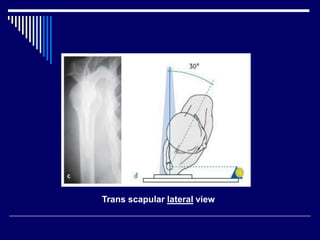
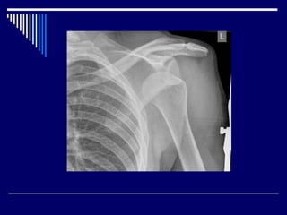
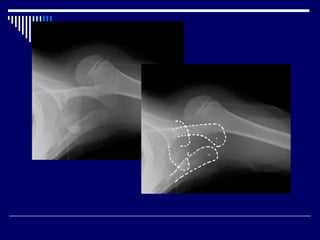
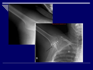
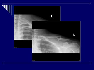
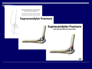
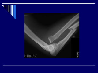
Regional trauma 
Shoulder and upper arm 
Dr.Aftab Qadir
Outline 
Shoulder dislocation 
Fractures 
Rotator cuff injury
Trauma-series x-rays 
Glenoid AP view
Trans scapular lateral view
Axillary view
1.Shoulder dislocation 
anterior > 95% 
posterior 2-4% 
inferior (luxatio erecta) < 1%
Plain Films 
X-rays are sufficient in most cases 
associated fractures/injuries 
Hill-Sachs lesion 
bony Bankart lesion 
proximal humeral fractures 
clavicular fracture 
acromioclavicular joint disruption 
acromial fracture
Hill-Sachs lesions 
Bankart lesion
Posterior Shoulder Dislocation 
Radiological signs on AP view: 
Internal rotation of the humerus 
Light bulb sign 
The ‘rim sign’ – Widening of the glenohumeral 
space
Inferior shoulder dislocation 
Radiographic features 
The humeral head is displaced directly below the 
glenoid fossa. 
arm often in marked abduction (luxatio erecta).
2.Acromioclavicular injury 
Features include : 
soft tissue swelling 
widening of the AC joint 
increased coracoclavicular distance 
superior displacement of the distal clavicle
4.Clavicular fracture 
vast majority occur in the midshaft 
fall onto an outstretched arm 
common in very young and very old patients.
5.Scapula fractures 
uncommon injuries, representing ~3% of all 
shoulder fractures. 
high energy trauma 
Requires trauma series views 
associated with other injuries
6.Humerus 
 fractured by excessive torsion or by a direct 
blow. 
The humeral neck is frequently fractured in 
elderly patients following a fall into the shoulder.
7.Supracondylar fracture 
90% are seen in children younger than 10 years 
commonly seen in boys 
due to accidental trauma 
Lateral and AP radiographs are usually sufficient 
iinnddiirreecctt ssiiggnnss
9.Medial epicondyle fracture 
represent almost all epicondyle fractures 
most common avulsion injury of the elbow 
typically seen in children and adolescents
10.Galeazzi fracture-dislocation 
fracture of the distal radius with dislocation of 
distal radioulnar joint 
children, peak incidence of 9-12 years of age 
Plain films , orthogonal views
11.Monteggia fracture-dislocation 
fracture of the ulna shaft and dislocation of the 
radial head 
 image both the elbow and the wrist, and good 
quality AP and lateral views are required.
12.Rotator cuff tear 
common causes of shoulder pain 
Prevalence of tear increases with age 
Acromiohumeral interval indicative of rotator cuff 
tear or tendinopathy. 
ultrasound may have up to 90% sensitivity and 
specificity 
Hyperintense signal area within the tendon on 
T2W, fat-suppressed and GRE sequences
Few Cases
Thank You

Shoulder and upper arm

Editor's Notes

  • #4 1.   clavicle 2.   surgical neck of humerus 3.   shaft of humerus 4.   acromion 6.   glenoid fossa 7.   scapula 8.   anatomical neck of humerus 9.   greater tuberosity 10. coracoid process                                    
  • #6 Trauma-series x-rays. With an acute fracture, all x-rays are taken with the patient standing or sitting and the arm supported to minimize pain. a–b True glenoid AP view. The patient must stand facing the x-ray source with the posterior aspect of the affected side against the x-ray plate. The opposite trunk is rotated at least 30°.
  • #7 c–d Transscapular lateral view. The patient stands with the x-ray source on the opposite side and the affected shoulder is placed against the x-ray plate. The trunk is turned 30° away from the x-ray beam, which is then directed posteriorly along the scapular spine.
  • #8 e–f Axillary view. The patient is supine with the x-ray plate placed above the shoulder. Abduction of about 30º is needed, which can be painful in an acute setting.
  • #9 Stryker Notch View Shoulder X ray Demonstrates: humeral head Helpful for: Hill-Sachs lesions (best view), Bankart Lesion. Position: Patient supine with cassette posterior to the shoulder. The hand placed on top of the head. The elbow should point straight upward. Beam directed 10° superiorly/toward the head, centered over the coracoid process. The acromion and coracoid form a "Y" or "peace sign" shape with the body of the scapula.  The head of the humerus should be normally centered to the middle of the "Y" shape as shown. The acromion and distal end of the clavicle form a "roof" over the shoulder joint and prevent superior displacement of the humeral head.
  • #10 shoulder dislocation, there is separation of the humerus from the glenoid of the scapula at the glenohumeral joint. most commonly dislocated large joint.  A number of biomechanical forces can produce shoulder dislocation, including : 1.forced extension, abduction and external rotation most commonly leads to anterior dislocation 2.direct blow from behind leads to anterior dislocation 3.forceful contraction of shoulder girdle muscles uncommon leads to posterior dislocation 4.forced hyperabduction leads to inferior dislocation
  • #11 AP and lateral +/- axillary view
  • #12 head of the humerus is driven forward from inside the glenoid cavity to a place under the coracoid process. This type of dislocation is sometimes referred to as a subcoracoid dislocation. The joint capsule is usually avulsed from the margin of the glenoid cavity. Anterior shoulder dislocation can also be the result of a detached labrum. When both the labrum and the capsule along the anterior margin of the glenoid cavity are avulsed, the injury is called a Bankart lesion. Compression fracture of the humeral head from the force of hitting the hard glenoid is called a Hill-Sach's lesion. Three-fourths of the patients with a Bankart lesion will also have a Hill-Sach's lesion.
  • #13 The humeral head is dislocated anteriorly and inferiorly with the anteroinferior margin of the glenoid, with some flattening of the corresponding part of the humerus. 
  • #14 An anteroinferior shoulder dislocation is present without associated fracture.
  • #15 Hill-Sachs lesions are a posterolateral humeral head compression fracture secondary to recurrent anterior shoulder dislocations, as the humeral head comes to rest against the anteroinferior part of the glenoid. It is often associated with a Bankart lesion of the glenoid. Bankart lesions are up to 11 times more common in patients with a Hill-Sachs lesion, with increasing incidence with increasing size A Bankart lesion results from detachment of the anterior inferior labrum from the underlying glenoid, and is usually as a result of previous anterior shoulder dislocation
  • #16 A large bony defect is noted in frontal view; involving posterior aspect of humeral head. No dislocation is noted. No lytic bony lesion is noted. previous history of anterior dislocation demonstrates a contour abnormality suggestive of a Hill-Sachs lesion.  X-ray Normal shoulder AP
  • #17 A small bone fragment is displaced from the inferior aspect of the glenoid consistent with a bony Bankart lesion. There is also subtle compression deformity of the superolateral humeral head consistent with a Hill-Sachs lesion. Both these lesions are the result of previous anterior shoulder dislocation. This patient had a history of recurrent dislocations.  A Bankart lesion results from detachment of the anterior inferior labrum from the underlying glenoid, and is usually as a result of previous anterior shoulder dislocation, and are associated with matching humeral lesion: the Hill-Sachs defect.
  • #18 Several radiological signs have been described on AP view, these include: Light bulb sign – The head of the humerus in the same axis as the shaft producing a light bulb shape Internal rotation of the humerus The ‘rim sign’ – Widening of the glenohumeral space The vacant glenoid sign – Where the anterior glenoid fossa looks empty The ‘trough’ sign – a vertical line made by the impression fracture of the anterior humeral head
  • #19 humeral head is forced posteriorly in internal rotation while the arm is abducted . In adults, convulsive disorder is the most common cause. Electrocution is a classic but uncommon cause of posterior shoulder dislocation Posterior dislocation may be missed initially on frontal radiographs in 50% of cases humeral head appears to be almost normally aligned with the glenoid the internally rotated humeral head takes on a rounded appearance known as the lightbulb sign 
  • #20 AP view of the shoulder demonstrates lack of overlap between the glenoid and the humeral head. The shoulder is rotated such that the humeral head has a 'light bulb' appearance.  glenohumeral joint space - 3-6mm increased - posterior dislocation decreased - degenerative joint disease, crystal arthropathies, inflammatory arthropathies rim sign
  • #22 Dislocation film demonstrates no congruency between the glenoid articular surface (blue) and that of the humeral head (yellow). In addition due to internal rotation the proximal humeral head has a rounded appearance, likened to a light bulb (light green). Post reduction film demonstrate the anterior rim of the glenoid (blue) and humeral (yellow) articular surfaces appearing congruent.
  • #23 An inferior shoulder dislocation is the least common form of shoulder dislocation. The condition is also called luxatio erecta because the arm appears to be permanently held upward or behind the head, in fixed abduction. It is caused by a hyper-abduction of the arm that forces the humeral head against the acromion. 
  • #24 The classic presentation of the inferior dislocation. The affected arm is hyperabducted, with the elbow flexed and forearm resting on top of the head.
  • #25 Frontal radiograph reveals the inferior dislocation of the humeral head (black arrow) from the glenoid fossa (white arrows). The humerus is parallel to the spine of the scapula, the common position in luxatio erecta.
  • #26 Acromioclavicular joint injuries are common and range from a mild sprain to complete disruption of the joint and injury to surrounding structures.  presence of soft tissue swelling widening of the AC joint  normal: 5-8 mm (narrower in the elderly) greater than 2-4 mm asymmetry (compared to radiographs of the contralateral side) increased coracoclavicular (CC) distance normal: 10-13 mm greater than 5 mm asymmetry (compared to radiographs of the contralateral side) superior displacement of the distal clavicle undersurface of the acromion should be level with the under surface of the clavicle degree of subluxation of the clavicle  grade II: inferior border of clavicle not elevated beyond the superior border of the acromion grade III: inferior border of clavicle is elevated beyond the superior border of the acromion, but coracoclavicular distance is not greatly increased (less than twice normal) grade V: marked superior elevation of the clavicle with coracoclavicular distance more than twice normal direction of dislocation (use axillary view) posteriorly into trapezius: grade IV inferiorly below the coracoid process: grade VI any fractures present glenohumeral joint alignment
  • #27 AC joint space and CC distances are widened. The distal clavicle is displaced superiorly compared to the acromion. Overlying soft tissue swelling. No fracture. Glenohumeral joint is normal.  CONCLUSION Grade II acromioclavicular joint injury.  ACJ Grade III
  • #28  account for 2.6-10% of all fractures. trauma situation, two views are better than one In most instances, the fracture is evident clinically and easily identified on radiographs.  fracture location of the fracture along the shaft angulation and fracture end displacement (including direction) comminution degree of overlap (measurement is useful) associated findings and relevant negatives acromioclavicular joint and sternoclavicular joint alignment coracoclavicular distance glenohumeral joint 
  • #29 Severely comminuted, displaced fracture of the mid-shaft of the left clavicle with depression of the lateral fracture fragment and comminution of the vertically orientated central fracture fragments.   The acromioclavicular joint appears enlocated.   Callus is noted around the fracture fragments.
  • #30 Two views of the clavicle demonstrate a fracture at the junction of middle and outer third. Displacement is best seen on the lordotic angled view. Cephalic tube angulation
  • #31  Associations Scpaular fractures are often associated with other injuries: clavicle fracture rib fracture sternal fracture spinal fracture pneumothorax and/or pulmonary contusion brachial plexus injury Plain film Requires trauma series views to demonstrate the fractures due to superimposition of shoulder girdle and thoracic cage. These include true anteroposterior view of the scapula, axillary view and true lateral view (Y-view) to demonstrate glenoid fracture. CT standard for diagnosis and evaluation of the fracture and its associated injuries axial scan with coronal, sagittal and 3D reconstructions are used in assessment of scapular injuries intra-articular glenoid fracture extra-articular glenoid fracture coracoid fracture acromial fracture
  • #32 Fracture of body of scapula is noted.   Humerus and clavicle are normal.  No dislocation is noted.
  • #33 humerus proximal humeral fracture humeral shaft fracture supracondylar fracture (extension) supracondylar fracture (flexion) internal epicondyle fracture external epicondylar fracture Flexion supracondylar humeral fractures account for only 2-4% of all supracondylar fractures . Unlike the much more common extension supracondylar fracture which are seen in children, flexion fractures are seen in older patients. They are usually the result of a fall directly onto a flexed elbow. 
  • #35 Typically supracondylar fractures occur as a result of a fall on a hyper-extended elbow.  posterior displacement of the distal component.  extra-articular fracture line indirect signs is essential:  anterior fat pad sign (sail sign) - the anterior fat pad is elevated by a joint effusion and appears as a lucent triangle on lateral projection posterior fat pad sign anterior humeral line should intersect the middle third of the capitellum in most children  although in children under 4, the anterior humeral line may pass through the anterior third without injury 
  • #37 In the setting of a painful elbow following a fall, if this line does not intersect the green area, but rather the red area then a supracondylar fracture should be suspected. 
  • #38 AP and lateral radiographs of the elbow demonstrate a lucency across the distal humerus consistent with a supracondylar fracture. A line drawn along the anterior border of the humerus does not intersect the middle third of the capitellum, indicating that the fracture is somewhat posteriorly angulated. 
  • #39 remembered using the mnemonic CRITOE is essential as an avulsed and displaced apophysis can mimic another centre.  Overall all centers are ossified by approximately 12 years of age Any missing ossification centre, or centres appearing in the wrong sequence should be viewed with a high suspicion of injury. A particularly devastating miss is that of an avulsed fragment displaced into the joint, mimicking the center of ossification of the trochlear
  • #40 Normal AP radiograph of the elbow in an 11 year old. All of the six ossification centers are present; the capitellum (C), radial head (R), internal epicondyle (I), trochlea (T), olecranon (O) and the external epicondyle (E). Two counting methods are taught to help remember the ages at which the ossification centers appear: 1-3-5-7-9-11 (simple) and 1-5-7-10-10-11 (more accurate).
  • #41 There is fracture / avulsion of the internal (medial) epicondyle ossification centre which is absent from it's normal position. It is displaced inferiorly and located just medial to the coronoid.  Features of a medial epicondylar avulsion injury include : soft tissue swelling this may be the only sign of an undisplaced injury this may be the only sign in children younger than ~7 years, in whom the medial apophysis is not calcified widening of the growth plate (comparison to the contralateral side may be useful) obvious displacement of the apophysis  fracture through the adjacent humeral metaphysis
  • #42 Galeazzi fracture-dislocations consist of fracture of the distal part of the radius with dislocation of distal radioulnar joint and an intact ulna. A Galeazzi equivalent fracture is a distal radial fracture with a distal ulnar physeal fracture Typically Galeazzi fracture-dislocations occur following a fall on an outstretched hand (FOOSH) with a flexed elbow. Galeazzi fractures are classified according to the position of the distal radius: type I: dorsal displacement type II: volar displacement Features include:  radial shaft fracture commonly at the junction of the middle and distal third dorsal angulation dislocation of the distal radioulnar joint radial shortening may occur and if greater than 10mm, suggests complete disruption of the interosseous membrane
  • #43 Fracture at the junction of the distal and middle 1/3rds of the radial diaphysis with distal radio-ulnar dislocation. Note that the radius is overlapped by 2-3 cm. 
  • #44 Monteggia fracture-dislocations comprise of a fracture of the ulna shaft and dislocation of the radial head. The ulna fracture is usually very obvious and the radial head dislocation can be overlooked Monteggia equivalent injuries can be present where the ulnar merely flexed or developed a subtle greenstick fracture. 
  • #45 fracture of the shaft of the ulna. The radial head is dislocated anteriorly. 
  • #46 Important causes of rotator cuff tear are trauma (acute, chronic repetitive) subacromial impingement tendon degeneration Hypovascularity Specific sub types The original Codman classification system published in 1930 was as  full thickness rotator cuff tear (FTRCT) partial thickness rotator cuff tear (PTRCT) bursal surface tear articular surface tear (rim rent tear) intratendinous tear  vertical, with connection from joint to bursa, not involving the whole breath of tendon Measurements of the AHI in the following intervals are suggestive of pathology 1-2: >12mm - shoulder dislocation, subluxation 9-10mm (range 8-12mm) - normal  6-7mm - thinning of supraspinatus tendon <6mm - supraspinatus tear Presence of tendon defect filled with fluid is most direct sign of rotator cuff tear. MRI Gradient echo (GRE) sequences
  • #48 Acromiohumeral interval (AHI) is a useful and reliable measurement on AP shoulder radiographs and when narrowed is indicative of rotator cuff tear or tendinopathy.  Pathology Measurements of the AHI in the following intervals are suggestive of pathology 1-2: >12mm - shoulder dislocation, subluxation range 8-12mm - normal  6-7mm - thinning of supraspinatus tendon <6mm - supraspinatus tear
  • #49 Full-thickness tears extend from bursal to articular surface, while partial thickness tears are focal defects in the tendon that involve either the bursal or articular surface. Full-thickness appear on ultrasound as hypoechoic/anechoic defects in the tendon. Due to the fluid replacing tendon direct signs are - non-visualization of supraspinatus tendon and hypoechoic discontinuity in tendon. Indirect signs are - double cortex sign, sagging peribursal fat sign, compressibility and muscle atrophy.
  • #52 Partial Supra Spinatus tear involving the articular side of the rotator cuff superficial layer of tendon is always present no fluid within the subacromial bursae
  • #53 Full-thickness tear.
  • #55 A joint effusion with sail sign and an anterior cortical supracondylar breach is present.  Anterior humeral line continues to intersect the mid point of the capitellum.  Completely displaced (posterormedial displacement) supracondylar fracture.
  • #56 There is an acute distal one-third radial diaphyseal fracture with dorsal lateral displacement and volar angulation of the distal fragment. There is also distal radioulnar dislocation with dorsal displacement of the ulna. Findings are keeping with type 1 Galeazzi fracture-dislocation. Remaining osseous structures are intact. No destructive bony lesions. 
  • #57 Pathological fracture of the humerus in a patient with metastasis of renal cell carcinoma
  • #58 A large bone fragment is noted on medial aspect of elbow, away from medial epicondyle region. A faint smaller elongated bone flake is noted adjacent to it. Elevation of anterior fat pad and visualization of posterior fat pad is seen. Avulsion fracture of medial epicondyle of humerus is represented by the larger fragment. There is a very likely associated avulsion from adjacent metaphysis represented by the smaller fragment. Elevation of anterior fat pad and visualization of posterior fat pad is indicative of an elbow effusion
  • #59 Intramuscular cyst and partial-thickness tear.
  • #60 Calcific tendinosis of the supraspinatus tendon with multiple small calcifications Supraspinatus tendon rupture and cortical irregularity longitudinal
  • #62 Bankart fracture This is a post-reduction view. Notice the very large fracture of the glenoid rim with displacement. Hill-Sachs is a posterolateral depression of the humeral head.