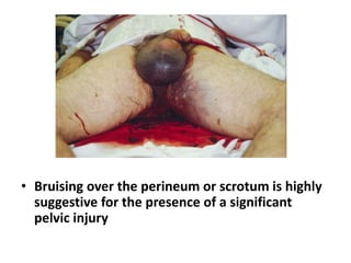
A Presentation on Injuries of Pelvis
Presentor : Pritam Pandey
Mentor : Dr. Sanjeeb Rijal
Surgical Anatomy
Pelvic support and stability
• Osseoligamentous structure
1. Posterior / anterior sacroiliac ligament
2. Iliolumbar ligament
3. Sacrospinous ligament
4. Sacrotuberous ligaments
Structures inside pelvis
• Major branches of common iiac arteries
• Sacral and lumbar plexus
• Urinary bladder
• Uretha
• Prostate
• pelvic colon
Introduction to Pelvic Trauma
Pelvic fractures represents
• 3% of all skeletal injuries annually in the United States
• 9% of trauma patients admitted to the hospital.
• An overall mortality of 10–16%
• Open pelvic fractures (2–4%) , have the highest mortality
rate at nearly 45%.
source :
http://www.jems.com/articles/print/volume-39/issue-
12/features/critical-management-deadly-pelvic-injuri.html
Mode of injuries
• motor vehicle accidents (60% of cases)
• falls from a height (30% of cases)
• crush injuries (10% of cases) .
Thus, displaced pelvic ring injuries are a marker for high-
energy trauma
source :
http://pubs.rsna.org/doi/full/10.1148/rg.345135113
Types of pelvis injuries
• Isolated Fracture with intact pelvic ring
• Fracture with broken pelvic ring ( Stable
• /unstable )
• Fracture of acetabulum
• Sacrococcygeal fracture
Classification of pelvic injuries
Tile’s classification
Type A, Stable
• A1, Without involvement of pelvic ring
• A2, With involvement of pelvic ring
Type B, Rotationally unstable
• B1, Open book
• B2, Ipsilateral lateral compression
• B3, Contralateral lateral compression
Type C, Rotationally and vertically unstable
• C1, Rotationally and vertically unstable
• C2, Bilateral
• C3, With associated acetabular fracture
The risk for mortality in pelvic
fractures
•
• Hemorrhagic Shock caused by rapid and
uncontrolled hemorrhage into the
retroperitoneal space
• ARDS
(Vascular bleed , 90 % being venous by osseous
fragments, disruption of vessels by shear
forces )
Approach to pelvic injuries
• Multidisciplinary Approach (head chest and
abdominal Injuries )
• Recognition of pelvic ring disruption and
determination of pelvic stability
• Suspect pelvic fracture in any case of RTA , Fall
in any patients with abdominal or lower limb
abnormalities
• Assess the vitals ( tachycardia , hypotension ,
increased respiratory rate may suggest
hypovolemia )
• Look for pallor , cold extremities, bleeding
from meatus , lacerated labia
• Rectal examination must be done (high
prostate)
• Bladder and bowel dysfunction may be
indication of nerve compression
• Blood at the urethral meatus is a classic sign
of urethral injury secondary to severe pelvic
trauma
• Bruising over the perineum or scrotum is highly
suggestive for the presence of a significant
pelvic injury
• Resuscitation
Circulation
1. IV line accessed (2 large gause )
2. Collect blood sample ( Hb%, hematocrit , PCV, Platelets ,
blood grouping ad cross matching )
3. IV fluid and blood transfusion
• Airways and breathing
• Urinary Catherization
• Adequate pain management (morphine )
Mechanically unstable injuries
• Fractures and dislocation with displacements
more than 1 cm
• injuries that compromise the posterior SI
ligaments
signs of instability is indicated by avulsion
fractures of
1. the sacrospinous or iliolumbar ligaments
2. L5 transverse process avulsion fractures
Methods of controlling bleeding
1. Rapping the pelvis with bedsheet tightly
2. Pneumatic antishock garments
3. commercial external pelvic compression devices (e.g.,
TPOD, SAM Pelvic Sling)
4. Early stabilization with external fixator (anterior/ C
clamp )in hypotensive patient with unstable fracture
5. Angiography and embolization
1. Laparotomy
2. Retroperitonal pelvic packing
3. ligate the internal iliac artery,
4. clamp the aorta
• Suspected Pelvic injuries ( AP radiograph)
Mechanically
stable
Mechanically
Unstable
Stable
hemodynamic
ally
Unstable
hemodynamic
ally
Stable
hemodynami
cally
Unstable
hemodynamicall
y
Pelvic CT inlet
and oulet view
And
observation
CT
angiograph
y
And
observation
Circumferential
pelvic rapping and
traction
Pelvic ct ,
observation ,
surgical fixation
when able
Circumferential
wrapping
Ct Angiography
Urgent surgical
fixation and pelvic
wrapping
Definitive Pelvic Stabilization
• Percutaneous Posterior fixation after
reduction using sacroiliac screws and
lumbopelvic fixation for comminuted
posterior injuries
• Iliac wing fractures can be fixed with plates
and screws
• rotationally unstable (APCII,LC-II) and/or
vertically unstable pelvic ring disruptions
(APC-III, LC-III, VS, CM )
• Selected lateral compression patterns with
rotational instability (LC-II, L-III)
• (external fixation + posterior pelvic ring
fixation)
• Pubic symphysis plating represents the
modality of choice for anterior fixation of
“open book” injuries with a pubic symphysis
diastasis > 2.5 cm (APC-II, APC-III)
• Spinopelvic fixation has the benefit of
immediate weight bearing in patients with
vertically unstable sacral fractures
Complication
• Deep vein Thrombosis
• ARDS
• Pulmonary embolisation
• Pressure sore
• Pulmonary infection
Reference
•
https://wjes.biomedcentral.com/articles/10.1186/s130
17-017-0117-6
• http://www.jems.com/articles/print/volume-39/issue-
12/features/critical-management-deadly-pelvic-
injuri.html
• http://medschool2.ucsf.edu/sfgh_surgery/education/sc
hecter-section/Publications/pelvic-fractures.pdf
• http://pubs.rsna.org/doi/full/10.1148/rg.345135113
• http://ccn.aacnjournals.org/
• Appleys system of orthopedics and fracture , 9th edition
Thank you

Pelvic injuries

  • 1.
    A Presentation onInjuries of Pelvis Presentor : Pritam Pandey Mentor : Dr. Sanjeeb Rijal
  • 2.
  • 6.
    Pelvic support andstability • Osseoligamentous structure 1. Posterior / anterior sacroiliac ligament 2. Iliolumbar ligament 3. Sacrospinous ligament 4. Sacrotuberous ligaments
  • 8.
    Structures inside pelvis •Major branches of common iiac arteries • Sacral and lumbar plexus • Urinary bladder • Uretha • Prostate • pelvic colon
  • 9.
    Introduction to PelvicTrauma Pelvic fractures represents • 3% of all skeletal injuries annually in the United States • 9% of trauma patients admitted to the hospital. • An overall mortality of 10–16% • Open pelvic fractures (2–4%) , have the highest mortality rate at nearly 45%. source : http://www.jems.com/articles/print/volume-39/issue- 12/features/critical-management-deadly-pelvic-injuri.html
  • 10.
    Mode of injuries •motor vehicle accidents (60% of cases) • falls from a height (30% of cases) • crush injuries (10% of cases) . Thus, displaced pelvic ring injuries are a marker for high- energy trauma source : http://pubs.rsna.org/doi/full/10.1148/rg.345135113
  • 11.
    Types of pelvisinjuries • Isolated Fracture with intact pelvic ring • Fracture with broken pelvic ring ( Stable • /unstable ) • Fracture of acetabulum • Sacrococcygeal fracture
  • 12.
  • 18.
    Tile’s classification Type A,Stable • A1, Without involvement of pelvic ring • A2, With involvement of pelvic ring Type B, Rotationally unstable • B1, Open book • B2, Ipsilateral lateral compression • B3, Contralateral lateral compression
  • 20.
    Type C, Rotationallyand vertically unstable • C1, Rotationally and vertically unstable • C2, Bilateral • C3, With associated acetabular fracture
  • 21.
    The risk formortality in pelvic fractures • • Hemorrhagic Shock caused by rapid and uncontrolled hemorrhage into the retroperitoneal space • ARDS (Vascular bleed , 90 % being venous by osseous fragments, disruption of vessels by shear forces )
  • 22.
    Approach to pelvicinjuries • Multidisciplinary Approach (head chest and abdominal Injuries ) • Recognition of pelvic ring disruption and determination of pelvic stability • Suspect pelvic fracture in any case of RTA , Fall in any patients with abdominal or lower limb abnormalities
  • 23.
    • Assess thevitals ( tachycardia , hypotension , increased respiratory rate may suggest hypovolemia ) • Look for pallor , cold extremities, bleeding from meatus , lacerated labia • Rectal examination must be done (high prostate) • Bladder and bowel dysfunction may be indication of nerve compression
  • 24.
    • Blood atthe urethral meatus is a classic sign of urethral injury secondary to severe pelvic trauma
  • 25.
    • Bruising overthe perineum or scrotum is highly suggestive for the presence of a significant pelvic injury
  • 27.
    • Resuscitation Circulation 1. IVline accessed (2 large gause ) 2. Collect blood sample ( Hb%, hematocrit , PCV, Platelets , blood grouping ad cross matching ) 3. IV fluid and blood transfusion • Airways and breathing • Urinary Catherization
  • 28.
    • Adequate painmanagement (morphine )
  • 29.
    Mechanically unstable injuries •Fractures and dislocation with displacements more than 1 cm • injuries that compromise the posterior SI ligaments
  • 30.
    signs of instabilityis indicated by avulsion fractures of 1. the sacrospinous or iliolumbar ligaments 2. L5 transverse process avulsion fractures
  • 31.
    Methods of controllingbleeding 1. Rapping the pelvis with bedsheet tightly 2. Pneumatic antishock garments 3. commercial external pelvic compression devices (e.g., TPOD, SAM Pelvic Sling) 4. Early stabilization with external fixator (anterior/ C clamp )in hypotensive patient with unstable fracture 5. Angiography and embolization
  • 32.
    1. Laparotomy 2. Retroperitonalpelvic packing 3. ligate the internal iliac artery, 4. clamp the aorta
  • 33.
    • Suspected Pelvicinjuries ( AP radiograph) Mechanically stable Mechanically Unstable Stable hemodynamic ally Unstable hemodynamic ally Stable hemodynami cally Unstable hemodynamicall y Pelvic CT inlet and oulet view And observation CT angiograph y And observation Circumferential pelvic rapping and traction Pelvic ct , observation , surgical fixation when able Circumferential wrapping Ct Angiography Urgent surgical fixation and pelvic wrapping
  • 34.
    Definitive Pelvic Stabilization •Percutaneous Posterior fixation after reduction using sacroiliac screws and lumbopelvic fixation for comminuted posterior injuries • Iliac wing fractures can be fixed with plates and screws
  • 35.
    • rotationally unstable(APCII,LC-II) and/or vertically unstable pelvic ring disruptions (APC-III, LC-III, VS, CM )
  • 36.
    • Selected lateralcompression patterns with rotational instability (LC-II, L-III) • (external fixation + posterior pelvic ring fixation)
  • 37.
    • Pubic symphysisplating represents the modality of choice for anterior fixation of “open book” injuries with a pubic symphysis diastasis > 2.5 cm (APC-II, APC-III)
  • 38.
    • Spinopelvic fixationhas the benefit of immediate weight bearing in patients with vertically unstable sacral fractures
  • 40.
    Complication • Deep veinThrombosis • ARDS • Pulmonary embolisation • Pressure sore • Pulmonary infection
  • 41.
  • 42.