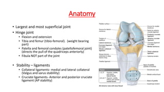
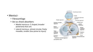
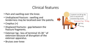
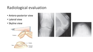
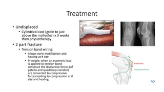
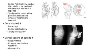
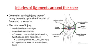
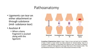
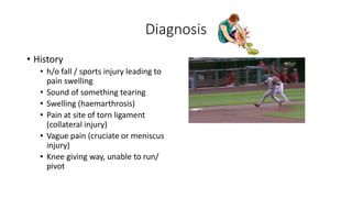
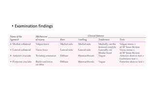
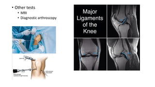
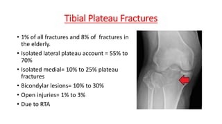
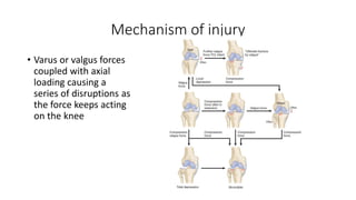
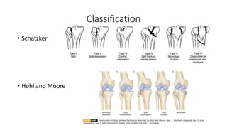
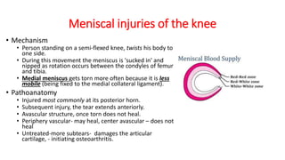
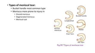
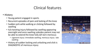
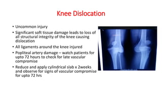
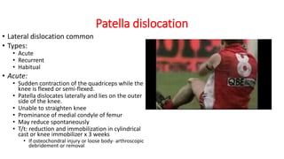
Injuries around the knee
Dr Siddhartha Sinha
Senior resident, Department of orthopaedics
Lady Hardinge Medical college
Anatomy
• Largest and most superficial joint
• Hinge joint
• Flexion and extension
• Tibia and femur (tibio-femoral). (weight bearing
part)
• Patella and femoral condyles (patellofemoral joint)
(directs the pull of the quadriceps anteriorly)
• Fibula NOT part of the joint
• Stability – ligaments
• Collateral ligaments- medial and lateral collateral
(Valgus and varus stablility)
• Cruciate ligaments- Anterior and posterior cruciate
ligament (AP stability)
• Menisci-
• Fibrocartilage
• Act as shock absorbers
• Medial meniscus- C shaped, broader
posteriorly than ant.
• Lateral meniscus- almost circular, freely
movable, smaller (less prone to injury)
• Movements around the knee
• Flexion-
• Primary: Hamstrings (semitendinosus,
semimembranosus, long head of biceps); short head of
biceps
• Secondary: Gracilis, sartorius, gastrocnemius, popliteus
• Extension
• Primary : Quadriceps femoris
• Secondary: TFL
• Some medial and lateral rotation
• Screw home mechanism:
• Knee passively “locks” because of medial rotation of
the femoral condyles on the tibial plateau
• Makes the lower limb a solid column and more
adapted for weight-bearing.
• When the knee is “locked,” the thigh and leg muscles
can relax briefly without making the knee joint too
unstable.
• To unlock the knee, the popliteus contracts, rotating
the femur laterally about 5° on the tibial plateau so
that flexion of the knee can occur
Mechanism of knee injuries
• Direct trauma
• Indirect trauma
• Valgus
• Varus
• Hyperextension
• Twisting
Condylar fractures of the femur
• 3 types
• Supracondylar fractures
• Unicondylar fractures – medial or
lateral. Hoffa # (frontal plane)
• Intercondylar fractures – T or Y
types
• Direct injury to distal femur
• Inter-condylar # patterns
• Indirect injury (valgus/ varus)
• Supracondylar
• Unicondylar patterns
• Diagnosis
• Pain
• Swelling
• Bruising around the
knee
• Difficulty walking
• Associated injuries
• Shaft of femur
• Tibia shaft
• Pelvis #
• X-ray
• Thigh with Knee –AP &
Lateral
• Thigh with hip- AP &
lateral
• Treatment
• Unicondylar fractures
• Undisplaced : long leg cast is given for 3-
6 weeks, followed by protected weight
bearing.
• Displaced: open reduction and internal
fixation
• cancellous screw or Herbert screws
• Buttress plate
• Intercondylar #
• Aim to restore articular congruity
and prevent knee stiffness
• ORIF with DCS/ Distal femoral
metaphyseal locking plate/ codylar
blade plate
• Conservative : rare using skeletal
traction
• Supracondylar fractures:
• Internal fixation. closed or
open techniques.
• Nail
• Antegrade or retrograde
• Plate
• Complications
• Knee stiffness- dense intra
and peri-articular adhesions
• Osteoarthritis-
malalignment of mechanical
axis or incorguent joint
surface causing change in
load bearing mechanics of
the knee
• Malunions- varus or valgus,
may require corrective
osteotomy
Patella #
• Largest sesamoid bone in the body.
• 7 articular facets; the lateral facet is the
largest (50% of the articular surface).
• Medial and lateral retinaculum
• Function
• Increase the mechanical advantage and
leverage of the quadriceps tendon
• Aid in nourishment of the femoral articular
surface,
• Protect the femoral condyles from direct
trauma.
Mechanism of injury
• Direct:
• Produce incomplete, simple, stellate, or
comminuted fracture patterns.
• Displacement minimal due to
preservation of the medial and lateral
retinacular expansions.
• Abrasions over the area or open injuries
are common.
• Active knee extension may be
preserved.
• Indirect (most common):
• Secondary to forcible quadriceps contraction while the
knee is in a semiflexed position (e.g., in a “stumble” or
“fall”).
• Intrinsic strength of the patella is exceeded by the pull of
the musculotendinous and ligamentous structures.
• Transverse fracture pattern is most commonly
• Degree of displacement of the fragments suggests the
degree of retinacular disruption.
• Active knee extension is usually lost.
• Combined direct/indirect mechanisms:
• Patient experiences direct and indirect trauma to the knee
(fall from a height)
Classification of patella #
Clinical features
• Pain and swelling over the knee.
• Undisplaced fracture : swelling and
tenderness may be localised over the patella.
• Crepitus (+)
• Displaced fractures : gap between the
fracture fragments.
• Extensor lag : loss of terminal 15-30 ° of
extension because of disruption of the
extensor apparatus.
• Bruises over knee
Radiological evaluation
• Antero-posterior view
• Lateral view
• Skyline view
Treatment
• Undisplaced
• Cylindrical cast (groin to just
above the malleolus) x 3 weeks
then physiotherapy
• 2 part fracture
• Tension band wiring:
• Allows early mobilization and
healing at # site
• Principle: when an eccentric load
is applied to tension band
construct the distractive forces (of
patella and quadriceps tendon)
are converted to compressive
forces leading to compression at #
site and healing
• Partial Patellectomy: part of
the patella is excised and
the extensor mechanism
repaired
• Total patellectomy: whole
patella is excised and
extensor mechanism
repaired
• Comminuted #
• Encirclage
• Partial patellectomy
• Total patellectomy
• Complications of patella #
• Knee stiffness
• Extensor mechanism
weakness
• Osteoartritis
Injuries of ligaments around the knee
• Common sporting injury, type of
injury depends upon the direction of
force and its severity.
• Mechanism of injury
• Medial collateral – Valgus
• Lateral collateral- Varus
• ACL –most commonly injured tendon,
twisting on a semi flexed knee
• O’ Donohuges triad- MCL , MM, ACL injury
• PCL –posterior force on a semi flexed
knee
Pathoanatomy
• Ligaments can tear on
either attachment or
through substance
(mid- substance tear)
• Avulsion #
• When a bony
fragment is avulsed
along with the
ligament
Diagnosis
• History
• h/o fall / sports injury leading to
pain swelling
• Sound of something tearing
• Swelling (haemarthrosis)
• Pain at site of torn ligament
(collateral injury)
• Vague pain (cruciate or meniscus
injury)
• Knee giving way, unable to run/
pivot
• Examination findings
• Valgus/Varus stress test for MCL and LCL in full extension and 30°
flexion
• Abnormal “opening up of the joint compared to normal side is positive)
• Cruciate ligaments
• Anterior cruciate
• Anterior drawer
• Knee at 90°, Hamstring relaxed
• Stabilize leg and foot
• Restore normal tibial step off
• Apply anterior directed force and
compare with the other side
• Can not be done in acute injury
• False negative due to doorstop effect of
meniscus
• Lachman
• Can be done in acute injury
• More sensitive than anterior drawer test
• Place knee in 30° flexion and apply
anterior force and assess end point
• Soft= injury
• Firm/ bony= no injury
• Pivot shift test
• Posterior cruciate
ligament
• Posterior drawer test
• Same perquisites as anterior
drawer but now direction of
force is posterior
• Posterior sag
• Dial test
• Radiological examination:
• Plain X-ray
• Normal usually or soft tissue swelling
• Chip of bone avulsed from the ligament attachment may be visible.
• Stress views in valgus/ varus: demonstrate an abnormal opening-up of the joint
• MRI is a non-invasive method of diagnosing ligament injuries, and may be of
use in doubtful cases.
• Segond fracture:
• Avulsion fracture of the proximal lateral tibia
• Pathognomonic for an ACL tear
• Represents bony avulsion by the anterolateral ligament (ALL)
• Associated with ACL tear 75-100% of the time
• Pellegrini-Steata lesion (calcification along the insertion of the medial
collateral ligament)
• Tibia Spine avulsion #
• ACL avulsion at tibial insertion
• Posterior cruciate ligament avulsion #
• Tibial insertion of PCL posteriorly
• Other tests
• MRI
• Diagnostic arthroscopy
• Treatment
• Immediate: RICE (REST , ICE, COMPRESSION,
ELEVATION) and analgesics
• Previously conservative management preferred due to
average results
• Operative treatment now preferred for patients with
instability and high daily demands like athletes
• Conservative Mx
• Cylindrical cast
• Appropriate for grade I or II
• Initiate physiotherapy after 2-3 weeks of immobilization in cast
• Surgical MX
• Performed 2-3 weeks after injury to allow soft tissue swelling to
subside and prevent knee stiffness
• Reconstruction of ligament using autograft or allograft (hamstring,
patella, TFL, peronei, quadriceps), synthetic materials
• Repair: not done now due to poor results
• Complications
• Knee instability
• Osteoarthritis
Tibial Plateau Fractures
• 1% of all fractures and 8% of fractures in
the elderly.
• Isolated lateral plateau account = 55% to
70%
• Isolated medial= 10% to 25% plateau
fractures
• Bicondylar lesions= 10% to 30%
• Open injuries= 1% to 3%
• Due to RTA
Mechanism of injury
• Varus or valgus forces
coupled with axial
loading causing a
series of disruptions as
the force keeps acting
on the knee
Classification
• Schatzker
• Hohl and Moore
• Clinical features
• Pain, swelling, deformity, inability to bear wt.
• Neurovascular examination
• Distal pulses of dorsalis pedis and posterior tibial- injury to popliteal
artery
• The peroneal nerve is tethered laterally as it courses around the
fibular neck.
• Hemarthrosis
• Compartment syndrome must be ruled out
• Assessment for ligament injury is essential. (but should be
done once pain has decreased)
• Associated injuries
• Meniscus
• Cruciate and collaterals
• Distal femur fractures
• Radiography
• Plain x rays
• CT scans
• MRI to evaluate soft
tissue and ligaments
• Arterial Doppler/
arteriogram for evaluation
of vascularity
• Treatment
• Non operative
• Undisplaced or minimally
displaced #
• Cast application followed
by hinge knee brace,
protected wt bearing for 8-
12 weeks and quadriceps
exercises
• Operative Mx
• Restore articular
congruency and
tibial alignment
• Choice of implant
related to fracture
patterns
• Plates, screws,
external fixator or
any combination
of these
• Assess for
ligamentous injury
and repair if
required
Complications
• Knee stiffness
• Infection
• Compartment syndrome
• Malunion or nonunion
• Posttraumatic osteoarthritis
• Peroneal nerve injury
• Popliteal artery laceration
• Avascular necrosis of small articular fragments
Meniscal injuries of the knee
• Mechanism
• Person standing on a semi-flexed knee, twists his body to
one side.
• During this movement the meniscus is 'sucked in' and
nipped as rotation occurs between the condyles of femur
and tibia.
• Medial meniscus gets torn more often because it is less
mobile (being fixed to the medial collateral ligament).
• Pathoanatomy
• Injured most commonly at its posterior horn.
• Subsequent injury, the tear extends anteriorly.
• Avascular structure, once torn does not heal.
• Periphery vascular- may heal, center avascular – does not
heal
• Untreated-more subtears- damages the articular
cartilage, - initiating osteoarthritis.
• Types of meniscal tear:
• Bucket handle most common type
• Meniscus more prone to injury in
• Discoid meniscus
• Degenerated meniscus
• Meniscal cyst
Clinical features
• History
• Young patient engaged in sports
• Recurrent episodes of pain and locking of the knee
• Sudden jerk while walking or clicking followed by
swelling
• h/o twisting injury followed by swelling appearing
overnight and once swelling subsides patient may not
be able to extend the knee fully d/t torn meniscus
• Ligament injury: immediate swelling, meniscus injury : late
swelling
• History of sudden locking and unlocking and click is
DIAGNOSTIC of meniscus injury
Examination of meniscus
• Tenderness in the region of the joint line
• Locked knee
• Attempts to force full extension produces a
sensation of elastic resistance and pain, localized
to the appropriate joint compartment
• Wasting of the quadriceps
• McMurray test
• Flex knee and IR to test for lateral meniscus
and extend
• Flex and ER to test for medial meniscus and
extend again
• Check for pain/ crepitus
• Apleys grinding test
• Distract knee to assess ligament injury
• Compress knee to assess meniscal injury
• Thessalays test
• Patient standing on affected limb with
support and asked to IR and ER lower limb
• Pain s/o injury
• Radiologial examination
• Xrays are normal
• MRI can detect tears non invasively and very sensitive
• Arthrography- injecting dye in joints – no longer used as MRI has high
sensitivity
• Diagnostic arthroscopy- invasive, direct visualization of the tear
Treatment
• Acute tear
• RICE + analgesics
• Locked knee: manipulation under
anaesthesia + immobilization x 2 weeks
then physiotherapy
• Chronic meniscal tear
• Partial menisectomy- remove the torn part
of the meniscus
• Repair- if tear in the vascular zone or
intermediate zone
• Total menisectomy- removal of whole
meniscus, rarely indicated, not preferred
Knee Dislocation
• Uncommon injury
• Significant soft tissue damage leads to loss of
all structural integrity of the knee causing
dislocation
• All ligaments around the knee injured
• Popliteal artery damage – watch patients for
upto 72 hours to check for late vascular
compromise
• Reduce and apply cylindrical slab x 2weeks
and observe for signs of vascular compromise
for upto 72 hrs
Extensor apparatus disruption
• Tearing of the quadriceps tendon from its attachment on the patella
• Tearing of the attachment of the patellar tendon from the tibial
tubercle.
• T/t: operative management
Patella dislocation
• Lateral dislocation common
• Types:
• Acute
• Recurrent
• Habitual
• Acute:
• Sudden contraction of the quadriceps while the
knee is flexed or semi-flexed.
• Patella dislocates laterally and lies on the outer
side of the knee.
• Unable to straighten knee
• Prominance of medial condyle of femur
• May reduce spontaneously
• T/t: reduction and immobilization in cylindrical
cast or knee immobilizer x 3 weeks
• If osteochondral injury or loose body- arthroscopic
debridement or removal
• Recurrent dislocation of the patella:
• Multiple and frequent episodes of dislocation after the first episode of
dislocation, more and more ease.
• Cause of recurrence
• Laxity of the medial capsule
• Defect in the anatomy of the knee.
• Excessive joint laxity
• Small patella
• Patella alta
• Genu valgum.
• Treatment
• Multiple procedures both bony and soft tissue
• Evaluate the cause of recurrence and treat
• Habitual dislocation of the patella:
• Patella dislocates laterally everytime the knee is flexed.
• Underlying defects are very similar to those in recurrent dislocation.
• Shortened quadriceps (vastus lateralis component) may result in an abnormal
lateral pull on the patella when the knee is flexed.
• Treatment :
• Release of the tight structures on the lateral side
• Repair of the lax structures on the medial side
• An additional ’checkrein’ mechanism of some sort is created to prevent re-dislocation.
Injuries around the knee

Injuries around the knee

  • 1.
    Injuries around theknee Dr Siddhartha Sinha Senior resident, Department of orthopaedics Lady Hardinge Medical college
  • 2.
    Anatomy • Largest andmost superficial joint • Hinge joint • Flexion and extension • Tibia and femur (tibio-femoral). (weight bearing part) • Patella and femoral condyles (patellofemoral joint) (directs the pull of the quadriceps anteriorly) • Fibula NOT part of the joint • Stability – ligaments • Collateral ligaments- medial and lateral collateral (Valgus and varus stablility) • Cruciate ligaments- Anterior and posterior cruciate ligament (AP stability)
  • 3.
    • Menisci- • Fibrocartilage •Act as shock absorbers • Medial meniscus- C shaped, broader posteriorly than ant. • Lateral meniscus- almost circular, freely movable, smaller (less prone to injury)
  • 4.
    • Movements aroundthe knee • Flexion- • Primary: Hamstrings (semitendinosus, semimembranosus, long head of biceps); short head of biceps • Secondary: Gracilis, sartorius, gastrocnemius, popliteus • Extension • Primary : Quadriceps femoris • Secondary: TFL • Some medial and lateral rotation • Screw home mechanism: • Knee passively “locks” because of medial rotation of the femoral condyles on the tibial plateau • Makes the lower limb a solid column and more adapted for weight-bearing. • When the knee is “locked,” the thigh and leg muscles can relax briefly without making the knee joint too unstable. • To unlock the knee, the popliteus contracts, rotating the femur laterally about 5° on the tibial plateau so that flexion of the knee can occur
  • 5.
    Mechanism of kneeinjuries • Direct trauma • Indirect trauma • Valgus • Varus • Hyperextension • Twisting
  • 6.
    Condylar fractures ofthe femur • 3 types • Supracondylar fractures • Unicondylar fractures – medial or lateral. Hoffa # (frontal plane) • Intercondylar fractures – T or Y types • Direct injury to distal femur • Inter-condylar # patterns • Indirect injury (valgus/ varus) • Supracondylar • Unicondylar patterns
  • 8.
    • Diagnosis • Pain •Swelling • Bruising around the knee • Difficulty walking • Associated injuries • Shaft of femur • Tibia shaft • Pelvis # • X-ray • Thigh with Knee –AP & Lateral • Thigh with hip- AP & lateral
  • 9.
    • Treatment • Unicondylarfractures • Undisplaced : long leg cast is given for 3- 6 weeks, followed by protected weight bearing. • Displaced: open reduction and internal fixation • cancellous screw or Herbert screws • Buttress plate
  • 10.
    • Intercondylar # •Aim to restore articular congruity and prevent knee stiffness • ORIF with DCS/ Distal femoral metaphyseal locking plate/ codylar blade plate • Conservative : rare using skeletal traction
  • 11.
    • Supracondylar fractures: •Internal fixation. closed or open techniques. • Nail • Antegrade or retrograde • Plate • Complications • Knee stiffness- dense intra and peri-articular adhesions • Osteoarthritis- malalignment of mechanical axis or incorguent joint surface causing change in load bearing mechanics of the knee • Malunions- varus or valgus, may require corrective osteotomy
  • 12.
    Patella # • Largestsesamoid bone in the body. • 7 articular facets; the lateral facet is the largest (50% of the articular surface). • Medial and lateral retinaculum • Function • Increase the mechanical advantage and leverage of the quadriceps tendon • Aid in nourishment of the femoral articular surface, • Protect the femoral condyles from direct trauma.
  • 13.
    Mechanism of injury •Direct: • Produce incomplete, simple, stellate, or comminuted fracture patterns. • Displacement minimal due to preservation of the medial and lateral retinacular expansions. • Abrasions over the area or open injuries are common. • Active knee extension may be preserved.
  • 14.
    • Indirect (mostcommon): • Secondary to forcible quadriceps contraction while the knee is in a semiflexed position (e.g., in a “stumble” or “fall”). • Intrinsic strength of the patella is exceeded by the pull of the musculotendinous and ligamentous structures. • Transverse fracture pattern is most commonly • Degree of displacement of the fragments suggests the degree of retinacular disruption. • Active knee extension is usually lost. • Combined direct/indirect mechanisms: • Patient experiences direct and indirect trauma to the knee (fall from a height)
  • 15.
  • 16.
    Clinical features • Painand swelling over the knee. • Undisplaced fracture : swelling and tenderness may be localised over the patella. • Crepitus (+) • Displaced fractures : gap between the fracture fragments. • Extensor lag : loss of terminal 15-30 ° of extension because of disruption of the extensor apparatus. • Bruises over knee
  • 17.
    Radiological evaluation • Antero-posteriorview • Lateral view • Skyline view
  • 18.
    Treatment • Undisplaced • Cylindricalcast (groin to just above the malleolus) x 3 weeks then physiotherapy • 2 part fracture • Tension band wiring: • Allows early mobilization and healing at # site • Principle: when an eccentric load is applied to tension band construct the distractive forces (of patella and quadriceps tendon) are converted to compressive forces leading to compression at # site and healing
  • 19.
    • Partial Patellectomy:part of the patella is excised and the extensor mechanism repaired • Total patellectomy: whole patella is excised and extensor mechanism repaired • Comminuted # • Encirclage • Partial patellectomy • Total patellectomy • Complications of patella # • Knee stiffness • Extensor mechanism weakness • Osteoartritis
  • 20.
    Injuries of ligamentsaround the knee • Common sporting injury, type of injury depends upon the direction of force and its severity. • Mechanism of injury • Medial collateral – Valgus • Lateral collateral- Varus • ACL –most commonly injured tendon, twisting on a semi flexed knee • O’ Donohuges triad- MCL , MM, ACL injury • PCL –posterior force on a semi flexed knee
  • 21.
    Pathoanatomy • Ligaments cantear on either attachment or through substance (mid- substance tear) • Avulsion # • When a bony fragment is avulsed along with the ligament
  • 22.
    Diagnosis • History • h/ofall / sports injury leading to pain swelling • Sound of something tearing • Swelling (haemarthrosis) • Pain at site of torn ligament (collateral injury) • Vague pain (cruciate or meniscus injury) • Knee giving way, unable to run/ pivot
  • 23.
  • 24.
    • Valgus/Varus stresstest for MCL and LCL in full extension and 30° flexion • Abnormal “opening up of the joint compared to normal side is positive)
  • 25.
    • Cruciate ligaments •Anterior cruciate • Anterior drawer • Knee at 90°, Hamstring relaxed • Stabilize leg and foot • Restore normal tibial step off • Apply anterior directed force and compare with the other side • Can not be done in acute injury • False negative due to doorstop effect of meniscus • Lachman • Can be done in acute injury • More sensitive than anterior drawer test • Place knee in 30° flexion and apply anterior force and assess end point • Soft= injury • Firm/ bony= no injury • Pivot shift test
  • 26.
    • Posterior cruciate ligament •Posterior drawer test • Same perquisites as anterior drawer but now direction of force is posterior • Posterior sag • Dial test
  • 27.
    • Radiological examination: •Plain X-ray • Normal usually or soft tissue swelling • Chip of bone avulsed from the ligament attachment may be visible. • Stress views in valgus/ varus: demonstrate an abnormal opening-up of the joint • MRI is a non-invasive method of diagnosing ligament injuries, and may be of use in doubtful cases. • Segond fracture: • Avulsion fracture of the proximal lateral tibia • Pathognomonic for an ACL tear • Represents bony avulsion by the anterolateral ligament (ALL) • Associated with ACL tear 75-100% of the time • Pellegrini-Steata lesion (calcification along the insertion of the medial collateral ligament) • Tibia Spine avulsion # • ACL avulsion at tibial insertion • Posterior cruciate ligament avulsion # • Tibial insertion of PCL posteriorly
  • 28.
    • Other tests •MRI • Diagnostic arthroscopy
  • 29.
    • Treatment • Immediate:RICE (REST , ICE, COMPRESSION, ELEVATION) and analgesics • Previously conservative management preferred due to average results • Operative treatment now preferred for patients with instability and high daily demands like athletes • Conservative Mx • Cylindrical cast • Appropriate for grade I or II • Initiate physiotherapy after 2-3 weeks of immobilization in cast • Surgical MX • Performed 2-3 weeks after injury to allow soft tissue swelling to subside and prevent knee stiffness • Reconstruction of ligament using autograft or allograft (hamstring, patella, TFL, peronei, quadriceps), synthetic materials • Repair: not done now due to poor results
  • 30.
    • Complications • Kneeinstability • Osteoarthritis
  • 31.
    Tibial Plateau Fractures •1% of all fractures and 8% of fractures in the elderly. • Isolated lateral plateau account = 55% to 70% • Isolated medial= 10% to 25% plateau fractures • Bicondylar lesions= 10% to 30% • Open injuries= 1% to 3% • Due to RTA
  • 32.
    Mechanism of injury •Varus or valgus forces coupled with axial loading causing a series of disruptions as the force keeps acting on the knee
  • 33.
  • 34.
    • Clinical features •Pain, swelling, deformity, inability to bear wt. • Neurovascular examination • Distal pulses of dorsalis pedis and posterior tibial- injury to popliteal artery • The peroneal nerve is tethered laterally as it courses around the fibular neck. • Hemarthrosis • Compartment syndrome must be ruled out • Assessment for ligament injury is essential. (but should be done once pain has decreased) • Associated injuries • Meniscus • Cruciate and collaterals • Distal femur fractures
  • 35.
    • Radiography • Plainx rays • CT scans • MRI to evaluate soft tissue and ligaments • Arterial Doppler/ arteriogram for evaluation of vascularity • Treatment • Non operative • Undisplaced or minimally displaced # • Cast application followed by hinge knee brace, protected wt bearing for 8- 12 weeks and quadriceps exercises
  • 36.
    • Operative Mx •Restore articular congruency and tibial alignment • Choice of implant related to fracture patterns • Plates, screws, external fixator or any combination of these • Assess for ligamentous injury and repair if required
  • 37.
    Complications • Knee stiffness •Infection • Compartment syndrome • Malunion or nonunion • Posttraumatic osteoarthritis • Peroneal nerve injury • Popliteal artery laceration • Avascular necrosis of small articular fragments
  • 38.
    Meniscal injuries ofthe knee • Mechanism • Person standing on a semi-flexed knee, twists his body to one side. • During this movement the meniscus is 'sucked in' and nipped as rotation occurs between the condyles of femur and tibia. • Medial meniscus gets torn more often because it is less mobile (being fixed to the medial collateral ligament). • Pathoanatomy • Injured most commonly at its posterior horn. • Subsequent injury, the tear extends anteriorly. • Avascular structure, once torn does not heal. • Periphery vascular- may heal, center avascular – does not heal • Untreated-more subtears- damages the articular cartilage, - initiating osteoarthritis.
  • 39.
    • Types ofmeniscal tear: • Bucket handle most common type • Meniscus more prone to injury in • Discoid meniscus • Degenerated meniscus • Meniscal cyst
  • 40.
    Clinical features • History •Young patient engaged in sports • Recurrent episodes of pain and locking of the knee • Sudden jerk while walking or clicking followed by swelling • h/o twisting injury followed by swelling appearing overnight and once swelling subsides patient may not be able to extend the knee fully d/t torn meniscus • Ligament injury: immediate swelling, meniscus injury : late swelling • History of sudden locking and unlocking and click is DIAGNOSTIC of meniscus injury
  • 41.
    Examination of meniscus •Tenderness in the region of the joint line • Locked knee • Attempts to force full extension produces a sensation of elastic resistance and pain, localized to the appropriate joint compartment • Wasting of the quadriceps
  • 42.
    • McMurray test •Flex knee and IR to test for lateral meniscus and extend • Flex and ER to test for medial meniscus and extend again • Check for pain/ crepitus • Apleys grinding test • Distract knee to assess ligament injury • Compress knee to assess meniscal injury • Thessalays test • Patient standing on affected limb with support and asked to IR and ER lower limb • Pain s/o injury
  • 43.
    • Radiologial examination •Xrays are normal • MRI can detect tears non invasively and very sensitive • Arthrography- injecting dye in joints – no longer used as MRI has high sensitivity • Diagnostic arthroscopy- invasive, direct visualization of the tear
  • 44.
    Treatment • Acute tear •RICE + analgesics • Locked knee: manipulation under anaesthesia + immobilization x 2 weeks then physiotherapy • Chronic meniscal tear • Partial menisectomy- remove the torn part of the meniscus • Repair- if tear in the vascular zone or intermediate zone • Total menisectomy- removal of whole meniscus, rarely indicated, not preferred
  • 45.
    Knee Dislocation • Uncommoninjury • Significant soft tissue damage leads to loss of all structural integrity of the knee causing dislocation • All ligaments around the knee injured • Popliteal artery damage – watch patients for upto 72 hours to check for late vascular compromise • Reduce and apply cylindrical slab x 2weeks and observe for signs of vascular compromise for upto 72 hrs
  • 46.
    Extensor apparatus disruption •Tearing of the quadriceps tendon from its attachment on the patella • Tearing of the attachment of the patellar tendon from the tibial tubercle. • T/t: operative management
  • 47.
    Patella dislocation • Lateraldislocation common • Types: • Acute • Recurrent • Habitual • Acute: • Sudden contraction of the quadriceps while the knee is flexed or semi-flexed. • Patella dislocates laterally and lies on the outer side of the knee. • Unable to straighten knee • Prominance of medial condyle of femur • May reduce spontaneously • T/t: reduction and immobilization in cylindrical cast or knee immobilizer x 3 weeks • If osteochondral injury or loose body- arthroscopic debridement or removal
  • 48.
    • Recurrent dislocationof the patella: • Multiple and frequent episodes of dislocation after the first episode of dislocation, more and more ease. • Cause of recurrence • Laxity of the medial capsule • Defect in the anatomy of the knee. • Excessive joint laxity • Small patella • Patella alta • Genu valgum. • Treatment • Multiple procedures both bony and soft tissue • Evaluate the cause of recurrence and treat
  • 49.
    • Habitual dislocationof the patella: • Patella dislocates laterally everytime the knee is flexed. • Underlying defects are very similar to those in recurrent dislocation. • Shortened quadriceps (vastus lateralis component) may result in an abnormal lateral pull on the patella when the knee is flexed. • Treatment : • Release of the tight structures on the lateral side • Repair of the lax structures on the medial side • An additional ’checkrein’ mechanism of some sort is created to prevent re-dislocation.