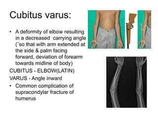
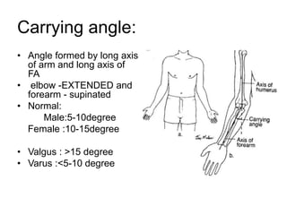
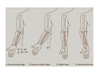
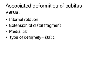
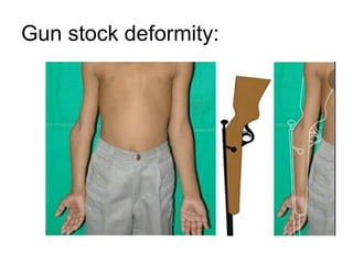
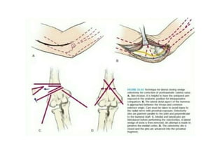
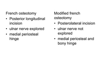
Cubitus varus
Ponnilavan
Cubitus varus:
• A deformity of elbow resulting
in a decreased carrying angle
(˘so that with arm extended at
the side & palm facing
forward, deviation of forearm
towards midline of body)
CUBITUS - ELBOW(LATIN)
VARUS - Angle inward
• Common complication of
supracondylar fracture of
humerus
Carrying angle:
• Angle formed by long axis
of arm and long axis of
FA
• elbow -EXTENDED and
forearm - supinated
• Normal:
Male:5-10degree
Female :10-15degree
• Valgus : >15 degree
• Varus :<5-10 degree
Associated deformities of cubitus
varus:
• Internal rotation
• Extension of distal fragment
• Medial tilt
• Type of deformity - static
Progressive cubitus varus
deformity after SCH#
• Growth disturbance in distal humerus
especially overgrowth of lateral condyle
can occur & sometimes avascular necrosis
and delayed growth of trochlea with
relative overgrowth of normal lateral side
of distal humerus epiphysis
Causes:
• Most common cause is malunited supracondylar
humerus fracture
• Congenital
• Malunited intercondylar #
• Malunited medial condyle#
• Malunited lateral condyle#
• Trochlear osteonecrosis
Malunited SCH #
• H/o significant trauma
• Thickening and irregularity of medial and
lateral supracondylar ridge
• Maintained 3 point relationship
• Elbow - Hyperextension
• Internal rotation increased with restricted
external rotation of shoulder
Gun stock deformity:
Displacements:
• Medial tilt
• Medial displacement
• Internal rotation
• Posterior tilt
• Posterior
displacement
• Proximal migration
Measurements on Xray: AP view
A line drawn parallel to
longitudinal axis of
humeral shaft as well as
a bisecting line parallel
to lateral condyle
• Normal:64-81 degree
Metaphyseal-diaphyseal angle:
• Transverse line is drawn
through axis of diaphysis.
angle measured between
lateral portion of
metaphyseal line and
proximal portion of
diaphyseal line
• Normal = 90 degree
• >90 degree = varus
angulation
• <90 degree = valgus
angulation
Lateral Xray:
• Humeral ulnar wrist
angle: crescent sign
• Presence of this imply
angulation and
rotation
Treatment:
• Observation with expectant remodelling
• Hemi epiphysiodesis and growth alteration
• Corrective osteotomy
Observation with expectant
remodelling
• Not appropriate because although
hyperextension may remodel to some
degree in a young child, in an older child
remodelling occurs even in joint's plane of
motion.
• Hence, it is not recommended
Hemi epiphysiodesis and growth
alteration:
• It is used to prevent cubitus varus
deformity in a patient with medial growth
arrest and progressive deformity,rather
than correcting it.
• It has a no role in child with a normal
physis
Corrective osteotomy:
• Medial open wedge osteotomy
• Lateral closing wedge(French osteotomy)
• Oblique osteotomy
• Dome osteotomy
• Step cut osteotomy
Approaches:
• Medial, Lateral and posterior
• Lateral: Good exposure with less
dissection
• Posterior: Complex osteotomy require
extensive exposure
Pre requisites:
• Atleast 1 year following fracture
• Patient demanding surgery
• Calculation of wedge to be removed by
normal side X ray
• Wedge angle = varus + normal
physiological valgus
French osteotomy:
• French, in 1959 first described a lateral
wedge osteotomy held with screws and a
figure of eight wire and this remains the
most popular method of correction.
• Lateral closed wedge osteotomy
Modified french osteotomy:
• Modification of French's osteotomy
appears to fulfill these criteria
• Easy procedure ,minimal dissection, little
possibility of nerve damage
French osteotomy
• Posterior longitudinal
incision
• ulnar nerve explored
• medial periosteal
hinge
Modified french
osteotomy
• Posterolateral incision
• ulnar nerve not
explored
• medial periosteal and
bony hinge
Step cut osteotomy:
• A standard posterior approach used
• Incision extended proximally distal 3rd
upper arm to a distance of 1-2 cm beyon
tip of olecranon distally
• Mobilize ulnar nerve anteriorly
• Triceps muscle split longitudinally
• Circumferential subperiosteal disection
done
• Osteotomy was
performed by first making
a proximal, transverse cut
perpendicular to
anatomical axis of
humerus
• Cut was made in a
proximal-medial to distal-
lateral direction
• Next cut perpendicular to
angular correction cut
was made at its lateral
margin creating a step cut
in distal humeral fragment
Complications of osteotomy:
• Stiffness
• Nerve injury (radial and ulnar nerve)
• Recurrent deformity
• Non union
• Osteomyelitis
• Malunion
THANK YOU

Cubitus varus

  • 1.
  • 2.
    Cubitus varus: • Adeformity of elbow resulting in a decreased carrying angle (˘so that with arm extended at the side & palm facing forward, deviation of forearm towards midline of body) CUBITUS - ELBOW(LATIN) VARUS - Angle inward • Common complication of supracondylar fracture of humerus
  • 3.
    Carrying angle: • Angleformed by long axis of arm and long axis of FA • elbow -EXTENDED and forearm - supinated • Normal: Male:5-10degree Female :10-15degree • Valgus : >15 degree • Varus :<5-10 degree
  • 5.
    Associated deformities ofcubitus varus: • Internal rotation • Extension of distal fragment • Medial tilt • Type of deformity - static
  • 6.
    Progressive cubitus varus deformityafter SCH# • Growth disturbance in distal humerus especially overgrowth of lateral condyle can occur & sometimes avascular necrosis and delayed growth of trochlea with relative overgrowth of normal lateral side of distal humerus epiphysis
  • 7.
    Causes: • Most commoncause is malunited supracondylar humerus fracture • Congenital • Malunited intercondylar # • Malunited medial condyle# • Malunited lateral condyle# • Trochlear osteonecrosis
  • 8.
    Malunited SCH # •H/o significant trauma • Thickening and irregularity of medial and lateral supracondylar ridge • Maintained 3 point relationship • Elbow - Hyperextension • Internal rotation increased with restricted external rotation of shoulder
  • 9.
  • 10.
    Displacements: • Medial tilt •Medial displacement • Internal rotation • Posterior tilt • Posterior displacement • Proximal migration
  • 11.
    Measurements on Xray:AP view A line drawn parallel to longitudinal axis of humeral shaft as well as a bisecting line parallel to lateral condyle • Normal:64-81 degree
  • 12.
    Metaphyseal-diaphyseal angle: • Transverseline is drawn through axis of diaphysis. angle measured between lateral portion of metaphyseal line and proximal portion of diaphyseal line • Normal = 90 degree • >90 degree = varus angulation • <90 degree = valgus angulation
  • 13.
    Lateral Xray: • Humeralulnar wrist angle: crescent sign • Presence of this imply angulation and rotation
  • 14.
    Treatment: • Observation withexpectant remodelling • Hemi epiphysiodesis and growth alteration • Corrective osteotomy
  • 15.
    Observation with expectant remodelling •Not appropriate because although hyperextension may remodel to some degree in a young child, in an older child remodelling occurs even in joint's plane of motion. • Hence, it is not recommended
  • 16.
    Hemi epiphysiodesis andgrowth alteration: • It is used to prevent cubitus varus deformity in a patient with medial growth arrest and progressive deformity,rather than correcting it. • It has a no role in child with a normal physis
  • 17.
    Corrective osteotomy: • Medialopen wedge osteotomy • Lateral closing wedge(French osteotomy) • Oblique osteotomy • Dome osteotomy • Step cut osteotomy
  • 18.
    Approaches: • Medial, Lateraland posterior • Lateral: Good exposure with less dissection • Posterior: Complex osteotomy require extensive exposure
  • 19.
    Pre requisites: • Atleast1 year following fracture • Patient demanding surgery • Calculation of wedge to be removed by normal side X ray • Wedge angle = varus + normal physiological valgus
  • 20.
    French osteotomy: • French,in 1959 first described a lateral wedge osteotomy held with screws and a figure of eight wire and this remains the most popular method of correction. • Lateral closed wedge osteotomy
  • 22.
    Modified french osteotomy: •Modification of French's osteotomy appears to fulfill these criteria • Easy procedure ,minimal dissection, little possibility of nerve damage
  • 23.
    French osteotomy • Posteriorlongitudinal incision • ulnar nerve explored • medial periosteal hinge Modified french osteotomy • Posterolateral incision • ulnar nerve not explored • medial periosteal and bony hinge
  • 24.
    Step cut osteotomy: •A standard posterior approach used • Incision extended proximally distal 3rd upper arm to a distance of 1-2 cm beyon tip of olecranon distally • Mobilize ulnar nerve anteriorly • Triceps muscle split longitudinally • Circumferential subperiosteal disection done
  • 25.
    • Osteotomy was performedby first making a proximal, transverse cut perpendicular to anatomical axis of humerus • Cut was made in a proximal-medial to distal- lateral direction • Next cut perpendicular to angular correction cut was made at its lateral margin creating a step cut in distal humeral fragment
  • 26.
    Complications of osteotomy: •Stiffness • Nerve injury (radial and ulnar nerve) • Recurrent deformity • Non union • Osteomyelitis • Malunion
  • 27.