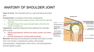
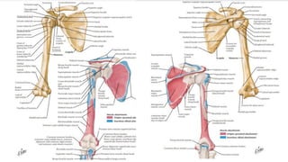
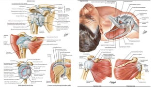
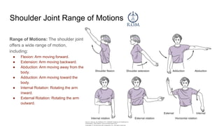
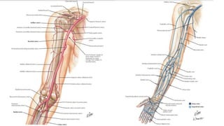
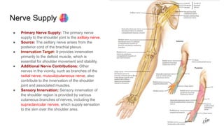
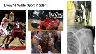
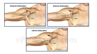
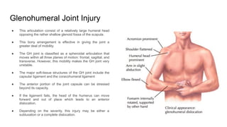
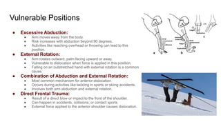
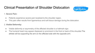
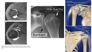
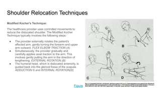
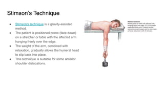
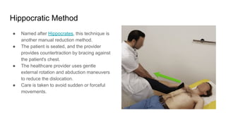
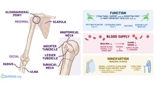
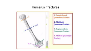
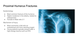
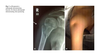
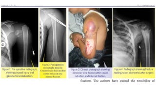
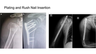
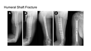
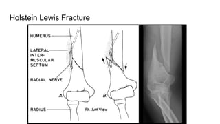
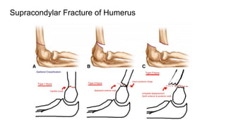
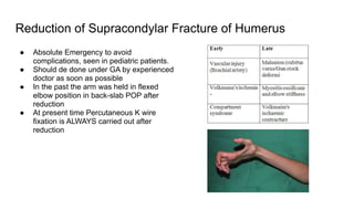
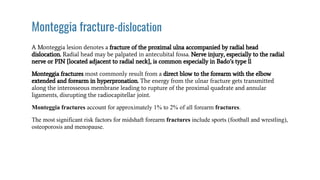
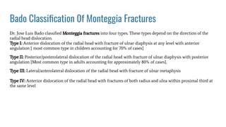
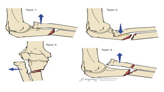
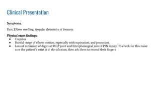
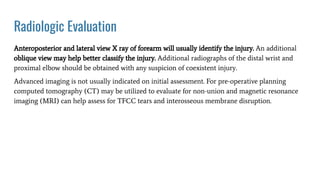
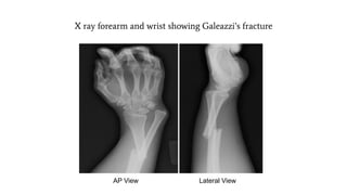
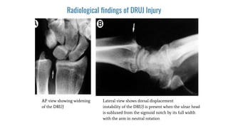
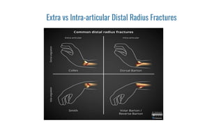
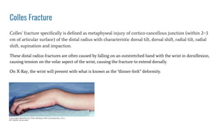
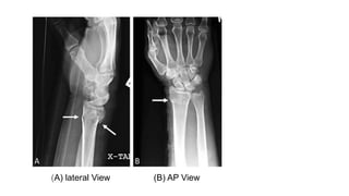
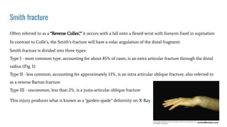
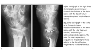
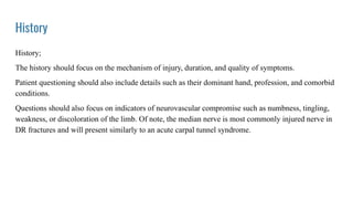
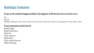
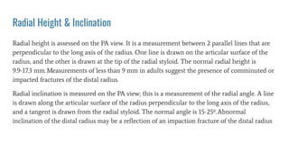
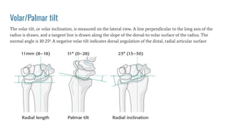
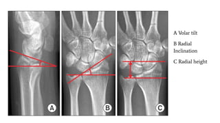
Fractures of the Upper Limb
Muhammad Wasil Khan
Shifa Amin
Learning Objectives
Shoulder Joint Anatomy:
● Understand the type of joint that forms the shoulder joint.
● Comprehend the extensive range of movements allowed by the shoulder joint.
● Recognize the importance of blood supply in the shoulder joint.
Stabilizing Fractures and Dislocations:
● Identify the most common type of shoulder dislocation and its mechanism of injury.
● Describe the vulnerable positions for shoulder dislocation.
● Explain the clinical presentation of shoulder dislocation.
● Summarize the key elements of the patient's history and physical examination.
● Interpret X-rays to confirm shoulder dislocation or fracture.
● Discuss the various techniques used for shoulder relocation and their names.
Post-Relocation Management:
● Outline the principles of post-relocation management.
● Understand the importance of follow-up care after shoulder relocation.
Proximal Humerus Fracture:
● Describe the clinical presentation of proximal humerus fractures.
● Recognize typical X-ray findings in proximal humerus fractures.
● Discuss the management options for proximal humerus fractures.
Learning Objectives
Monteggia Fracture-Dislocation:
● Define Monteggia fracture-dislocation.
● Explain the clinical presentation of Monteggia fractures.
● Discuss the management approach for Monteggia fractures.
Galeazzi Fracture-Dislocation:
● Define Galeazzi fracture-dislocation.
● Describe the clinical presentation of Galeazzi fractures.
● Discuss the management approach for Galeazzi fractures.
Distal Radius Fractures (DRF):
● Define distal radius fractures.
● Understand the anatomy of the wrist joint, including radial inclination and palmar tilt.
● Classify DRFs based on extraarticular and intraarticular categories.
● Explain the concept of radial height and its importance in DRF management.
● Differentiate between extraarticular fractures (e.g., Colles and Smith fractures) and their clinical presentations
and management.
● Describe the components of an intraarticular fracture like Barton fracture and its management.
Upper Limb
ANATOMY OF SHOULDER JOINT
Type of Joint: The shoulder joint is a synovial ball-and-socket
joint.
Components: It consists of two main components:
● The head of the humerus (upper arm bone), which forms the "ball" part
of the joint.
● The glenoid fossa of the scapula (shoulder blade), which forms the
"socket" or shallow cup.
Ligaments: Important ligaments that stabilize the shoulder
joint include:
● Glenohumeral ligaments: Reinforce the anterior, posterior, and inferior
aspects.
● Coracohumeral ligament: Provides additional stability.
Muscles: Muscles surrounding the shoulder joint play a crucial
role in its function, including the rotator cuff muscles
(supraspinatus, infraspinatus, teres minor, and subscapularis)
and the deltoid muscle.
Shoulder Joint Range of Motions
Range of Motions: The shoulder joint
offers a wide range of motion,
including:
● Flexion: Arm moving forward.
● Extension: Arm moving backward.
● Abduction: Arm moving away from the
body.
● Adduction: Arm moving toward the
body.
● Internal Rotation: Rotating the arm
inward.
● External Rotation: Rotating the arm
outward.
Blood Supply
The blood supply to the shoulder joint primarily comes from branches of the axillary artery. The axillary artery is a
continuation of the subclavian artery and extends into the axilla (armpit) region, giving rise to several branches that
supply blood to the shoulder joint and surrounding structures.
These branches include:
● Anterior Humeral Circumflex Artery: This branch runs anteriorly around the surgical neck of the humerus,
providing blood supply to the front of the shoulder joint.
● Posterior Humeral Circumflex Artery: This branch runs posteriorly around the surgical neck of the humerus,
supplying the posterior aspect of the shoulder joint.
● Suprascapular Artery: While not a direct branch of the axillary artery, the suprascapular artery originates from
the thyrocervical trunk, a branch of the subclavian artery. The suprascapular artery provides blood supply to
the superior portion of the shoulder joint and the supraspinatus muscle.
Nerve Supply
● Primary Nerve Supply: The primary nerve
supply to the shoulder joint is the axillary nerve.
● Source: The axillary nerve arises from the
posterior cord of the brachial plexus.
● Innervation Target: It provides innervation
primarily to the deltoid muscle, which is
essential for shoulder movement and stability.
● Additional Nerve Contributions: Other
nerves in the vicinity, such as branches of the
radial nerve, musculocutaneous nerve, also
contribute to the innervation of the shoulder
joint and associated muscles.
● Sensory Innervation: Sensory innervation of
the shoulder region is provided by various
cutaneous branches of nerves, including the
supraclavicular nerves, which supply sensation
to the skin over the shoulder area.
Dwayne Wade Sport Incident!
Stabilizing Fractures and Dislocations
● Shoulder dislocations are common orthopedic
injuries.
● The most frequently encountered type is anterior
shoulder dislocation.
Anterior Shoulder Dislocation:
● Anterior shoulder dislocation involves the
displacement of the humeral head out of the front
(anterior) of the glenoid fossa of the scapula.
● It is also known as "subcoracoid dislocation."
Mechanism of Injury:
● Primary Mechanism: Traumatic force applied to an
abducted and externally rotated arm.
● Abduction: Moving the arm away from the body, often to
the side or above the head.
● External Rotation: Rotating the arm outward, as if trying
to turn the palm up or away from the body.
Glenohumeral Joint Injury
● This articulation consist of a relatively large humeral head
opposing the rather shallow glenoid fossa of the scapula.
● This bony arrangement is effective in giving the joint a
greater deal of mobility.
● The GH joint is classified as a spheroidal articulation that
moves within all three planes of motion: frontal, sagittal, and
transverse. However, this mobility makes the GH joint very
unstable.
● The major soft-tissue structures of the GH joint include the
capsular ligament and the coracohumeral ligament
● The anterior portion of the joint capsule can be stressed
beyond its capacity.
● If the ligament fails, the head of the humerus can move
forward and out of place which leads to an anterior
dislocation.
● Depending on the severity, this injury may be either a
subluxation or a complete dislocation.
Vulnerable Positions
● Excessive Abduction:
● Arm moves away from the body.
● Risk increases with abduction beyond 90 degrees.
● Activities like reaching overhead or throwing can lead to this
position.
● External Rotation:
● Arm rotates outward, palm facing upward or away.
● Vulnerable to dislocation when force is applied in this position.
● Falling on an outstretched hand with external rotation is a common
cause.
● Combination of Abduction and External Rotation:
● Most common mechanism for anterior dislocation.
● Occurs during activities like tackling in sports or skiing accidents.
● Involves both arm abduction and external rotation.
● Direct Frontal Trauma:
● Result of a direct blow or impact to the front of the shoulder.
● Can happen in accidents, collisions, or contact sports.
● External force applied to the anterior shoulder causes dislocation.
Clinical Presentation of Shoulder Dislocation
1. Severe Pain:
● Patients experience severe pain localized to the shoulder region.
● This pain often results from ligamentous and soft tissue damage during the dislocation.
2. Visible Deformity:
● Visible deformity or asymmetry of the affected shoulder is a hallmark sign.
● The humeral head may appear displaced or prominent in the front or back of the shoulder.The
athlete will be supporting the arm on the affected side with the opposite arm.
3. Limited or Complete Loss of Motion:
● Limited or complete loss of the ability to move the affected arm is common.
● The patient may be unable to perform even basic movements due to pain and instability.
4. Potential Numbness or Tingling:
● Nerve involvement may lead to numbness or tingling in the arm or hand.
● This can occur due to nerve compression or stretching during the dislocation.
Clinical Relevance:
● Recognizing these clinical features is crucial for timely diagnosis and treatment of shoulder
dislocation.
Patient History
● Gathering a comprehensive patient history is essential for diagnosing shoulder
dislocation:
1. Mechanism of Injury:
● Inquire about the mechanism of injury to understand how the dislocation occurred.
● Was there a fall, direct trauma, or another event that led to the injury?
2. Previous Shoulder Issues:
● Ask about any previous shoulder issues or injuries, as they can influence treatment
decisions.
● Have there been prior dislocations or chronic instability?
3. Level of Pain and Disability:
● Assess the patient's level of pain and disability related to the shoulder.
● This information guides pain management and treatment planning.
Clinical Relevance:
● A thorough history helps establish the context and potential risk factors for
shoulder dislocation.
Physical Examination
1. Range of Motion:
● Assess range of motion in the affected shoulder, including flexion, extension,
abduction, adduction, internal rotation, and external rotation.
● Note any limitations, pain, or apprehension during movement. Apprehension
Test
2. Strength Assessment:
● Evaluate strength in the affected arm by assessing the ability to perform basic
movements.
● Identify any muscle weakness or loss of function. Examination
3. Neurovascular Status:
● Examine neurovascular status, checking for sensory deficits, numbness,
tingling, or motor deficits.
● Assess radial, ulnar, and median nerve function.
4. Signs of Instability or Deformity:
● Look for signs of instability or deformity in the shoulder region. Shoulder
AROM PROM
● Note any visible asymmetry, deformity, or joint laxity.
Radiological Confirmation of Shoulder Dislocation
Significance of X-ray Imaging:
● X-ray imaging plays a critical role in confirming
shoulder dislocation and identifying associated
fractures.
Importance:
● X-rays provide essential diagnostic information
for healthcare professionals managing shoulder
injuries.
● They help determine the type of dislocation,
assess fracture patterns, and guide treatment
decisions.
Specific Features to Look For:
1. Position of Humeral Head:
● Examine the position of the humeral head relative to the
glenoid fossa.
● In anterior shoulder dislocation, the humeral head will
typically appear displaced anteriorly from its normal
anatomical position within the glenoid fossa.
● In posterior dislocation, the humeral head may be displaced
posteriorly.
2. Associated Fractures:
● Look for associated fractures in the shoulder region.
● Fractures may involve the humerus, scapula, or clavicle.
● Evaluate the integrity of the glenoid rim, as fractures here
may affect joint stability.
3. Soft Tissue Changes:
● Assess for soft tissue changes on X-rays, such as:
● Swelling or soft tissue emphysema (air in soft tissues), indicating
potential injury.
● Abnormalities in the labrum, ligaments, or tendons.
Shoulder Relocation Techniques
Modified Kocher's Technique:
The healthcare provider uses controlled movements to
reduce the dislocated shoulder. The Modified Kocher
Technique typically involves the following steps:
● The provider externally rotates the patient's
affected arm, gently turning the forearm and upper
arm outward. FLEX ELBOW TRACTION (A)
● Simultaneously, the provider gradually and
carefully applies axial traction to the arm. This
involves gently pulling the arm in the direction of
lengthening. EXTERNAL ROTATION (B)
● The humeral head, which is dislocated anteriorly, is
guided back into the glenoid fossa of the scapula.
ADDUCTION © and INTERNAL ROTATION(D)
Figure
Stimson’s Technique
● Stimson's technique is a gravity-assisted
method.
● The patient is positioned prone (face down)
on a stretcher or table with the affected arm
hanging freely over the edge.
● The weight of the arm, combined with
relaxation, gradually allows the humeral head
to slip back into place.
● This technique is suitable for some anterior
shoulder dislocations.
Hippocratic Method
● Named after Hippocrates, this technique is
another manual reduction method.
● The patient is seated, and the provider
provides countertraction by bracing against
the patient's chest.
● The healthcare provider uses gentle
external rotation and abduction maneuvers
to reduce the dislocation.
● Care is taken to avoid sudden or forceful
movements.
● The Milch technique is a modification of the Hippocratic
method.
● It involves external rotation and abduction but also
incorporates scapular manipulation to facilitate reduction.
● The patient is usually seated.
● It requires careful coordination and experience.
Importance of Professional Involvement:
● It is essential that shoulder relocation techniques are
performed by trained healthcare professionals.
● These maneuvers demand precision, knowledge of
anatomy, and an understanding of patient comfort and
safety.
● Inappropriate techniques or excessive force can lead to
complications or injury.
Milch Technique
Post-Relocation Management and Follow-up Care
Key Principles:
● Immobilization: The affected shoulder is typically immobilized for 3
to 4 weeks using a sling or brace. This provides support, minimizes
movement, and reduces strain on healing structures.
● Pain Management: Effective pain management is essential to
enhance patient comfort. Non-steroidal anti-inflammatory drugs
(NSAIDs) or analgesics may be prescribed.
● Physical Therapy: Early involvement of physical therapy is often
recommended to:
○ Restore range of motion.
○ Strengthen the shoulder muscles.
○ Improve joint stability.
○ Prevent stiffness and muscle atrophy.
● Follow-up Imaging: Periodic X-rays or imaging studies may be
conducted to assess the healing progress and check for any
complications.
Importance of Follow-up Care:
Follow-up care after shoulder relocation is vital for several reasons:
1. Assessment of Healing: It allows healthcare providers to monitor the progress of
tissue healing, bone alignment, and joint stability.
2. Complication Detection: Follow-up appointment enable early detection of potential
complications like recurrent dislocation, nerve injury, or infection.
3. Rehabilitation Progress: Physical therapy and exercises are adjusted based on
patient progress and challenges faced during rehabilitation.
4. Educational Support: Patients receive ongoing education and guidance on
shoulder care, exercises, and injury prevention.
5. Prevention of Recurrence: Careful management and monitoring help reduce the
risk of recurrent dislocations.
Anatomy of Humerus
Humerus Fractures
Proximal Humerus Fractures
Epidemiology
● Most common fracture of the humerus
● Higher incidence in the elderly, related to
osteoporosis
● Female to Male ratio 2:1
Mechanism of Injury
● Most commonly a fall onto an
outstretched arm from standing height
● Young patients typically present after a
high energy trauma such as RTA
Clinical Presentation
● Proximal humerus fractures can present with a range of symptoms and signs:
1. Pain and Swelling:
● Patients typically experience pain and swelling in the shoulder region.
● Pain severity may vary from mild to severe.
2. Limited Range of Motion:
● There is often a noticeable reduction in range of motion of the affected shoulder.
● This limitation can impact daily activities.
3. Deformity or Displacement:
● In some cases, visible deformity or displacement of the shoulder may be evident.
● Displaced fractures may result in a noticeable change in the shoulder's appearance.
Management Options
● The choice of management depends on several factors:
1. Non-Operative Management:
● Stable fractures with minimal displacement may be managed non-operatively.
● This often involves immobilization with a sling or brace.
● Early rehabilitation for range of motion is crucial.
2. Surgical Intervention:
● Surgery is considered for displaced or complex fractures.
● Options include open reduction and internal fixation (ORIF) with plates and screws, intramedullary
nailing, or shoulder arthroplasty.
● Surgical choice is based on fracture type, patient age, and overall health.
3. Rehabilitation:
● Physical therapy and rehabilitation are essential in all cases to restore strength and range of motion.
● Rehabilitation protocols are tailored to the type of fracture and surgical intervention.
Treatment
Minimally displaced fractures- Sling immobilization, early motion
■Two-part fractures-
Anatomic neck fractures likely require ORIF. High incidence of osteonecrosis
Surgical neck fractures that are minimally displaced can be treated conservatively. Displacement usually
requires ORIF
■Three-part fractures
Due to disruption of opposing muscle forces, these are unstable so closed treatment is difficult. Displacement
requires ORIF.
■ Four-part fractures
In general for displacement or unstable injuries ORIF in the young and hemiarthroplasty in the elderly and
those with severe comminution. High rate of AVN (13-34%)
Generally recovery takes atleast one year but union is expected at 6 to 8 weeks.
Plating and Rush Nail Insertion
Humeral Shaft Fracture
Holstein Lewis Fracture
Supracondylar Fracture of Humerus
Reduction of Supracondylar Fracture of Humerus
● Absolute Emergency to avoid
complications, seen in pediatric patients.
● Should de done under GA by experienced
doctor as soon as possible
● In the past the arm was held in flexed
elbow position in back-slab POP after
reduction
● At present time Percutaneous K wire
fixation is ALWAYS carried out after
reduction
Monteggia fracture-dislocation
A Monteggia lesion denotes a fracture of the proximal ulna accompanied by radial head
dislocation. Radial head may be palpated in antecubital fossa. Nerve injury, especially to the radial
nerve or PIN {located adjacent to radial neck}, is common especially in Bado’s type ll
Monteggia fractures most commonly result from a direct blow to the forearm with the elbow
extended and forearm in hyperpronation. The energy from the ulnar fracture gets transmitted
along the interosseous membrane leading to rupture of the proximal quadrate and annular
ligaments, disrupting the radiocapitellar joint.
Monteggia fractures account for approximately 1% to 2% of all forearm fractures.
The most significant risk factors for midshaft forearm fractures include sports (football and wrestling),
osteoporosis and menopause.
Bado Classification 0f Monteggia Fractures
Dr. Jose Luis Bado classified Monteggia fractures into four types. These types depend on the direction of the
radial head dislocation.
Type I: Anterior dislocation of the radial head with fracture of ulnar diaphysis at any level with anterior
angulation { most common type in children accounting for 70% of cases}
Type II: Posterior/posterolateral dislocation of the radial head with fracture of ulnar diaphysis with posterior
angulation {Most common type in adults accounting for approximately 80% of cases}.
Type III: Lateral/anterolateral dislocation of the radial head with fracture of ulnar metaphysis
Type IV: Anterior dislocation of the radial head with fractures of both radius and ulna within proximal third at
the same level
Clinical Presentation
Symptoms;
Pain, Elbow swelling, Angular deformity of forearm
Physical exam findings;
● Crepitus
● Painful range of elbow motion, especially with supination, and pronation.
● Loss of extension of digits at MCP joint and Interphalangeal joint if PIN injury. To check for this make
sure the patient’s wrist is in dorsiflexion, then ask them to extend their fingers
Radiologic Evaluation
Anteroposterior and lateral view X ray of forearm will usually identify the injury. An additional
oblique view may help better classify the injury. Additional radiographs of the distal wrist and
proximal elbow should be obtained with any suspicion of coexistent injury.
Advanced imaging is not usually indicated on initial assessment. For pre-operative planning
computed tomography (CT) may be utilized to evaluate for non-union and magnetic resonance
imaging (MRI) can help assess for TFCC tears and interosseous membrane disruption.
Radiocapitellar Relation;
A line drawn along the long axis
of radius bisects the capitellum
regardless of the degree of
flexion or extension.
PA (a) and lateral (b) radiographs of
the forearm demonstrate angulated
fracture of the proximal ulna (arrow)
and dislocation of the radial head
from the capitellum
Management
All Monteggia fractures are considered unstable and require intervention.
Emergent orthopedic consultation is essential for open fractures and vascular compromise and Urgent
orthopedic consultation is indicated for neurologic deficits without vascular compromise.
Initial management for a suspected fracture; Rest, ice, immobilization, and elevation. Patients with a Monteggia
fracture should be placed in a sugar-tong splint with urgent referral to an orthopedist.
Pediatrics:
Children usually have better overall outcomes than adults, due to multiple influences, including the remodeling
ability of small angle deformities, shorter healing time, and overall solidity of Monteggia fractures in children.
Management is determined by the characteristics of the ulna fracture.
● Non-operative management is successful in;
1. Plastic deformation (bending or bowing without fracture) OR
2. Incomplete fracture (greenstick); should be treated with a closed reduction and splinted with the elbow
flexed at approximately 110-degrees in full supination for 6 weeks.
● Operative management is required by;
1. Complete ulnar fractures where Short, oblique fractures should be stabilized with elastic intramedullary
titanium nail fixation VS Comminuted or long oblique ulna fractures are fixed by ORIF using plates and
screws.
Adults
Operative management is crucial as adults are more prone to the persistent angulation and
shortening despite closed reduction techniques.
The most common operative repair is an ORIF; In most cases, a single compression plate is placed
with cortical screws anchored proximally and distally. The radial head dislocation usually reduces
easily after the ulna fracture is realigned.
After surgery, the extremity is placed in a long-arm splint with full supination and elbow flexion
around 100-degrees for Babo types 1, 3, and 4 fractures. For Babo type 2 fractures the elbow
should be splinted at 70-degrees.
The length of recovery depends on multiple variables including; the severity of the injury,
intended use of the extremity, and the individual’s ability to heal.
Rehabilitation usually begins at 2 weeks after surgical fixation.
Total return to activity depends upon the severity of the injury as well as the patient’s intended
use of their upper extremities; Patients with low physical demands require ~ 8 to 12 weeks VS
those with high demand activity (athletes and manual workers) may require up to 12 to 16 weeks
of rehab.
Galeazzi’s fracture-dislocation
A Galeazzi fracture is defined as a fracture of the distal radial shaft with subluxation or dislocation of the Distal
Radioulnar Joint {DRUJ}. It is an uncommon injury with the incidence varying from 3% to 6% of all forearm
fractures.
Mechanism of injury;
The mechanism is a direct blow to the dorsoradial wrist or a fall onto an outstretched hand with forced
pronation of the forearm.
Classified by position of radius
Type I: Dorsal displacement of distal radius, caused by supination force. Reduce with forced pronation and
dorsal to volar force on the distal radius.
Type II: Volar displacement, caused by pronation. Reduce with supination and volar to dorsal force on the distal
radius.
Clinical features
Symptoms;
Pain, swelling, deformity
Physical exam findings;
● Point tenderness over fracture site
● ROM; test forearm supination and pronation for instability
● DRUJ stress; causes wrist or midline forearm pain
● Weakness of the index and thumb finger pincer movement if Anterior Interosseous Nerve Syndrome
AIN Damage
Radiologic Evaluation
Anteroposterior and lateral view X ray of forearm, elbow, and wrist
If a distal to mid-shaft radial fracture is seen on the radiograph, close examination of the DRUJ is merited.
Signs of DRUJ disruption include:
● Widening of the DRUJ on the AP view
● Ulnar styloid fracture
● Displacement of the ulna dorsally on the lateral view
● Radial shortening greater than 5 mm (would need to compare with unaffected limb)
X ray forearm and wrist showing Galeazzi’s fracture
AP View Lateral View
Radiological findings of DRUJ Injury
AP view showing widening
of the DRUJ
Lateral view shows dorsal displacement
instability of the DRUJ is present when the ulnar head
is subluxed from the sigmoid notch by its full width
with the arm in neutral rotation
Management
In most cases, conservative management is indicated in children while surgical intervention is warranted in
adults
Initial Management
Initial management for a presumed fracture includes rest, ice, immobilization, and elevation. In most cases,
closed reduction of the radius followed by reduction of the ulna in the DRUJ should be attempted in the acute
setting.
Pediatrics
Children tend to have overall better long-term outcomes compared to adults. The approach is usually
conservative with closed reduction and splinting. Above-elbow casting in supination is the preferred
immobilization. Irreducible and unstable injuries, as well as variants of the Galeazzi fracture, may require
surgical intervention with open reduction and internal fixation (ORIF).
Adults ; ORIF of radius with reduction and stabilization of DRUJ in all cases, as anatomic reduction of DRUJ
is required
1. ORIF of radius
Approach
Volar (Henry) approach to radius
Plate fixation
perform anatomic plate fixation of radial shaft
radial bow must be restored/maintained
2. Reduction & stabilization of DRUJ
Approach
Dorsal capsulotomy
Reduction technique
Immobilization in supination (6 weeks)
Percutaneous pin fixation
Open surgical reduction
Open reduction internal fixation
Distal Radius fracture
The term “distal radial fracture” is a catch-all for any fracture of the distal radius that occurs through the metaphysis and
may involve the articular surfaces. These fractures are the most common orthopaedic injury and generally result from
fall on an outstretched hand. The description of these fractures is based on distal radial angulation and displacement,
intra-articular or extra-articular involvement, and associated anomalies of the ulnar or carpal bones.
Diagnosis is made clinically and radiographically with orthogonal radiographs of the wrist
Demographics;
>More common in females (2-3:1)
>Bimodal distribution
younger patients due to high energy mechanisms
older patients due to low energy mechanisms (i.e. FOOSH)
Risk factors
>OSTEOPOROSIS
● high incidence of distal radius fractures in women > 50 years old
● distal radius fractures are a predictor of subsequent fractures
● DEXA scan is recommended for women with distal radius fractures
ANATOMY OF THE WRIST JOINT
The wrist joint aka the radiocarpal joint connects and serves as a transition point between the forearm and hand.
1. Type of Joint; A Condyloid Synovial Joint i.e. a modified ball and socket joint that allows for flexion,
extension, abduction, and adduction movements.
2. Articulations; The joint itself is formed through the articulations between the distal radius and the scaphoid,
lunate, and triquetrum. The distal radius is responsible for 80% of axial load & articulates with scaphoid via
scaphoid fossa & lunate via lunate fossa. The ulna is not part of the wrist joint itself, as it articulates with the
distal radius via the distal radioulnar joint (DRUJ) at the ulnar/sigmoid notch.
3. Ligaments; The four ligaments responsible for maintaining the stability of the joint are
● Palmar and dorsal radiocarpal ligaments
● Ulnar and Radial collateral ligaments.
4. Innervation;
● Median nerve: Via the anterior interosseous branch
● Radial nerve: Via the posterior interosseous branch
● Ulnar nerve: Directly through its deep motor branches
5. Blood Supply; Via ulnar and radial arteries, through penetrating branches from the dorsal and palmar carpal
arches
Classification of DRF’s
There are numerous classification systems that describe fractures of the distal radius, traditionally
chosen by the clinician based on preference. Classification systems should be based on the following 2
principles:
● The classification should dictate the treatment.
● The classification should suggest the long-term, functional results of treatment or be
correlated with these anticipated results.
Cooney’s Universal Classification; divides into extra and intra articular fractures
Fernandez; based on mechanism of injury
Frykman; based on joint involvement (radiocarpal and/or radioulnar) +/- ulnar styloid fracture
Melone; divides intra-articular fractures into 4 types based on displacement
AO/ASIF; divides into extra, partial and complete articular fractures
Older’s
Mayo
Universal/Clooney Classification
Fernandez Classification
Frykman Classification
Melone Classification
Extra vs Intra-articular Distal Radius Fractures
Colles Fracture
Colles’ fracture specifically is defined as metaphyseal injury of cortico-cancellous junction (within 2−3
cm of articular surface) of the distal radius with characteristic dorsal tilt, dorsal shift, radial tilt, radial
shift, supination and impaction.
These distal radius fractures are often caused by falling on an outstretched hand with the wrist in dorsiflexion,
causing tension on the volar aspect of the wrist, causing the fracture to extend dorsally.
On X-Ray, the wrist will present with what is known as the “dinner-fork” deformity.
(A) lateral View (B) AP View
Smith fracture
Often referred to as a “Reverse Colles’,” it occurs with a fall onto a flexed wrist with forearm fixed in supination
In contrast to Colle’s, the Smith’s fracture will have a volar angulation of the distal fragment.
Smith fracture is divided into three types:
Type I - most common type, accounting for about 85% of cases, is an extra articular fracture through the distal
radius (Fig. 1)
Type II - less common, accounting for approximately 13%, is an intra articular oblique fracture, also referred to
as a reverse Barton fracture
Type III - uncommon, less than 2%, is a juxta-articular oblique fracture
This injury produces what is known as a “garden-spade” deformity on X-Ray.
AP View Lateral View
Smith Vs Colles
Barton’s Fracture
A Barton’s fracture is an intra-articular rim fracture of the distal radius which extends through the dorsal aspect
of the articular surface with associated dislocation of the radiocarpal joint.
There is no disruption of the radiocarpal ligaments, and the articular surface of the fractured distal radius
remains in contact with the proximal carpal row.
This preserved relationship between the radius and carpus is what distinguishes the Barton fracture from other
types of distal radius fracture/dislocations.
The distal radius fracture may involve either the volar or dorsal cortex. Volar and dorsal barton fractures are
subclassified based on the fracture pattern. As compared to the dorsal rim fracture, the volar barton fracture
occurs more frequently.
These fractures are unstable and often present with a dislocation of the carpal bones.
The most common mechanisms of injury vary depending on the patient population.
In the pediatric and young adult population, most Barton fractures result from sporting activities and motor
vehicle accidents.
The most common reason for it to happen is a direct, traumatic wrist injury. Young male workers or motorcycle
riders account for 70% of Barton's fracture cases.
However, in the elderly, particularly women, decreased bone density from osteoporosis means that less force is
needed to cause this injury. Therefore, the majority of these fractures are a result of a fall while standing.
(a) PA radiograph of the right wrist
demonstrates a comminuted
intraarticular fracture of the distal
radius with the distal fracture
fragment migrated proximally and
radially.
(b)Lateral radiograph of the same
wrist demonstrates an
intraarticular fracture of the distal
radius with the volar fragment
(arrows) maintaining its
relationship with the carpus. The
volar fracture fragment and
carpus have migrated proximally
relative to the dorsal fracture
fragment and shaft of the radius.
History
History;
The history should focus on the mechanism of injury, duration, and quality of symptoms.
Patient questioning should also include details such as their dominant hand, profession, and comorbid
conditions.
Questions should also focus on indicators of neurovascular compromise such as numbness, tingling,
weakness, or discoloration of the limb. Of note, the median nerve is most commonly injured nerve in
DR fractures and will present similarly to an acute carpal tunnel syndrome.
Clinical Features
Symptoms;
Wrist pain,swelling & deformity
Physical exam findings;
● Skin tenting or lacerations {could indicate an open fracture as this will quickly change injury
● management}.
● Crepitus overlying the fracture site.
● Visible deformity if displaced
● Limited Range of motion
The physical exam should also focus on ruling out complications such as compartment syndrome or other neurovascular
issues.
Check if the ;
1. Forearm compartments are soft & radial pulse is strong and equal between upper extremities.
2. Capillary refill distal to the fracture should be < 2-3 seconds.
3. Motor function;
● Radial nerve function analysed by having the patient extend the metacarpophalangeal joints of the fingers against
resistance.
● Median nerve function analysed by Palmar abduction of the thumb and holding the “A-OK sign” against
resistance
● Ulnar nerve strength testing is via abduction of the fingers against resistance.
4. Sensory function;
● Median and ulnar nerves evaluated by two-point discrimination on the pads of the 2nd and 5th fingers,
respectively.
● Radial Nerve tested just proximal to the 2nd MCP.
● Normal two-point discrimination is < 5mm, or 6mm in the elderly.
Radiologic Evaluation
X-rays are the standard imaging modality in the diagnosis of DR fractures. Recommended views:
AP
Lateral
Oblique; Oblique views may reveal intra-articular involvement that is not apparent on the other views.
X-rays examination should look for
Radial height,
Radial inclination,
Volar tilt,
Ulnar variance,
Radial shift,
Ulnar styloid fracture,
DRUJ widening.
Radial Height & Inclination
Radial height is assessed on the PA view. It is a measurement between 2 parallel lines that are
perpendicular to the long axis of the radius. One line is drawn on the articular surface of the
radius, and the other is drawn at the tip of the radial styloid. The normal radial height is
9.9-17.3 mm.Measurements of less than 9 mm in adults suggest the presence of comminuted or
impacted fractures of the distal radius.
Radial inclination is measured on the PA view; this is a measurement of the radial angle. A line
is drawn along the articular surface of the radius perpendicular to the long axis of the radius,
and a tangent is drawn from the radial styloid. The normal angle is 15-25º.Abnormal
inclination of the distal radius may be a reflection of an impaction fracture of the distal radius
Volar/Palmar tilt
The volar tilt, or volar inclination, is measured on the lateral view. A line perpendicular to the long axis of the
radius is drawn, and a tangent line is drawn along the slope of the dorsal-to-volar surface of the radius. The
normal angle is 10-25º. A negative volar tilt indicates dorsal angulation of the distal, radial articular surface
A Volar tilt
B Radial
Inclination
C Radial height
Ulnar Variance
Ulnar variance (also known as Hulten variance) refers to the relative lengths of the distal articular
surfaces Ulnar variance may be:
● neutral (both the ulnar and radial articular surfaces at the same level)
● positive (ulna projects more distally)
● negative (ulna projects more proximally)
Normal ulnar variance is 9-12 mm & ulnar variance does not depend on the length of the ulnar
styloid but on the positioning of the forearm, as well as on the radiographic technique.
CT
Indications
evaluate intra-articular involvement
surgical planning
MRI
Indications
evaluate for soft tissue injury;
TFCC injuries
scapholunate ligament injuries (DISI)
lunotriquetral injuries (VISI)
Management
NON OPERATIVE
Closed reduction and splint/cast immobilization
Indications
extra-articular
< 5mm radial shortening and < 2 mm of articular step-off
< 5-degrees change in radial inclination
dorsal angulation < 5° or within 20° of contralateral distal radius
Closed Reduction and Percutaneous Pinning (CRPP)
Indications
extra-articular fracture with stable volar cortex
Outcomes
82-90% good results if used appropriately
OPERATIVE
ORIF
Indications
Radiographic findings indicating instability (pre-reduction radiographs best predictor of stability)
dorsal angulation > 5° or > 20° of contralateral distal radius
volar or dorsal comminution
displaced intra-articular fractures > 2mm
radial shortening > 5mm
associated ulnar fracture {Associated ulnar styloid fractures do not require fixation}
severe osteoporosis
articular margin fractures (dorsal and volar Barton's fractures)
comminuted and displaced extra-articular fractures (Smith's fractures)
die-punch fractures
progressive loss of volar tilt and radial length following closed reduction and casting
External fixation
Indications
open fractures
highly comminuted fractures
medically unstable patients unable to undergo a lengthy procedure
Outcomes
important adjunct with 80-90% good/excellent results
alone cannot reliably restore 10° palmar tilt therefore usually combined with percutaneous
pinning technique or plate fixation
REFERENCES
● Bailey & Love's Short Practice of Surgery - 28th Edition
● https://www.ncbi.nlm.nih.gov/pmc/articles/PMC5392888/
● https://www.aafp.org/pubs/afp/issues/2016/0715/p119.html
● https://cambridgeshoulder.co.uk/shoulder/instabilitydislocation/
● https://emedicine.medscape.com/article/398406-overview#a1
● https://www.orthobullets.com/trauma/1027/distal-radius-fractures
● https://www.ncbi.nlm.nih.gov/books/NBK536916/
● https://www.ncbi.nlm.nih.gov/books/NBK470575/
●
● Atlas of Human Anatomy 8th Edition
● Current Therapy in Trauma and Critical Care
THANK YOU!

Fractures of the Upper Limb

  • 1.
    Fractures of theUpper Limb Muhammad Wasil Khan Shifa Amin
  • 2.
    Learning Objectives Shoulder JointAnatomy: ● Understand the type of joint that forms the shoulder joint. ● Comprehend the extensive range of movements allowed by the shoulder joint. ● Recognize the importance of blood supply in the shoulder joint. Stabilizing Fractures and Dislocations: ● Identify the most common type of shoulder dislocation and its mechanism of injury. ● Describe the vulnerable positions for shoulder dislocation. ● Explain the clinical presentation of shoulder dislocation. ● Summarize the key elements of the patient's history and physical examination. ● Interpret X-rays to confirm shoulder dislocation or fracture. ● Discuss the various techniques used for shoulder relocation and their names. Post-Relocation Management: ● Outline the principles of post-relocation management. ● Understand the importance of follow-up care after shoulder relocation. Proximal Humerus Fracture: ● Describe the clinical presentation of proximal humerus fractures. ● Recognize typical X-ray findings in proximal humerus fractures. ● Discuss the management options for proximal humerus fractures.
  • 3.
    Learning Objectives Monteggia Fracture-Dislocation: ●Define Monteggia fracture-dislocation. ● Explain the clinical presentation of Monteggia fractures. ● Discuss the management approach for Monteggia fractures. Galeazzi Fracture-Dislocation: ● Define Galeazzi fracture-dislocation. ● Describe the clinical presentation of Galeazzi fractures. ● Discuss the management approach for Galeazzi fractures. Distal Radius Fractures (DRF): ● Define distal radius fractures. ● Understand the anatomy of the wrist joint, including radial inclination and palmar tilt. ● Classify DRFs based on extraarticular and intraarticular categories. ● Explain the concept of radial height and its importance in DRF management. ● Differentiate between extraarticular fractures (e.g., Colles and Smith fractures) and their clinical presentations and management. ● Describe the components of an intraarticular fracture like Barton fracture and its management.
  • 4.
  • 5.
    ANATOMY OF SHOULDERJOINT Type of Joint: The shoulder joint is a synovial ball-and-socket joint. Components: It consists of two main components: ● The head of the humerus (upper arm bone), which forms the "ball" part of the joint. ● The glenoid fossa of the scapula (shoulder blade), which forms the "socket" or shallow cup. Ligaments: Important ligaments that stabilize the shoulder joint include: ● Glenohumeral ligaments: Reinforce the anterior, posterior, and inferior aspects. ● Coracohumeral ligament: Provides additional stability. Muscles: Muscles surrounding the shoulder joint play a crucial role in its function, including the rotator cuff muscles (supraspinatus, infraspinatus, teres minor, and subscapularis) and the deltoid muscle.
  • 8.
    Shoulder Joint Rangeof Motions Range of Motions: The shoulder joint offers a wide range of motion, including: ● Flexion: Arm moving forward. ● Extension: Arm moving backward. ● Abduction: Arm moving away from the body. ● Adduction: Arm moving toward the body. ● Internal Rotation: Rotating the arm inward. ● External Rotation: Rotating the arm outward.
  • 9.
    Blood Supply The bloodsupply to the shoulder joint primarily comes from branches of the axillary artery. The axillary artery is a continuation of the subclavian artery and extends into the axilla (armpit) region, giving rise to several branches that supply blood to the shoulder joint and surrounding structures. These branches include: ● Anterior Humeral Circumflex Artery: This branch runs anteriorly around the surgical neck of the humerus, providing blood supply to the front of the shoulder joint. ● Posterior Humeral Circumflex Artery: This branch runs posteriorly around the surgical neck of the humerus, supplying the posterior aspect of the shoulder joint. ● Suprascapular Artery: While not a direct branch of the axillary artery, the suprascapular artery originates from the thyrocervical trunk, a branch of the subclavian artery. The suprascapular artery provides blood supply to the superior portion of the shoulder joint and the supraspinatus muscle.
  • 11.
    Nerve Supply ● PrimaryNerve Supply: The primary nerve supply to the shoulder joint is the axillary nerve. ● Source: The axillary nerve arises from the posterior cord of the brachial plexus. ● Innervation Target: It provides innervation primarily to the deltoid muscle, which is essential for shoulder movement and stability. ● Additional Nerve Contributions: Other nerves in the vicinity, such as branches of the radial nerve, musculocutaneous nerve, also contribute to the innervation of the shoulder joint and associated muscles. ● Sensory Innervation: Sensory innervation of the shoulder region is provided by various cutaneous branches of nerves, including the supraclavicular nerves, which supply sensation to the skin over the shoulder area.
  • 12.
  • 13.
    Stabilizing Fractures andDislocations ● Shoulder dislocations are common orthopedic injuries. ● The most frequently encountered type is anterior shoulder dislocation. Anterior Shoulder Dislocation: ● Anterior shoulder dislocation involves the displacement of the humeral head out of the front (anterior) of the glenoid fossa of the scapula. ● It is also known as "subcoracoid dislocation." Mechanism of Injury: ● Primary Mechanism: Traumatic force applied to an abducted and externally rotated arm. ● Abduction: Moving the arm away from the body, often to the side or above the head. ● External Rotation: Rotating the arm outward, as if trying to turn the palm up or away from the body.
  • 15.
    Glenohumeral Joint Injury ●This articulation consist of a relatively large humeral head opposing the rather shallow glenoid fossa of the scapula. ● This bony arrangement is effective in giving the joint a greater deal of mobility. ● The GH joint is classified as a spheroidal articulation that moves within all three planes of motion: frontal, sagittal, and transverse. However, this mobility makes the GH joint very unstable. ● The major soft-tissue structures of the GH joint include the capsular ligament and the coracohumeral ligament ● The anterior portion of the joint capsule can be stressed beyond its capacity. ● If the ligament fails, the head of the humerus can move forward and out of place which leads to an anterior dislocation. ● Depending on the severity, this injury may be either a subluxation or a complete dislocation.
  • 16.
    Vulnerable Positions ● ExcessiveAbduction: ● Arm moves away from the body. ● Risk increases with abduction beyond 90 degrees. ● Activities like reaching overhead or throwing can lead to this position. ● External Rotation: ● Arm rotates outward, palm facing upward or away. ● Vulnerable to dislocation when force is applied in this position. ● Falling on an outstretched hand with external rotation is a common cause. ● Combination of Abduction and External Rotation: ● Most common mechanism for anterior dislocation. ● Occurs during activities like tackling in sports or skiing accidents. ● Involves both arm abduction and external rotation. ● Direct Frontal Trauma: ● Result of a direct blow or impact to the front of the shoulder. ● Can happen in accidents, collisions, or contact sports. ● External force applied to the anterior shoulder causes dislocation.
  • 17.
    Clinical Presentation ofShoulder Dislocation 1. Severe Pain: ● Patients experience severe pain localized to the shoulder region. ● This pain often results from ligamentous and soft tissue damage during the dislocation. 2. Visible Deformity: ● Visible deformity or asymmetry of the affected shoulder is a hallmark sign. ● The humeral head may appear displaced or prominent in the front or back of the shoulder.The athlete will be supporting the arm on the affected side with the opposite arm.
  • 18.
    3. Limited orComplete Loss of Motion: ● Limited or complete loss of the ability to move the affected arm is common. ● The patient may be unable to perform even basic movements due to pain and instability. 4. Potential Numbness or Tingling: ● Nerve involvement may lead to numbness or tingling in the arm or hand. ● This can occur due to nerve compression or stretching during the dislocation. Clinical Relevance: ● Recognizing these clinical features is crucial for timely diagnosis and treatment of shoulder dislocation.
  • 19.
    Patient History ● Gatheringa comprehensive patient history is essential for diagnosing shoulder dislocation: 1. Mechanism of Injury: ● Inquire about the mechanism of injury to understand how the dislocation occurred. ● Was there a fall, direct trauma, or another event that led to the injury? 2. Previous Shoulder Issues: ● Ask about any previous shoulder issues or injuries, as they can influence treatment decisions. ● Have there been prior dislocations or chronic instability?
  • 20.
    3. Level ofPain and Disability: ● Assess the patient's level of pain and disability related to the shoulder. ● This information guides pain management and treatment planning. Clinical Relevance: ● A thorough history helps establish the context and potential risk factors for shoulder dislocation.
  • 21.
    Physical Examination 1. Rangeof Motion: ● Assess range of motion in the affected shoulder, including flexion, extension, abduction, adduction, internal rotation, and external rotation. ● Note any limitations, pain, or apprehension during movement. Apprehension Test 2. Strength Assessment: ● Evaluate strength in the affected arm by assessing the ability to perform basic movements. ● Identify any muscle weakness or loss of function. Examination
  • 22.
    3. Neurovascular Status: ●Examine neurovascular status, checking for sensory deficits, numbness, tingling, or motor deficits. ● Assess radial, ulnar, and median nerve function. 4. Signs of Instability or Deformity: ● Look for signs of instability or deformity in the shoulder region. Shoulder AROM PROM ● Note any visible asymmetry, deformity, or joint laxity.
  • 23.
    Radiological Confirmation ofShoulder Dislocation Significance of X-ray Imaging: ● X-ray imaging plays a critical role in confirming shoulder dislocation and identifying associated fractures. Importance: ● X-rays provide essential diagnostic information for healthcare professionals managing shoulder injuries. ● They help determine the type of dislocation, assess fracture patterns, and guide treatment decisions.
  • 24.
    Specific Features toLook For: 1. Position of Humeral Head: ● Examine the position of the humeral head relative to the glenoid fossa. ● In anterior shoulder dislocation, the humeral head will typically appear displaced anteriorly from its normal anatomical position within the glenoid fossa. ● In posterior dislocation, the humeral head may be displaced posteriorly. 2. Associated Fractures: ● Look for associated fractures in the shoulder region. ● Fractures may involve the humerus, scapula, or clavicle. ● Evaluate the integrity of the glenoid rim, as fractures here may affect joint stability. 3. Soft Tissue Changes: ● Assess for soft tissue changes on X-rays, such as: ● Swelling or soft tissue emphysema (air in soft tissues), indicating potential injury. ● Abnormalities in the labrum, ligaments, or tendons.
  • 27.
    Shoulder Relocation Techniques ModifiedKocher's Technique: The healthcare provider uses controlled movements to reduce the dislocated shoulder. The Modified Kocher Technique typically involves the following steps: ● The provider externally rotates the patient's affected arm, gently turning the forearm and upper arm outward. FLEX ELBOW TRACTION (A) ● Simultaneously, the provider gradually and carefully applies axial traction to the arm. This involves gently pulling the arm in the direction of lengthening. EXTERNAL ROTATION (B) ● The humeral head, which is dislocated anteriorly, is guided back into the glenoid fossa of the scapula. ADDUCTION © and INTERNAL ROTATION(D) Figure
  • 28.
    Stimson’s Technique ● Stimson'stechnique is a gravity-assisted method. ● The patient is positioned prone (face down) on a stretcher or table with the affected arm hanging freely over the edge. ● The weight of the arm, combined with relaxation, gradually allows the humeral head to slip back into place. ● This technique is suitable for some anterior shoulder dislocations.
  • 29.
    Hippocratic Method ● Namedafter Hippocrates, this technique is another manual reduction method. ● The patient is seated, and the provider provides countertraction by bracing against the patient's chest. ● The healthcare provider uses gentle external rotation and abduction maneuvers to reduce the dislocation. ● Care is taken to avoid sudden or forceful movements.
  • 30.
    ● The Milchtechnique is a modification of the Hippocratic method. ● It involves external rotation and abduction but also incorporates scapular manipulation to facilitate reduction. ● The patient is usually seated. ● It requires careful coordination and experience. Importance of Professional Involvement: ● It is essential that shoulder relocation techniques are performed by trained healthcare professionals. ● These maneuvers demand precision, knowledge of anatomy, and an understanding of patient comfort and safety. ● Inappropriate techniques or excessive force can lead to complications or injury. Milch Technique
  • 31.
    Post-Relocation Management andFollow-up Care Key Principles: ● Immobilization: The affected shoulder is typically immobilized for 3 to 4 weeks using a sling or brace. This provides support, minimizes movement, and reduces strain on healing structures. ● Pain Management: Effective pain management is essential to enhance patient comfort. Non-steroidal anti-inflammatory drugs (NSAIDs) or analgesics may be prescribed. ● Physical Therapy: Early involvement of physical therapy is often recommended to: ○ Restore range of motion. ○ Strengthen the shoulder muscles. ○ Improve joint stability. ○ Prevent stiffness and muscle atrophy. ● Follow-up Imaging: Periodic X-rays or imaging studies may be conducted to assess the healing progress and check for any complications.
  • 32.
    Importance of Follow-upCare: Follow-up care after shoulder relocation is vital for several reasons: 1. Assessment of Healing: It allows healthcare providers to monitor the progress of tissue healing, bone alignment, and joint stability. 2. Complication Detection: Follow-up appointment enable early detection of potential complications like recurrent dislocation, nerve injury, or infection. 3. Rehabilitation Progress: Physical therapy and exercises are adjusted based on patient progress and challenges faced during rehabilitation. 4. Educational Support: Patients receive ongoing education and guidance on shoulder care, exercises, and injury prevention. 5. Prevention of Recurrence: Careful management and monitoring help reduce the risk of recurrent dislocations.
  • 33.
  • 35.
  • 36.
    Proximal Humerus Fractures Epidemiology ●Most common fracture of the humerus ● Higher incidence in the elderly, related to osteoporosis ● Female to Male ratio 2:1 Mechanism of Injury ● Most commonly a fall onto an outstretched arm from standing height ● Young patients typically present after a high energy trauma such as RTA
  • 37.
    Clinical Presentation ● Proximalhumerus fractures can present with a range of symptoms and signs: 1. Pain and Swelling: ● Patients typically experience pain and swelling in the shoulder region. ● Pain severity may vary from mild to severe. 2. Limited Range of Motion: ● There is often a noticeable reduction in range of motion of the affected shoulder. ● This limitation can impact daily activities. 3. Deformity or Displacement: ● In some cases, visible deformity or displacement of the shoulder may be evident. ● Displaced fractures may result in a noticeable change in the shoulder's appearance.
  • 38.
    Management Options ● Thechoice of management depends on several factors: 1. Non-Operative Management: ● Stable fractures with minimal displacement may be managed non-operatively. ● This often involves immobilization with a sling or brace. ● Early rehabilitation for range of motion is crucial. 2. Surgical Intervention: ● Surgery is considered for displaced or complex fractures. ● Options include open reduction and internal fixation (ORIF) with plates and screws, intramedullary nailing, or shoulder arthroplasty. ● Surgical choice is based on fracture type, patient age, and overall health. 3. Rehabilitation: ● Physical therapy and rehabilitation are essential in all cases to restore strength and range of motion. ● Rehabilitation protocols are tailored to the type of fracture and surgical intervention.
  • 41.
    Treatment Minimally displaced fractures-Sling immobilization, early motion ■Two-part fractures- Anatomic neck fractures likely require ORIF. High incidence of osteonecrosis Surgical neck fractures that are minimally displaced can be treated conservatively. Displacement usually requires ORIF ■Three-part fractures Due to disruption of opposing muscle forces, these are unstable so closed treatment is difficult. Displacement requires ORIF. ■ Four-part fractures In general for displacement or unstable injuries ORIF in the young and hemiarthroplasty in the elderly and those with severe comminution. High rate of AVN (13-34%) Generally recovery takes atleast one year but union is expected at 6 to 8 weeks.
  • 44.
    Plating and RushNail Insertion
  • 45.
  • 46.
  • 47.
  • 48.
    Reduction of SupracondylarFracture of Humerus ● Absolute Emergency to avoid complications, seen in pediatric patients. ● Should de done under GA by experienced doctor as soon as possible ● In the past the arm was held in flexed elbow position in back-slab POP after reduction ● At present time Percutaneous K wire fixation is ALWAYS carried out after reduction
  • 50.
    Monteggia fracture-dislocation A Monteggialesion denotes a fracture of the proximal ulna accompanied by radial head dislocation. Radial head may be palpated in antecubital fossa. Nerve injury, especially to the radial nerve or PIN {located adjacent to radial neck}, is common especially in Bado’s type ll Monteggia fractures most commonly result from a direct blow to the forearm with the elbow extended and forearm in hyperpronation. The energy from the ulnar fracture gets transmitted along the interosseous membrane leading to rupture of the proximal quadrate and annular ligaments, disrupting the radiocapitellar joint. Monteggia fractures account for approximately 1% to 2% of all forearm fractures. The most significant risk factors for midshaft forearm fractures include sports (football and wrestling), osteoporosis and menopause.
  • 51.
    Bado Classification 0fMonteggia Fractures Dr. Jose Luis Bado classified Monteggia fractures into four types. These types depend on the direction of the radial head dislocation. Type I: Anterior dislocation of the radial head with fracture of ulnar diaphysis at any level with anterior angulation { most common type in children accounting for 70% of cases} Type II: Posterior/posterolateral dislocation of the radial head with fracture of ulnar diaphysis with posterior angulation {Most common type in adults accounting for approximately 80% of cases}. Type III: Lateral/anterolateral dislocation of the radial head with fracture of ulnar metaphysis Type IV: Anterior dislocation of the radial head with fractures of both radius and ulna within proximal third at the same level
  • 53.
    Clinical Presentation Symptoms; Pain, Elbowswelling, Angular deformity of forearm Physical exam findings; ● Crepitus ● Painful range of elbow motion, especially with supination, and pronation. ● Loss of extension of digits at MCP joint and Interphalangeal joint if PIN injury. To check for this make sure the patient’s wrist is in dorsiflexion, then ask them to extend their fingers
  • 54.
    Radiologic Evaluation Anteroposterior andlateral view X ray of forearm will usually identify the injury. An additional oblique view may help better classify the injury. Additional radiographs of the distal wrist and proximal elbow should be obtained with any suspicion of coexistent injury. Advanced imaging is not usually indicated on initial assessment. For pre-operative planning computed tomography (CT) may be utilized to evaluate for non-union and magnetic resonance imaging (MRI) can help assess for TFCC tears and interosseous membrane disruption.
  • 55.
    Radiocapitellar Relation; A linedrawn along the long axis of radius bisects the capitellum regardless of the degree of flexion or extension.
  • 56.
    PA (a) andlateral (b) radiographs of the forearm demonstrate angulated fracture of the proximal ulna (arrow) and dislocation of the radial head from the capitellum
  • 57.
  • 58.
    All Monteggia fracturesare considered unstable and require intervention. Emergent orthopedic consultation is essential for open fractures and vascular compromise and Urgent orthopedic consultation is indicated for neurologic deficits without vascular compromise. Initial management for a suspected fracture; Rest, ice, immobilization, and elevation. Patients with a Monteggia fracture should be placed in a sugar-tong splint with urgent referral to an orthopedist. Pediatrics: Children usually have better overall outcomes than adults, due to multiple influences, including the remodeling ability of small angle deformities, shorter healing time, and overall solidity of Monteggia fractures in children. Management is determined by the characteristics of the ulna fracture. ● Non-operative management is successful in; 1. Plastic deformation (bending or bowing without fracture) OR 2. Incomplete fracture (greenstick); should be treated with a closed reduction and splinted with the elbow flexed at approximately 110-degrees in full supination for 6 weeks. ● Operative management is required by; 1. Complete ulnar fractures where Short, oblique fractures should be stabilized with elastic intramedullary titanium nail fixation VS Comminuted or long oblique ulna fractures are fixed by ORIF using plates and screws.
  • 59.
    Adults Operative management iscrucial as adults are more prone to the persistent angulation and shortening despite closed reduction techniques. The most common operative repair is an ORIF; In most cases, a single compression plate is placed with cortical screws anchored proximally and distally. The radial head dislocation usually reduces easily after the ulna fracture is realigned. After surgery, the extremity is placed in a long-arm splint with full supination and elbow flexion around 100-degrees for Babo types 1, 3, and 4 fractures. For Babo type 2 fractures the elbow should be splinted at 70-degrees. The length of recovery depends on multiple variables including; the severity of the injury, intended use of the extremity, and the individual’s ability to heal. Rehabilitation usually begins at 2 weeks after surgical fixation. Total return to activity depends upon the severity of the injury as well as the patient’s intended use of their upper extremities; Patients with low physical demands require ~ 8 to 12 weeks VS those with high demand activity (athletes and manual workers) may require up to 12 to 16 weeks of rehab.
  • 60.
    Galeazzi’s fracture-dislocation A Galeazzifracture is defined as a fracture of the distal radial shaft with subluxation or dislocation of the Distal Radioulnar Joint {DRUJ}. It is an uncommon injury with the incidence varying from 3% to 6% of all forearm fractures. Mechanism of injury; The mechanism is a direct blow to the dorsoradial wrist or a fall onto an outstretched hand with forced pronation of the forearm. Classified by position of radius Type I: Dorsal displacement of distal radius, caused by supination force. Reduce with forced pronation and dorsal to volar force on the distal radius. Type II: Volar displacement, caused by pronation. Reduce with supination and volar to dorsal force on the distal radius.
  • 61.
    Clinical features Symptoms; Pain, swelling,deformity Physical exam findings; ● Point tenderness over fracture site ● ROM; test forearm supination and pronation for instability ● DRUJ stress; causes wrist or midline forearm pain ● Weakness of the index and thumb finger pincer movement if Anterior Interosseous Nerve Syndrome
  • 62.
  • 63.
    Radiologic Evaluation Anteroposterior andlateral view X ray of forearm, elbow, and wrist If a distal to mid-shaft radial fracture is seen on the radiograph, close examination of the DRUJ is merited. Signs of DRUJ disruption include: ● Widening of the DRUJ on the AP view ● Ulnar styloid fracture ● Displacement of the ulna dorsally on the lateral view ● Radial shortening greater than 5 mm (would need to compare with unaffected limb)
  • 64.
    X ray forearmand wrist showing Galeazzi’s fracture AP View Lateral View
  • 65.
    Radiological findings ofDRUJ Injury AP view showing widening of the DRUJ Lateral view shows dorsal displacement instability of the DRUJ is present when the ulnar head is subluxed from the sigmoid notch by its full width with the arm in neutral rotation
  • 66.
  • 67.
    In most cases,conservative management is indicated in children while surgical intervention is warranted in adults Initial Management Initial management for a presumed fracture includes rest, ice, immobilization, and elevation. In most cases, closed reduction of the radius followed by reduction of the ulna in the DRUJ should be attempted in the acute setting. Pediatrics Children tend to have overall better long-term outcomes compared to adults. The approach is usually conservative with closed reduction and splinting. Above-elbow casting in supination is the preferred immobilization. Irreducible and unstable injuries, as well as variants of the Galeazzi fracture, may require surgical intervention with open reduction and internal fixation (ORIF).
  • 68.
    Adults ; ORIFof radius with reduction and stabilization of DRUJ in all cases, as anatomic reduction of DRUJ is required 1. ORIF of radius Approach Volar (Henry) approach to radius Plate fixation perform anatomic plate fixation of radial shaft radial bow must be restored/maintained 2. Reduction & stabilization of DRUJ Approach Dorsal capsulotomy Reduction technique Immobilization in supination (6 weeks) Percutaneous pin fixation Open surgical reduction Open reduction internal fixation
  • 69.
    Distal Radius fracture Theterm “distal radial fracture” is a catch-all for any fracture of the distal radius that occurs through the metaphysis and may involve the articular surfaces. These fractures are the most common orthopaedic injury and generally result from fall on an outstretched hand. The description of these fractures is based on distal radial angulation and displacement, intra-articular or extra-articular involvement, and associated anomalies of the ulnar or carpal bones. Diagnosis is made clinically and radiographically with orthogonal radiographs of the wrist Demographics; >More common in females (2-3:1) >Bimodal distribution younger patients due to high energy mechanisms older patients due to low energy mechanisms (i.e. FOOSH) Risk factors >OSTEOPOROSIS ● high incidence of distal radius fractures in women > 50 years old ● distal radius fractures are a predictor of subsequent fractures ● DEXA scan is recommended for women with distal radius fractures
  • 70.
    ANATOMY OF THEWRIST JOINT The wrist joint aka the radiocarpal joint connects and serves as a transition point between the forearm and hand. 1. Type of Joint; A Condyloid Synovial Joint i.e. a modified ball and socket joint that allows for flexion, extension, abduction, and adduction movements. 2. Articulations; The joint itself is formed through the articulations between the distal radius and the scaphoid, lunate, and triquetrum. The distal radius is responsible for 80% of axial load & articulates with scaphoid via scaphoid fossa & lunate via lunate fossa. The ulna is not part of the wrist joint itself, as it articulates with the distal radius via the distal radioulnar joint (DRUJ) at the ulnar/sigmoid notch. 3. Ligaments; The four ligaments responsible for maintaining the stability of the joint are ● Palmar and dorsal radiocarpal ligaments ● Ulnar and Radial collateral ligaments. 4. Innervation; ● Median nerve: Via the anterior interosseous branch ● Radial nerve: Via the posterior interosseous branch ● Ulnar nerve: Directly through its deep motor branches 5. Blood Supply; Via ulnar and radial arteries, through penetrating branches from the dorsal and palmar carpal arches
  • 72.
    Classification of DRF’s Thereare numerous classification systems that describe fractures of the distal radius, traditionally chosen by the clinician based on preference. Classification systems should be based on the following 2 principles: ● The classification should dictate the treatment. ● The classification should suggest the long-term, functional results of treatment or be correlated with these anticipated results. Cooney’s Universal Classification; divides into extra and intra articular fractures Fernandez; based on mechanism of injury Frykman; based on joint involvement (radiocarpal and/or radioulnar) +/- ulnar styloid fracture Melone; divides intra-articular fractures into 4 types based on displacement AO/ASIF; divides into extra, partial and complete articular fractures Older’s Mayo
  • 73.
  • 74.
  • 75.
  • 76.
  • 77.
    Extra vs Intra-articularDistal Radius Fractures
  • 78.
    Colles Fracture Colles’ fracturespecifically is defined as metaphyseal injury of cortico-cancellous junction (within 2−3 cm of articular surface) of the distal radius with characteristic dorsal tilt, dorsal shift, radial tilt, radial shift, supination and impaction. These distal radius fractures are often caused by falling on an outstretched hand with the wrist in dorsiflexion, causing tension on the volar aspect of the wrist, causing the fracture to extend dorsally. On X-Ray, the wrist will present with what is known as the “dinner-fork” deformity.
  • 79.
    (A) lateral View(B) AP View
  • 80.
    Smith fracture Often referredto as a “Reverse Colles’,” it occurs with a fall onto a flexed wrist with forearm fixed in supination In contrast to Colle’s, the Smith’s fracture will have a volar angulation of the distal fragment. Smith fracture is divided into three types: Type I - most common type, accounting for about 85% of cases, is an extra articular fracture through the distal radius (Fig. 1) Type II - less common, accounting for approximately 13%, is an intra articular oblique fracture, also referred to as a reverse Barton fracture Type III - uncommon, less than 2%, is a juxta-articular oblique fracture This injury produces what is known as a “garden-spade” deformity on X-Ray.
  • 81.
  • 82.
  • 83.
    Barton’s Fracture A Barton’sfracture is an intra-articular rim fracture of the distal radius which extends through the dorsal aspect of the articular surface with associated dislocation of the radiocarpal joint. There is no disruption of the radiocarpal ligaments, and the articular surface of the fractured distal radius remains in contact with the proximal carpal row. This preserved relationship between the radius and carpus is what distinguishes the Barton fracture from other types of distal radius fracture/dislocations. The distal radius fracture may involve either the volar or dorsal cortex. Volar and dorsal barton fractures are subclassified based on the fracture pattern. As compared to the dorsal rim fracture, the volar barton fracture occurs more frequently. These fractures are unstable and often present with a dislocation of the carpal bones.
  • 84.
    The most commonmechanisms of injury vary depending on the patient population. In the pediatric and young adult population, most Barton fractures result from sporting activities and motor vehicle accidents. The most common reason for it to happen is a direct, traumatic wrist injury. Young male workers or motorcycle riders account for 70% of Barton's fracture cases. However, in the elderly, particularly women, decreased bone density from osteoporosis means that less force is needed to cause this injury. Therefore, the majority of these fractures are a result of a fall while standing.
  • 85.
    (a) PA radiographof the right wrist demonstrates a comminuted intraarticular fracture of the distal radius with the distal fracture fragment migrated proximally and radially. (b)Lateral radiograph of the same wrist demonstrates an intraarticular fracture of the distal radius with the volar fragment (arrows) maintaining its relationship with the carpus. The volar fracture fragment and carpus have migrated proximally relative to the dorsal fracture fragment and shaft of the radius.
  • 86.
    History History; The history shouldfocus on the mechanism of injury, duration, and quality of symptoms. Patient questioning should also include details such as their dominant hand, profession, and comorbid conditions. Questions should also focus on indicators of neurovascular compromise such as numbness, tingling, weakness, or discoloration of the limb. Of note, the median nerve is most commonly injured nerve in DR fractures and will present similarly to an acute carpal tunnel syndrome.
  • 87.
    Clinical Features Symptoms; Wrist pain,swelling& deformity Physical exam findings; ● Skin tenting or lacerations {could indicate an open fracture as this will quickly change injury ● management}. ● Crepitus overlying the fracture site. ● Visible deformity if displaced ● Limited Range of motion
  • 88.
    The physical examshould also focus on ruling out complications such as compartment syndrome or other neurovascular issues. Check if the ; 1. Forearm compartments are soft & radial pulse is strong and equal between upper extremities. 2. Capillary refill distal to the fracture should be < 2-3 seconds. 3. Motor function; ● Radial nerve function analysed by having the patient extend the metacarpophalangeal joints of the fingers against resistance. ● Median nerve function analysed by Palmar abduction of the thumb and holding the “A-OK sign” against resistance ● Ulnar nerve strength testing is via abduction of the fingers against resistance. 4. Sensory function; ● Median and ulnar nerves evaluated by two-point discrimination on the pads of the 2nd and 5th fingers, respectively. ● Radial Nerve tested just proximal to the 2nd MCP. ● Normal two-point discrimination is < 5mm, or 6mm in the elderly.
  • 89.
    Radiologic Evaluation X-rays arethe standard imaging modality in the diagnosis of DR fractures. Recommended views: AP Lateral Oblique; Oblique views may reveal intra-articular involvement that is not apparent on the other views. X-rays examination should look for Radial height, Radial inclination, Volar tilt, Ulnar variance, Radial shift, Ulnar styloid fracture, DRUJ widening.
  • 90.
    Radial Height &Inclination Radial height is assessed on the PA view. It is a measurement between 2 parallel lines that are perpendicular to the long axis of the radius. One line is drawn on the articular surface of the radius, and the other is drawn at the tip of the radial styloid. The normal radial height is 9.9-17.3 mm.Measurements of less than 9 mm in adults suggest the presence of comminuted or impacted fractures of the distal radius. Radial inclination is measured on the PA view; this is a measurement of the radial angle. A line is drawn along the articular surface of the radius perpendicular to the long axis of the radius, and a tangent is drawn from the radial styloid. The normal angle is 15-25º.Abnormal inclination of the distal radius may be a reflection of an impaction fracture of the distal radius
  • 91.
    Volar/Palmar tilt The volartilt, or volar inclination, is measured on the lateral view. A line perpendicular to the long axis of the radius is drawn, and a tangent line is drawn along the slope of the dorsal-to-volar surface of the radius. The normal angle is 10-25º. A negative volar tilt indicates dorsal angulation of the distal, radial articular surface
  • 92.
    A Volar tilt BRadial Inclination C Radial height
  • 93.
    Ulnar Variance Ulnar variance(also known as Hulten variance) refers to the relative lengths of the distal articular surfaces Ulnar variance may be: ● neutral (both the ulnar and radial articular surfaces at the same level) ● positive (ulna projects more distally) ● negative (ulna projects more proximally) Normal ulnar variance is 9-12 mm & ulnar variance does not depend on the length of the ulnar styloid but on the positioning of the forearm, as well as on the radiographic technique.
  • 95.
    CT Indications evaluate intra-articular involvement surgicalplanning MRI Indications evaluate for soft tissue injury; TFCC injuries scapholunate ligament injuries (DISI) lunotriquetral injuries (VISI)
  • 96.
  • 97.
    NON OPERATIVE Closed reductionand splint/cast immobilization Indications extra-articular < 5mm radial shortening and < 2 mm of articular step-off < 5-degrees change in radial inclination dorsal angulation < 5° or within 20° of contralateral distal radius Closed Reduction and Percutaneous Pinning (CRPP) Indications extra-articular fracture with stable volar cortex Outcomes 82-90% good results if used appropriately OPERATIVE
  • 98.
    ORIF Indications Radiographic findings indicatinginstability (pre-reduction radiographs best predictor of stability) dorsal angulation > 5° or > 20° of contralateral distal radius volar or dorsal comminution displaced intra-articular fractures > 2mm radial shortening > 5mm associated ulnar fracture {Associated ulnar styloid fractures do not require fixation} severe osteoporosis articular margin fractures (dorsal and volar Barton's fractures) comminuted and displaced extra-articular fractures (Smith's fractures) die-punch fractures progressive loss of volar tilt and radial length following closed reduction and casting
  • 99.
    External fixation Indications open fractures highlycomminuted fractures medically unstable patients unable to undergo a lengthy procedure Outcomes important adjunct with 80-90% good/excellent results alone cannot reliably restore 10° palmar tilt therefore usually combined with percutaneous pinning technique or plate fixation
  • 100.
    REFERENCES ● Bailey &Love's Short Practice of Surgery - 28th Edition ● https://www.ncbi.nlm.nih.gov/pmc/articles/PMC5392888/ ● https://www.aafp.org/pubs/afp/issues/2016/0715/p119.html ● https://cambridgeshoulder.co.uk/shoulder/instabilitydislocation/ ● https://emedicine.medscape.com/article/398406-overview#a1 ● https://www.orthobullets.com/trauma/1027/distal-radius-fractures ● https://www.ncbi.nlm.nih.gov/books/NBK536916/ ● https://www.ncbi.nlm.nih.gov/books/NBK470575/ ● ● Atlas of Human Anatomy 8th Edition ● Current Therapy in Trauma and Critical Care
  • 101.