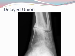
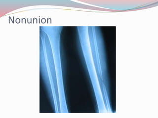
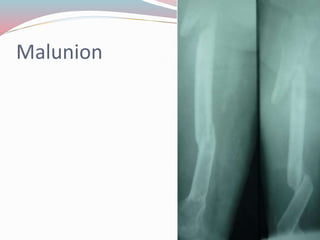
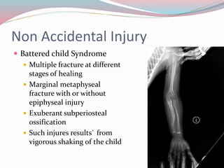
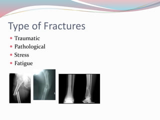
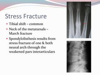
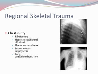
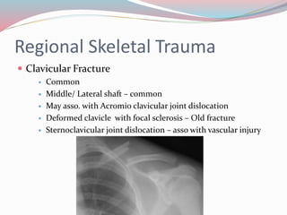
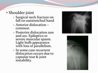
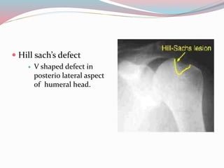
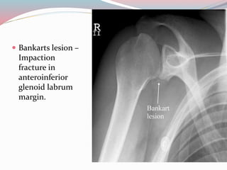
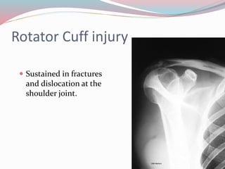
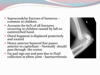
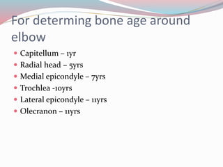
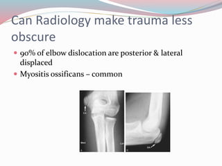
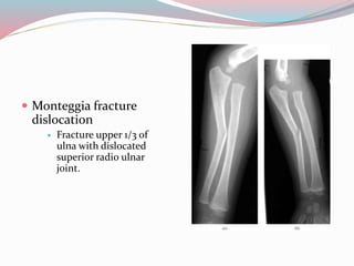
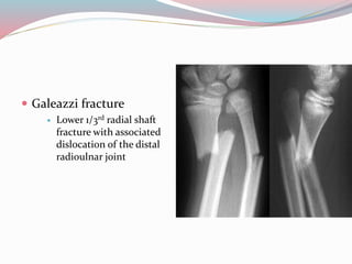
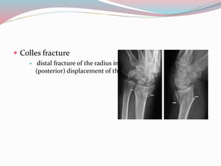
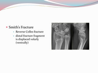
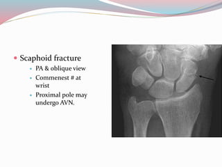
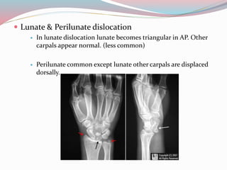
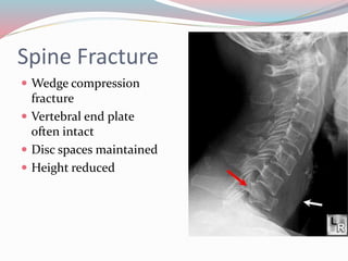
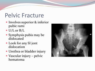
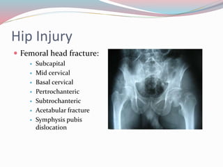
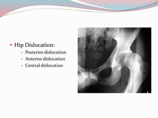
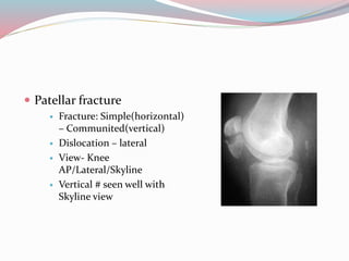
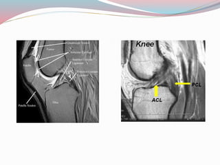
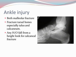
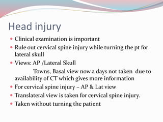
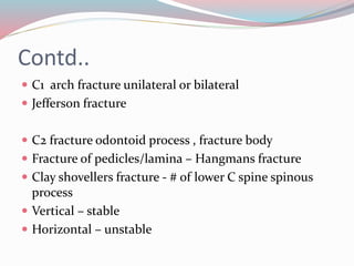
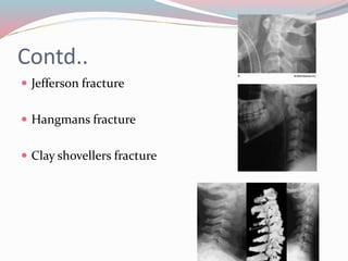
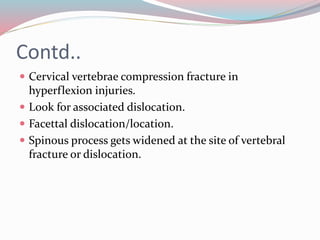
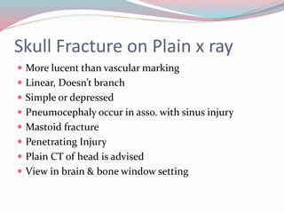
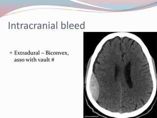
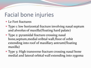
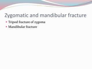
Dr G.Jayaraman
Introduction
๏‚— Proper history &
thorough examination,
do not ignore patients
symptoms
๏‚— Trauma imaging
constitute major bulk
of the of work
๏‚— Diagnosis starts with
plain x ray -most cases
๏‚— At least 2views
required
๏‚— Radiological examination permits assessment of
๏‚— Presence of Fracture
๏‚— Type & position of fracture
๏‚— Simple, comminuted, segmental, etc
๏‚— Asso. Joint injury
๏‚— Epiphyseal injury
๏‚— Asso. Soft tissue involvement
๏‚— Age & healing of fracture
COMPLICATION OF FRACTURE
๏‚— Delayed union
๏‚— Nonunion
๏‚— Malunion
๏‚— AVN
๏‚— Traumatic subperiosteal ossification
๏‚— Myositis osificans
๏‚— Sudeckโ€™s osteodystrophy
Delayed Union
Nonunion
Malunion
AVN
Traumatic subperiosteal reaction
Myositis ossificans
Sudecks osteodystrophy
TRAUMATIC LESION IN CHILDREN
๏‚— Fracture of lower arm & forearm- more common than
adults
๏‚— Greenstick fracture common
๏‚— Heals fast
๏‚— Remodeling effects leads to good alignment without
deformity
๏‚— Compressed vertebral body in children may get fully
reconstructed as the child grows
Greenstick fracture
Salter & Harris type of epiphseal injury
Slipped Femoral capital Epiphysis
๏‚— Common โ€“ children & young adolescents
having limited internal rotation
๏‚— Age โ€“ 10 to 14
๏‚— Obese person
๏‚— H/o fracture may be present or not
๏‚— Early diagnosis is important
๏‚— Radiological Signs
๏‚— Blurring of metaphysis
๏‚— Dislocation of femoral head from acetabulum
๏‚— Growth plate widening
๏‚— Prolongated superior neck line
๏‚— Reduction of epiphyseal height
Non Accidental Injury
๏‚— Battered child Syndrome
๏‚— Multiple fracture at different
stages of healing
๏‚— Marginal metaphyseal
fracture with or without
epiphyseal injury
๏‚— Exuberant subperiosteal
ossification
๏‚— Such injures results` from
vigorous shaking of the child
Type of Fractures
๏‚— Traumatic
๏‚— Pathological
๏‚— Stress
๏‚— Fatigue
Stress Fracture
๏‚— Tibial shift โ€“ common
๏‚— Neck of the metatarsals โ€“
March fracture
๏‚— Spondylolisthesis results from
stress fracture of one & both
neural arch through the
weakened pars interarticulars
Regional Skeletal Trauma
๏‚— Chest injury
๏‚— Rib fracture
๏‚— Hemothorax(Pleural
effusion)
๏‚— Hemopneumothorax
๏‚— Subcutaneous
emphysema
๏‚— Lung
contusion/laceration
Regional Skeletal Trauma
๏‚— Clavicular Fracture
๏‚— Common
๏‚— Middle/ Lateral shaft โ€“ common
๏‚— May asso. with Acromio clavicular joint dislocation
๏‚— Deformed clavicle with focal sclerosis โ€“ Old fracture
๏‚— Sternoclavicular joint dislocation โ€“ asso with vascular injury
๏‚— Shoulder joint
๏‚— Surgical neck fracture on
fall on outstretched hand
๏‚— Anterior dislocation โ€“
common
๏‚— Posterior dislocation rare
and ass. Epileptics or
severe muscular spasm.
Light bulb appearance
with loss of parallelism.
๏‚— In some case recurrent
dislocation occurs due to
capsular tear & joint
instability.
๏‚— Hill sachโ€™s defect
๏‚— V shaped defect in
posterio lateral aspect
of humeral head.
๏‚— Bankarts lesion โ€“
Impaction
fracture in
anteroinferior
glenoid labrum
margin.
Rotator Cuff injury
๏‚— Sustained in fractures
and dislocation at the
shoulder joint.
๏‚— Supracondylar fracture of humerus โ€“
common in children.
๏‚— Accounts for 60% of all fractures
occurring in children caused by fall on
outstretched hand.
๏‚— Distal fragment is displaced posteriorly
and rotated
๏‚— Hence anterior humeral line passes
anterior to capitellum โ€“ Normally should
pass through the centre.
๏‚— Fat pad sign ant and post due to fluid
collection in elbow joint - haemarrthrosis
For determing bone age around
elbow
๏‚— Capitellum โ€“ 1yr
๏‚— Radial head โ€“ 5yrs
๏‚— Medial epicondyle โ€“ 7yrs
๏‚— Trochlea -10yrs
๏‚— Lateral epicondyle โ€“ 11yrs
๏‚— Olecranon โ€“ 11yrs
Can Radiology make trauma less
obscure
๏‚— 90% of elbow dislocation are posterior & lateral
displaced
๏‚— Myositis ossificans โ€“ common
๏‚— Monteggia fracture
dislocation
๏‚— Fracture upper 1/3 of
ulna with dislocated
superior radio ulnar
joint.
๏‚— Galeazzi fracture
๏‚— Lower 1/3rd radial shaft
fracture with associated
dislocation of the distal
radioulnar joint
๏‚— Colles fracture
๏‚— distal fracture of the radius in the forearm with dorsal
(posterior) displacement of the wrist and hand
๏‚— Smithโ€™s Fracture
๏‚— Reverse Colles fracture
๏‚— distal fracture fragment
is displaced volarly
(ventrally)
๏‚— Scaphoid fracture
๏‚— PA & oblique view
๏‚— Commenest # at
wrist
๏‚— Proximal pole may
undergo AVN.
๏‚— Lunate & Perilunate dislocation
๏‚— In lunate dislocation lunate becomes triangular in AP. Other
carpals appear normal. (less common)
๏‚— Perilunate common except lunate other carpals are displaced
dorsally.
Spine Fracture
๏‚— Wedge compression
fracture
๏‚— Vertebral end plate
often intact
๏‚— Disc spaces maintained
๏‚— Height reduced
Pelvic Fracture
๏‚— Involves superior & inferior
pubic rami
๏‚— U/L or B/L
๏‚— Symphysis pubis may be
dislocated
๏‚— Look for any SI jont
dislocation
๏‚— Urethra or bladder injury
๏‚— Vascular injury โ€“ pelvic
hematoma
Hip Injury
๏‚— Femoral head fracture:
๏‚— Subcapital
๏‚— Mid cervical
๏‚— Basal cervical
๏‚— Pertrochanteric
๏‚— Subtrochanteric
๏‚— Acetabular fracture
๏‚— Symphysis pubis
dislocation
๏‚— Hip Dislocation:
๏‚— Posterior dislocation
๏‚— Anterior dislocation
๏‚— Central dislocation
๏‚— Femoral shaft fracture
๏‚— Mid shaft โ€“ common
๏‚— Supracondylar region
with intracondylar
extension
๏‚— Transcondylar
fracture
๏‚— Patellar fracture
๏‚— Fracture: Simple(horizontal)
โ€“ Communited(vertical)
๏‚— Dislocation โ€“ lateral
๏‚— View- Knee
AP/Lateral/Skyline
๏‚— Vertical # seen well with
Skyline view
Knee injury
๏‚— MRI indicated to study the integrity of
cruciate & collateral ligaments, hyaline
cartilage menisci capsule any marrow edema
๏‚— Useful when plain x rays are non contributory
but patient has pain during locking and
unlocking movements
Ankle injury
๏‚— Both malleolar fracture
๏‚— Fracture tarsal bones
especially talus and
calcaneum.
๏‚— Any H/O fall from a
height look for calcaneal
fracture
Head injury
๏‚— Clinical examination is important
๏‚— Rule out cervical spine injury while turning the pt for
lateral skull
๏‚— Views: AP /Lateral Skull
Towns, Basal view now a days not taken due to
availability of CT which gives more information
๏‚— For cervical spine injury โ€“ AP & Lat view
๏‚— Translateral view is taken for cervical spine injury.
๏‚— Taken without turning the patient
Contd..
๏‚— C1 arch fracture unilateral or bilateral
๏‚— Jefferson fracture
๏‚— C2 fracture odontoid process , fracture body
๏‚— Fracture of pedicles/lamina โ€“ Hangmans fracture
๏‚— Clay shovellers fracture - # of lower C spine spinous
process
๏‚— Vertical โ€“ stable
๏‚— Horizontal โ€“ unstable
Contd..
๏‚— Jefferson fracture
๏‚— Hangmans fracture
๏‚— Clay shovellers fracture
Contd..
๏‚— Cervical vertebrae compression fracture in
hyperflexion injuries.
๏‚— Look for associated dislocation.
๏‚— Facettal dislocation/location.
๏‚— Spinous process gets widened at the site of vertebral
fracture or dislocation.
Skull Fracture on Plain x ray
๏‚— More lucent than vascular marking
๏‚— Linear, Doesnโ€™t branch
๏‚— Simple or depressed
๏‚— Pneumocephaly occur in asso. with sinus injury
๏‚— Mastoid fracture
๏‚— Penetrating Injury
๏‚— Plain CT of head is advised
๏‚— View in brain & bone window setting
Intracranial bleed
๏‚— Extradural โ€“ Biconvex,
asso with vault #
Intracranial bleed
๏‚— Subdural โ€“ Semilunar in shape
๏‚— Midline shift & mass effect.
Intracranial bleed
๏‚— Intracerbral โ€“ irregular
dense collection
Intracranial bleed
๏‚— Subarachnoid โ€“ between sulci
, cistern
Intracranial bleed
Intraventricular bleed
Facial bone injuries
๏‚— Le Fort fractures
๏‚— Type 1: low horizontal fracture involving nasal septum
and alveolus of maxilla(floating hard palate)
๏‚— Type 2: pyramidal fracture crossing nasal
bone,septum,medial oribtal wall,floor of orbit
extending into roof of maxillary antrum(floating
maxilla)
๏‚— Type 3: High transverse fracture crossing nasal bone
medial and lateral orbital wall extending into zygoma
Zygomatic and mandibular fracture
๏‚— Tripod fracture of zygoma
๏‚— Mandibular fracture
THANK
YOU!!

Skeletal trauma imaging

  • 1.
  • 2.
    Introduction ๏‚— Proper history& thorough examination, do not ignore patients symptoms ๏‚— Trauma imaging constitute major bulk of the of work ๏‚— Diagnosis starts with plain x ray -most cases ๏‚— At least 2views required
  • 3.
    ๏‚— Radiological examinationpermits assessment of ๏‚— Presence of Fracture ๏‚— Type & position of fracture ๏‚— Simple, comminuted, segmental, etc ๏‚— Asso. Joint injury ๏‚— Epiphyseal injury ๏‚— Asso. Soft tissue involvement ๏‚— Age & healing of fracture
  • 4.
    COMPLICATION OF FRACTURE ๏‚—Delayed union ๏‚— Nonunion ๏‚— Malunion ๏‚— AVN ๏‚— Traumatic subperiosteal ossification ๏‚— Myositis osificans ๏‚— Sudeckโ€™s osteodystrophy
  • 5.
  • 6.
  • 7.
  • 8.
  • 9.
  • 10.
  • 11.
  • 12.
    TRAUMATIC LESION INCHILDREN ๏‚— Fracture of lower arm & forearm- more common than adults ๏‚— Greenstick fracture common ๏‚— Heals fast ๏‚— Remodeling effects leads to good alignment without deformity ๏‚— Compressed vertebral body in children may get fully reconstructed as the child grows
  • 13.
  • 14.
    Salter & Harristype of epiphseal injury
  • 15.
    Slipped Femoral capitalEpiphysis ๏‚— Common โ€“ children & young adolescents having limited internal rotation ๏‚— Age โ€“ 10 to 14 ๏‚— Obese person ๏‚— H/o fracture may be present or not ๏‚— Early diagnosis is important ๏‚— Radiological Signs ๏‚— Blurring of metaphysis ๏‚— Dislocation of femoral head from acetabulum ๏‚— Growth plate widening ๏‚— Prolongated superior neck line ๏‚— Reduction of epiphyseal height
  • 16.
    Non Accidental Injury ๏‚—Battered child Syndrome ๏‚— Multiple fracture at different stages of healing ๏‚— Marginal metaphyseal fracture with or without epiphyseal injury ๏‚— Exuberant subperiosteal ossification ๏‚— Such injures results` from vigorous shaking of the child
  • 17.
    Type of Fractures ๏‚—Traumatic ๏‚— Pathological ๏‚— Stress ๏‚— Fatigue
  • 18.
    Stress Fracture ๏‚— Tibialshift โ€“ common ๏‚— Neck of the metatarsals โ€“ March fracture ๏‚— Spondylolisthesis results from stress fracture of one & both neural arch through the weakened pars interarticulars
  • 19.
    Regional Skeletal Trauma ๏‚—Chest injury ๏‚— Rib fracture ๏‚— Hemothorax(Pleural effusion) ๏‚— Hemopneumothorax ๏‚— Subcutaneous emphysema ๏‚— Lung contusion/laceration
  • 20.
    Regional Skeletal Trauma ๏‚—Clavicular Fracture ๏‚— Common ๏‚— Middle/ Lateral shaft โ€“ common ๏‚— May asso. with Acromio clavicular joint dislocation ๏‚— Deformed clavicle with focal sclerosis โ€“ Old fracture ๏‚— Sternoclavicular joint dislocation โ€“ asso with vascular injury
  • 21.
    ๏‚— Shoulder joint ๏‚—Surgical neck fracture on fall on outstretched hand ๏‚— Anterior dislocation โ€“ common ๏‚— Posterior dislocation rare and ass. Epileptics or severe muscular spasm. Light bulb appearance with loss of parallelism. ๏‚— In some case recurrent dislocation occurs due to capsular tear & joint instability.
  • 22.
    ๏‚— Hill sachโ€™sdefect ๏‚— V shaped defect in posterio lateral aspect of humeral head.
  • 23.
    ๏‚— Bankarts lesionโ€“ Impaction fracture in anteroinferior glenoid labrum margin.
  • 24.
    Rotator Cuff injury ๏‚—Sustained in fractures and dislocation at the shoulder joint.
  • 25.
    ๏‚— Supracondylar fractureof humerus โ€“ common in children. ๏‚— Accounts for 60% of all fractures occurring in children caused by fall on outstretched hand. ๏‚— Distal fragment is displaced posteriorly and rotated ๏‚— Hence anterior humeral line passes anterior to capitellum โ€“ Normally should pass through the centre. ๏‚— Fat pad sign ant and post due to fluid collection in elbow joint - haemarrthrosis
  • 26.
    For determing boneage around elbow ๏‚— Capitellum โ€“ 1yr ๏‚— Radial head โ€“ 5yrs ๏‚— Medial epicondyle โ€“ 7yrs ๏‚— Trochlea -10yrs ๏‚— Lateral epicondyle โ€“ 11yrs ๏‚— Olecranon โ€“ 11yrs
  • 27.
    Can Radiology maketrauma less obscure ๏‚— 90% of elbow dislocation are posterior & lateral displaced ๏‚— Myositis ossificans โ€“ common
  • 28.
    ๏‚— Monteggia fracture dislocation ๏‚—Fracture upper 1/3 of ulna with dislocated superior radio ulnar joint.
  • 29.
    ๏‚— Galeazzi fracture ๏‚—Lower 1/3rd radial shaft fracture with associated dislocation of the distal radioulnar joint
  • 30.
    ๏‚— Colles fracture ๏‚—distal fracture of the radius in the forearm with dorsal (posterior) displacement of the wrist and hand
  • 31.
    ๏‚— Smithโ€™s Fracture ๏‚—Reverse Colles fracture ๏‚— distal fracture fragment is displaced volarly (ventrally)
  • 32.
    ๏‚— Scaphoid fracture ๏‚—PA & oblique view ๏‚— Commenest # at wrist ๏‚— Proximal pole may undergo AVN.
  • 33.
    ๏‚— Lunate &Perilunate dislocation ๏‚— In lunate dislocation lunate becomes triangular in AP. Other carpals appear normal. (less common) ๏‚— Perilunate common except lunate other carpals are displaced dorsally.
  • 34.
    Spine Fracture ๏‚— Wedgecompression fracture ๏‚— Vertebral end plate often intact ๏‚— Disc spaces maintained ๏‚— Height reduced
  • 35.
    Pelvic Fracture ๏‚— Involvessuperior & inferior pubic rami ๏‚— U/L or B/L ๏‚— Symphysis pubis may be dislocated ๏‚— Look for any SI jont dislocation ๏‚— Urethra or bladder injury ๏‚— Vascular injury โ€“ pelvic hematoma
  • 36.
    Hip Injury ๏‚— Femoralhead fracture: ๏‚— Subcapital ๏‚— Mid cervical ๏‚— Basal cervical ๏‚— Pertrochanteric ๏‚— Subtrochanteric ๏‚— Acetabular fracture ๏‚— Symphysis pubis dislocation
  • 37.
    ๏‚— Hip Dislocation: ๏‚—Posterior dislocation ๏‚— Anterior dislocation ๏‚— Central dislocation
  • 38.
    ๏‚— Femoral shaftfracture ๏‚— Mid shaft โ€“ common ๏‚— Supracondylar region with intracondylar extension ๏‚— Transcondylar fracture
  • 39.
    ๏‚— Patellar fracture ๏‚—Fracture: Simple(horizontal) โ€“ Communited(vertical) ๏‚— Dislocation โ€“ lateral ๏‚— View- Knee AP/Lateral/Skyline ๏‚— Vertical # seen well with Skyline view
  • 40.
    Knee injury ๏‚— MRIindicated to study the integrity of cruciate & collateral ligaments, hyaline cartilage menisci capsule any marrow edema ๏‚— Useful when plain x rays are non contributory but patient has pain during locking and unlocking movements
  • 42.
    Ankle injury ๏‚— Bothmalleolar fracture ๏‚— Fracture tarsal bones especially talus and calcaneum. ๏‚— Any H/O fall from a height look for calcaneal fracture
  • 43.
    Head injury ๏‚— Clinicalexamination is important ๏‚— Rule out cervical spine injury while turning the pt for lateral skull ๏‚— Views: AP /Lateral Skull Towns, Basal view now a days not taken due to availability of CT which gives more information ๏‚— For cervical spine injury โ€“ AP & Lat view ๏‚— Translateral view is taken for cervical spine injury. ๏‚— Taken without turning the patient
  • 44.
    Contd.. ๏‚— C1 archfracture unilateral or bilateral ๏‚— Jefferson fracture ๏‚— C2 fracture odontoid process , fracture body ๏‚— Fracture of pedicles/lamina โ€“ Hangmans fracture ๏‚— Clay shovellers fracture - # of lower C spine spinous process ๏‚— Vertical โ€“ stable ๏‚— Horizontal โ€“ unstable
  • 45.
    Contd.. ๏‚— Jefferson fracture ๏‚—Hangmans fracture ๏‚— Clay shovellers fracture
  • 46.
    Contd.. ๏‚— Cervical vertebraecompression fracture in hyperflexion injuries. ๏‚— Look for associated dislocation. ๏‚— Facettal dislocation/location. ๏‚— Spinous process gets widened at the site of vertebral fracture or dislocation.
  • 47.
    Skull Fracture onPlain x ray ๏‚— More lucent than vascular marking ๏‚— Linear, Doesnโ€™t branch ๏‚— Simple or depressed ๏‚— Pneumocephaly occur in asso. with sinus injury ๏‚— Mastoid fracture ๏‚— Penetrating Injury ๏‚— Plain CT of head is advised ๏‚— View in brain & bone window setting
  • 48.
    Intracranial bleed ๏‚— Extraduralโ€“ Biconvex, asso with vault #
  • 49.
    Intracranial bleed ๏‚— Subduralโ€“ Semilunar in shape ๏‚— Midline shift & mass effect.
  • 50.
    Intracranial bleed ๏‚— Intracerbralโ€“ irregular dense collection
  • 51.
    Intracranial bleed ๏‚— Subarachnoidโ€“ between sulci , cistern
  • 52.
  • 53.
    Facial bone injuries ๏‚—Le Fort fractures ๏‚— Type 1: low horizontal fracture involving nasal septum and alveolus of maxilla(floating hard palate) ๏‚— Type 2: pyramidal fracture crossing nasal bone,septum,medial oribtal wall,floor of orbit extending into roof of maxillary antrum(floating maxilla) ๏‚— Type 3: High transverse fracture crossing nasal bone medial and lateral orbital wall extending into zygoma
  • 54.
    Zygomatic and mandibularfracture ๏‚— Tripod fracture of zygoma ๏‚— Mandibular fracture
  • 55.