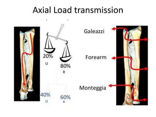
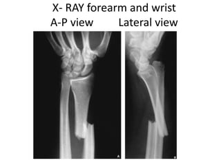
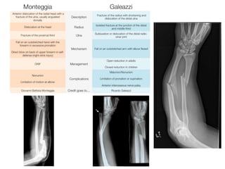
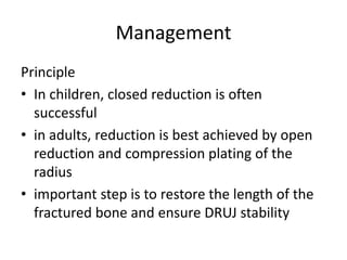
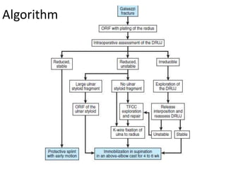
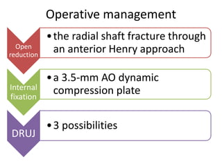
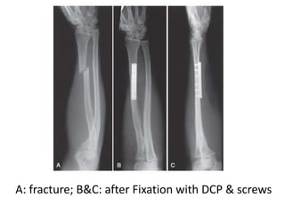
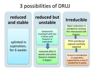
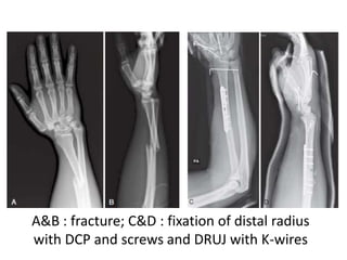
Galeazzi fracture-dislocation is a fracture of the distal or middle third of the radius shaft combined with dislocation of the distal radioulnar joint. It most often occurs in males due to indirect trauma from a fall on an outstretched hand with rotation. Radiographs show the radial fracture and dislocation of the distal radioulnar joint. Treatment involves open reduction and internal fixation of the radial fracture with a plate while restoring length and stability of the distal radioulnar joint. The forearm is then immobilized in supination for 4-6 weeks to heal.
5. Definition
• The combination of fracture of the distal or middle
third of the shaft of the radius and dislocation of the
distal radioulnar joint.
• counterpart of the Monteggia fracture-dislocation
• also known as a reverse Monteggia fracture.
6. History
• first described by Cooper in
1842
• Ricardo Galeazzi (1866-1952)
• an Italian surgeon in Milan, he
reported on his experience with
18 fractures with the above-
described pattern
• In 1941, Campbell termed the
"fracture of necessity,"
necessitates surgical treatment
• Hughston outlined the definitive
management in 1957
8. Mechanism of injury
• as indirect trauma : due to a fall on an outstretched
hand (FOOSH) with a superimposed rotation force
• Rotation determines direction of angulation
– Pronation flexion injury ( dorsal angulation )
– Supination extension injury (volar angulation)
• direct trauma to the wrist, typically on the
dorsolateral aspect
10. Types
• Type I
• apex volar
• Caused by axial loading
of forearm in supination
• dorsal displacement of
radius and volar
dislocation of distal ulna
11. Types
• Type II
• apex dorsal
• fractures are caused by
axial loading of forearm
in pronation
• anterior displacement of
radius and dorsal
dislocation of distal ulna
12. Clinical features
History : FOOSH
Symptoms :
– Pain and swelling in forearm and wrist
– painful Forearm rotation
Signs
• Prominence or tenderness over the lower end of ulna
• evaluate for compartment syndrome
• NV exam : Anterior interosseous nerve (AIN) palsy
18. Plain Radiographs
• radial shaft fracture
– commonly at the junction of the middle and distal
third
– dorsal or volar angulation
• dislocation of the distal radioulnar joint
• radial shortening may occur
24. Variants / Differentials
• Piedmont fractures
– An isolated radial fracture without distal radioulnar
dissociation
– named by the Piedmont Orthopaedic Society.
– distal radioulnar dislocation described only as a
secondary complication of maltreatment
– occur following a direct blow to the dorsoradial aspect
of the forearm
• Fracture anywhere along the radius or associated
with fractures of both bones with DRUJ
disruption
25.
26. Management
Principle
• In children, closed reduction is often
successful
• in adults, reduction is best achieved by open
reduction and compression plating of the
radius
• important step is to restore the length of the
fractured bone and ensure DRUJ stability
29. Approaches-Henry (volar)
• incision begins 1 cm lateral to
the biceps insertion
• extends distally to the radial
styloid
• Interval between
brachioradialis and FCR
• Identify radial artery and
superficial radial n.
• Protect PIN proximally
32. 3 possibilities of DRUJ
reduced
and stable
splinted in
supination,
for 6 weeks
reduced but
unstable
temporarily
transfixed with two
K Wires with
forearm in
supination
removed after 6
weeks and active
forearm rotation
is begun.
Irreducible
Open reduction is
needed to remove
the interposed soft
tissues.
TFCC and dorsal
capsule carefully
repaired
forearm immobilized
in supination
supported by a wire if
needed for 6 weeks
33. A&B : fracture; C&D : fixation of distal radius
with DCP and screws and DRUJ with K-wires
34. Post operative care
• Check neurologic and vascular status
– Specifically, evaluate for function of the anterior
interosseous nerve (AIN)
– presence of compartment syndrome
• Immobilize the forearm in supination for 4
weeks
• obtain radiographs to recheck alignment and
reduction of the radius and the DRUJ
35. Post operative care
• removal of any percutaneous pins at 4 weeks
• recheck radiographs to confirm maintenance
of reduction, and replace the cast brace in
supination
• institute Physiotherapy for elbow, digital and
shoulder range of motion
• Reexamine radiographs at 6-week intervals
until healing is apparent.
36. Post operative Complications
• Nonunion
• Malunion
• Infection
• Refracture following plate removal
• Posterior interosseous nerve (PIN) injury
• Instability of the DRUJ
37. References
• Apley’s System of Orthopaedics and Fractures
9th Edition
• Campbells Orthopedics, 13th edition
• Medscape.com
• PubMed
Editor's Notes
2 bone radius and ulnar conncted with interosseous membrane in between
Joints
Elbow joint
Proximal Radio-ulnar joint
Distal Radio-ulnar joint
Mobile wad of henry also known as the lateral comparment
A reverse Galeazzi denotes a fracture of the distal ulna with disruption of radioulnar joint
Cooper : , 92 years before Galeazzi reported his results.
Eponymous fracture to galeazzi’s name
; in adults, nonsurgical treatment of the injury results in persistent or recurrent dislocations of the distal ulna.
Reverse Galeazzi results from fall with hand in supination
on the basis of direction of radial displacement
Ulnar styloid # : 60% of galeazzi
AIN. branch to FDP; branch to FPL; branch to pronator quadratus. (volar interosseous nerve) is a branch of the median nerve that supplies the deep muscles on the anterior of the forearm, except the ulnar (medial) half of the flexor digitorum profundus.
The posterior interosseous nerve (or dorsal interosseous nerve) is a nerve in the forearm. It is the continuation of the deep branch of the radial nerve, after this has crossed the supinator muscle.
The triangular fibrocartilage complex (TFCC) is a cartilage structure located on the small finger side of the wrist. TFCC consists of five parts; the articular disc, the superficial and deep (ligamentum Subcruentum) radioulnar fibers, and the two disc-carpal ligaments. it cushions and supports the small carpal bones in the wrist. The TFCC keeps the forearm bones (radius and ulna) stable when the hand grasps or the forearm rotates.
It can be demostrated with piano key sign by pressing down ulnar styloid and is positive if it pushes back like a piano key.
radial shortening may occur, and if greater than 10 mm, suggests complete disruption of the interosseous membrane
A forearm series is usually sufficient for diagnosis and management planning.
However, good quality orthogonal views are needed to identify and characterise displacement correctly.
more than 5 mm of shortening of the radius relative to the ulna when compared with the contralateral wrist
This means widening of the distal radioulnar joint on the anteroposterior view
And lastly
dislocation of the ulna relative to the radius on a true lateral view of the wrist
Ulnar variance (also known as Hulten variance) refers to the relative lengths of the distal articular surfaces of the radius and ulna.
Ulnar variance may be:
neutral (both the ulnar and radial articular surfaces at the same level)
positive (ulna projects more distally)
negative (ulna projects more proximally)
positive ulnar variance describes where the distal articular surface of the ulna is more distal when compared to the articular surface of the radius.
It plays important role in wrist pathology such as ulnar impaction syndromes and thinning of the triangular fibrocartilage complex.
Many people consider the Galeazzi and Piedmont fractures as the same injury. However, some state that the latter is an isolated radial fracture without distal radioulnar dissociation. The Piedmont fracture was so named by the Piedmont Orthopaedic Society.
fracture of the radial shaft at the junction of the middle and distal thirds
Galeazzi fractures in skeletally immature patients (children and early adolescents) are typically treated with closed reduction and casting because of the enhanced viscoelastic nature of pediatric bone, as well as the presence of a stout periosteal sleeve
However, proper reduction of the radius with concomitant reduction of the distal radioulnar joint and cast immobilization provides good to excellent outcome even if the Galeazzi lesion is primarily not recognized.
Geissler fernandez instability classification after radius reduction :
1 : stable, 2 unstable and 3 potentially unstable
Thompsons approach it is a dorsal approach
Incision begins just anterior to the lateral epicondyle
Extends distally towards the ulnar side of Lister’s tubercle
interval is developed between the ECRB and the EDC, exposing the supinator muscle
Identify PIN
1cm proximal to its distal edge of supinator
-Treatment with closed reduction and cast immobilization has a high rate of unsatisfactory results.
- DCP or the newer limited contact dynamic compression plate (LCDCP)
with 3.5-mm AO dynamic compression plate and screws.
Temporary stabilization of distal radioulnar joint with transverse Kirschner wire was unnecessary
An irreducible distal radioulnar joint usually indicates soft-tissue
interposition and requires open treatment.
Galeazzi fractures: Is DRUJ instability predicted by current guidelines?
The presence of an ulnar styloid fracture can be helpful. Surgeons should be aware of these associations but rely primarily on intraoperative assessment of the DRUJ after radial fixation to determine treatment.
The radial shaft fracture usually is too distal to allow fixation with an intramedullary device
Remove suture in 10-14 days
Nonunion and malunion are primarily associated with closed reduction, plaster immobilization, intramedullary nails, and inadequate plate fixation.
Pin tract infections are also a possibility of complication
PIN PALSY : The PIN, branch of the radial nerve, also is vulnerable (during the dorsal Thompson approach),
Nerve injury occur during either the volar or dorsal forearm approach to a Galeazzi fracture as well. The radial sensory nerve is reported to be the most frequently injured branch, with damage occurring in association with the Henry (volar) approach.
tardy ulnar tunnel syndrome has been reported in the closed treatment of a Galeazzi fracture; this resulted from a malunion and the compression of a stretched vascular branch situated over the ulnar head
Instability of the DRUJ may occur because of a failure to recognize the injury, a failure to reduce the dislocation intraoperatively, nonanatomic radial reduction, or interposed soft-tissue that blocks reduction. Most often, the ECU is the interposed structure