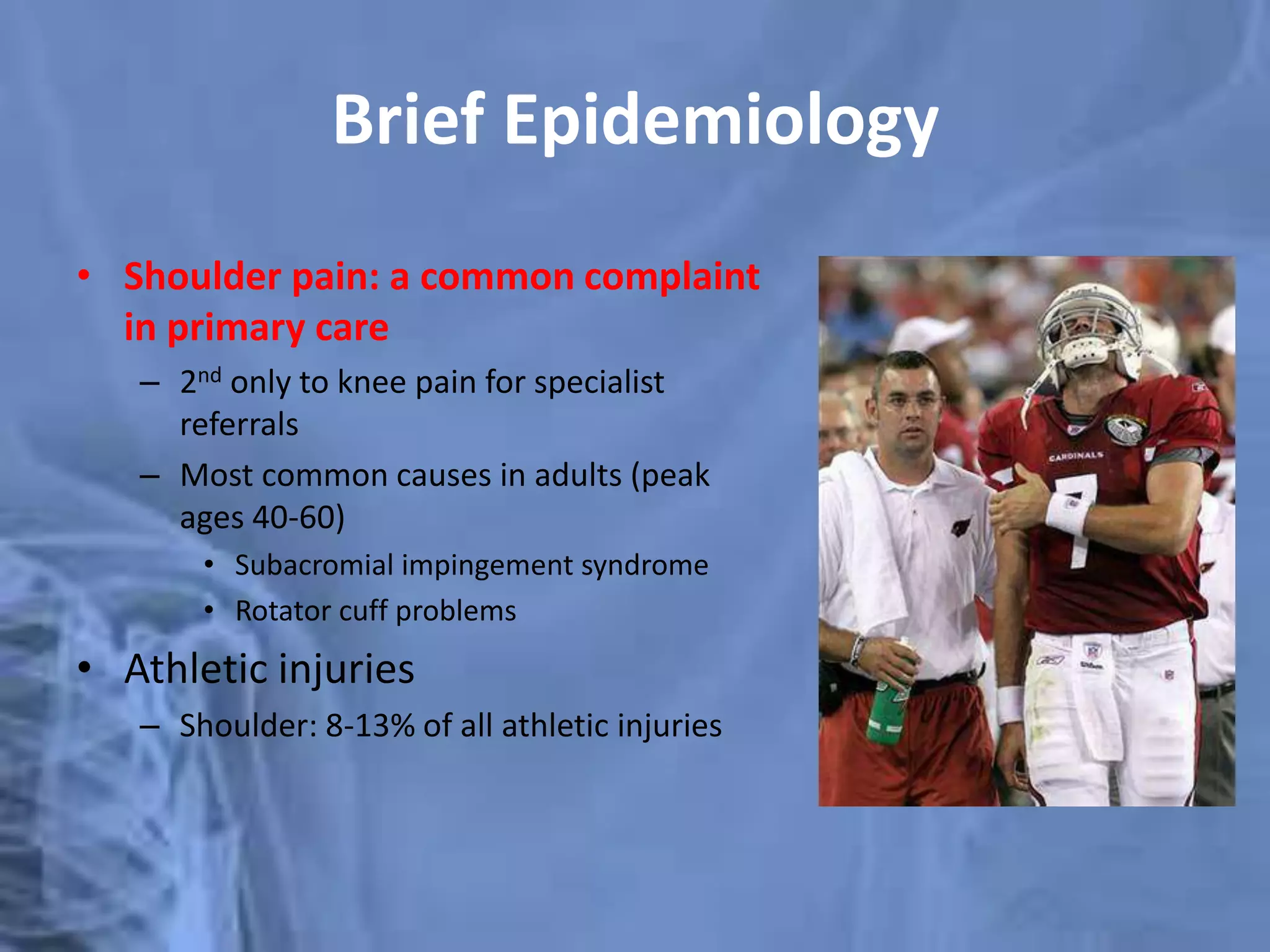
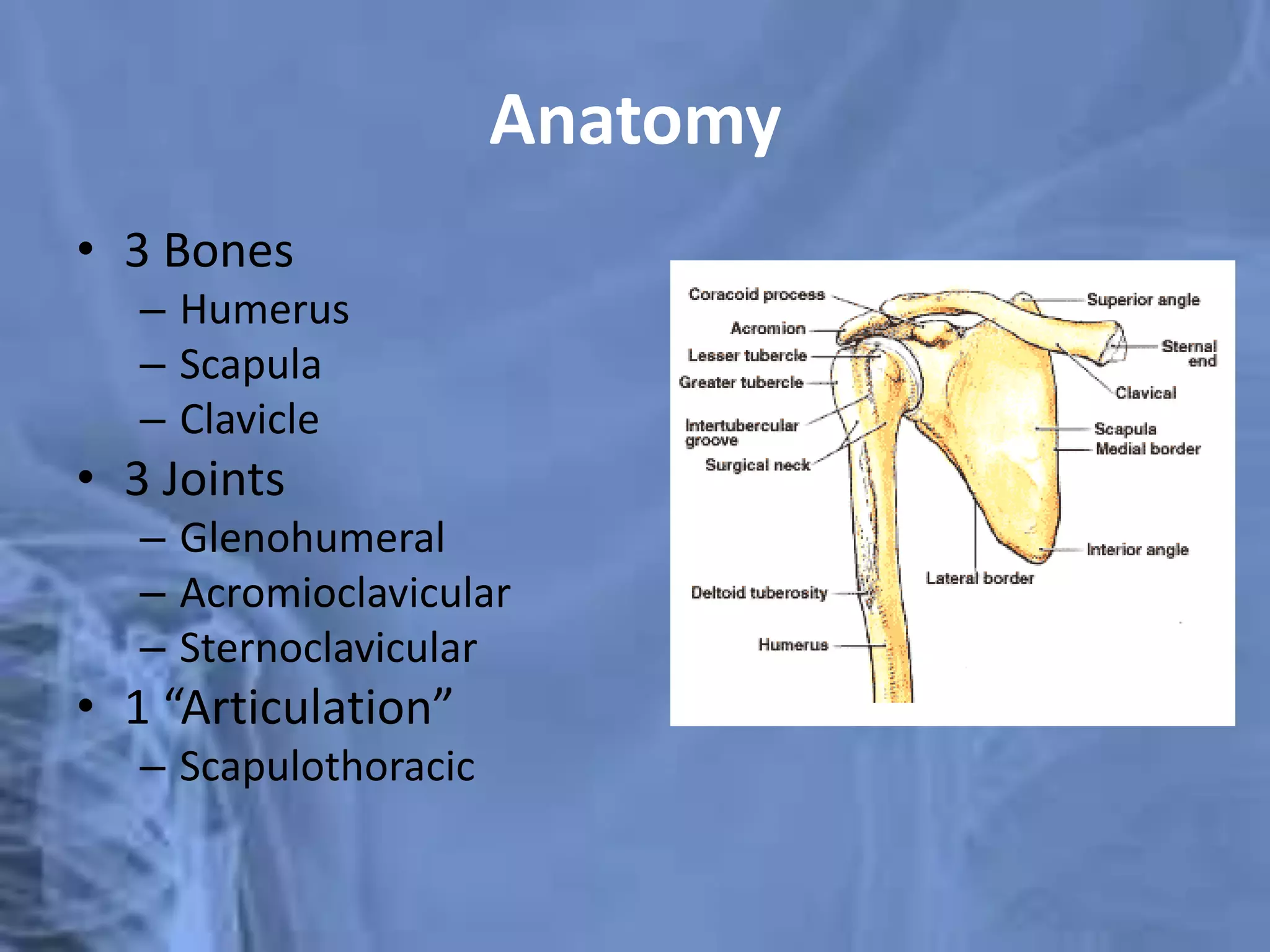
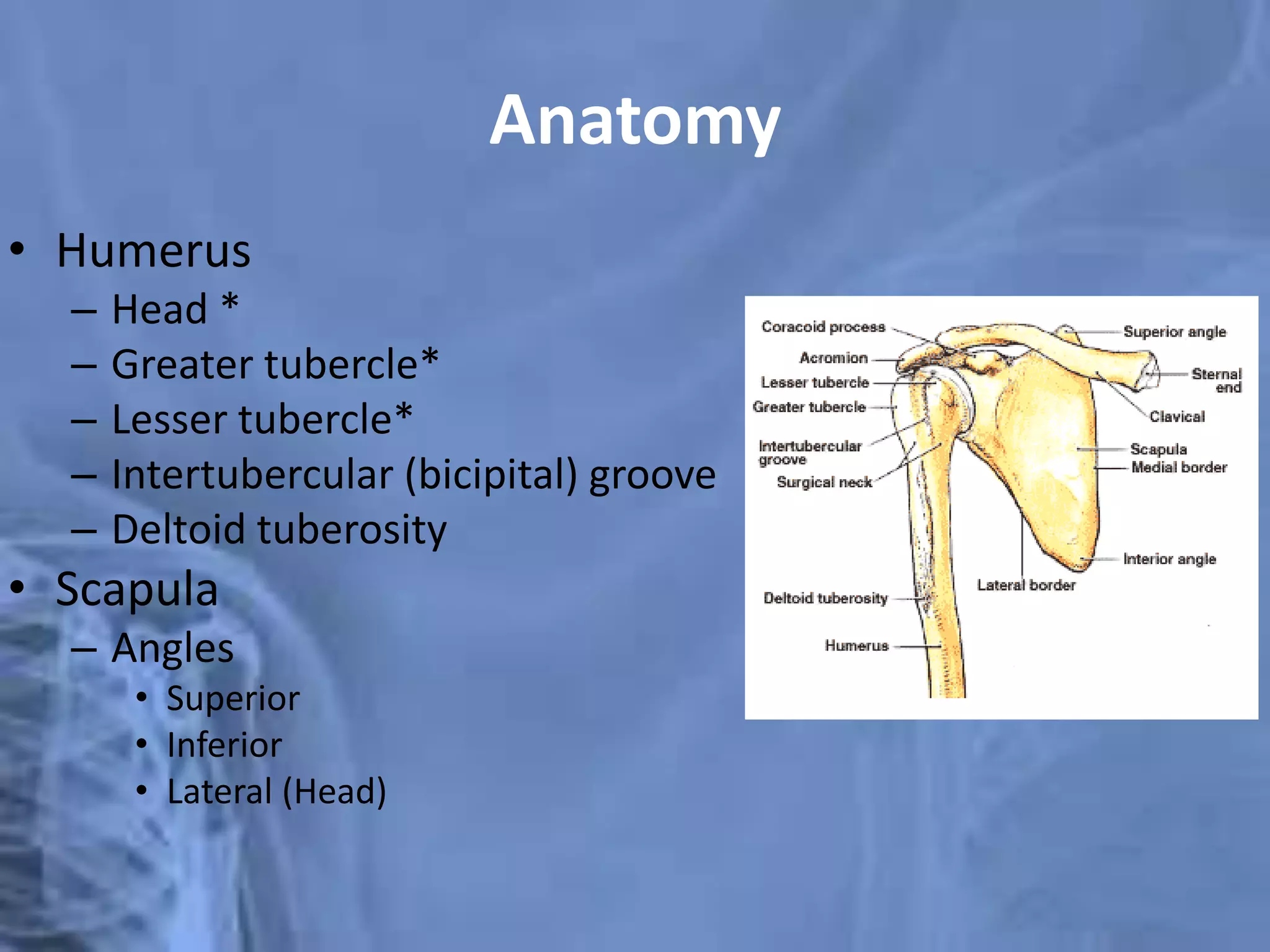
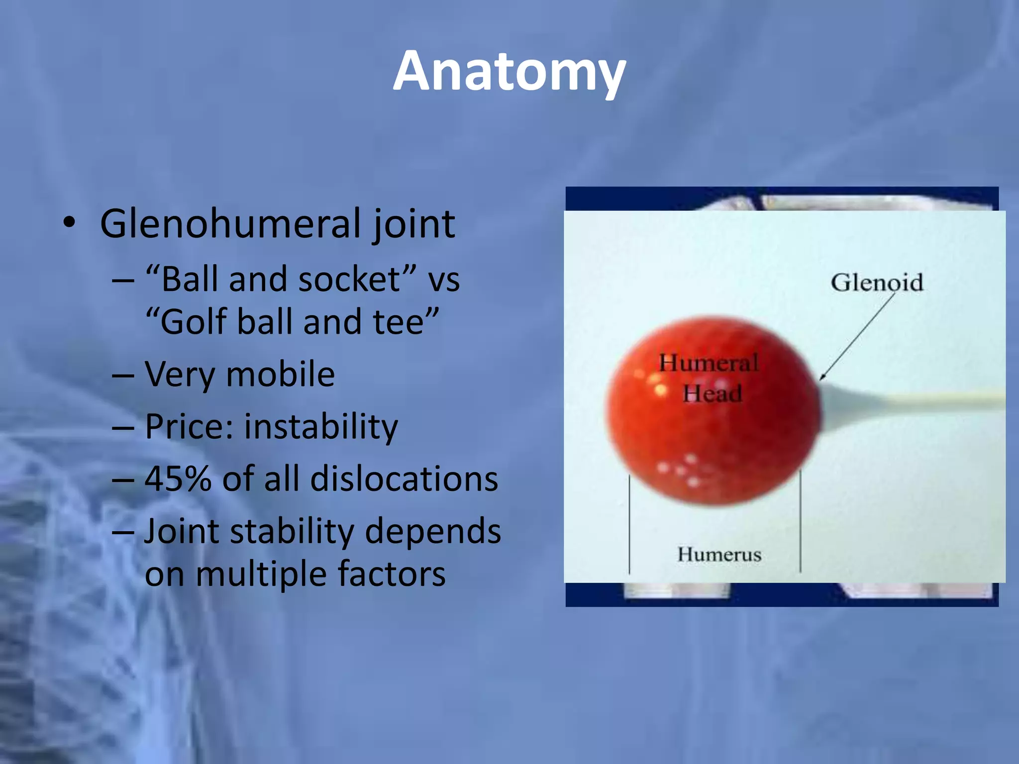
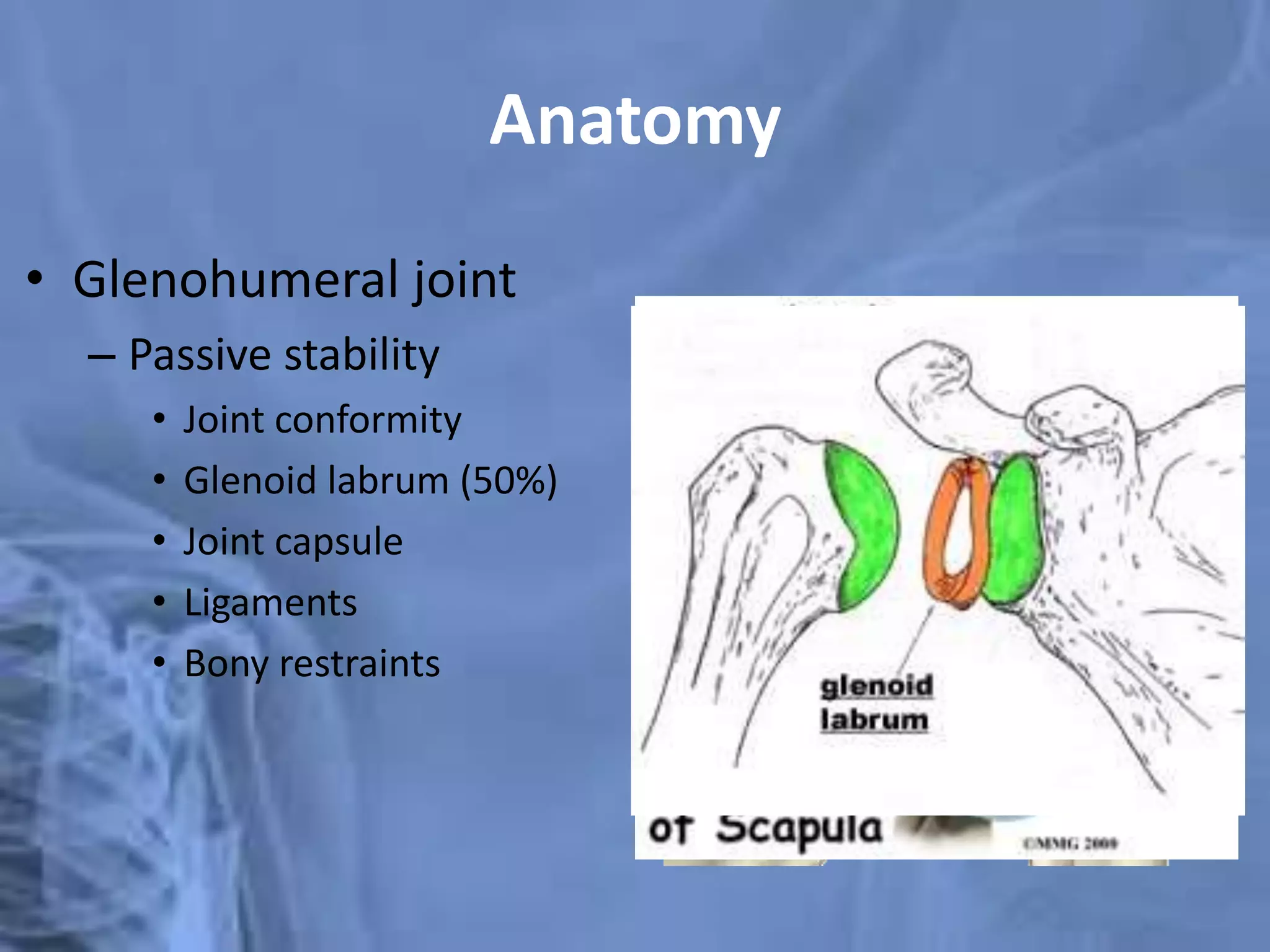
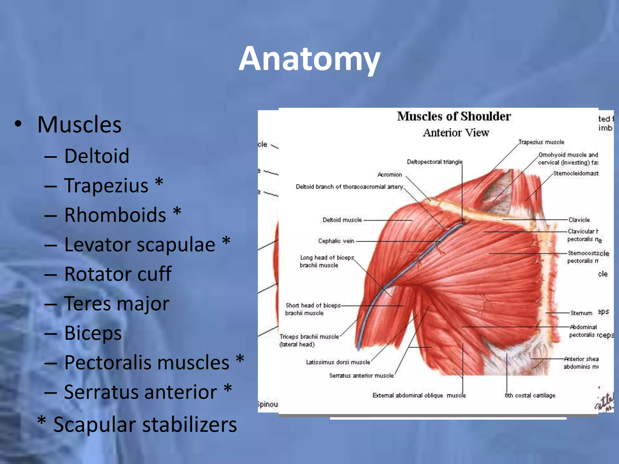
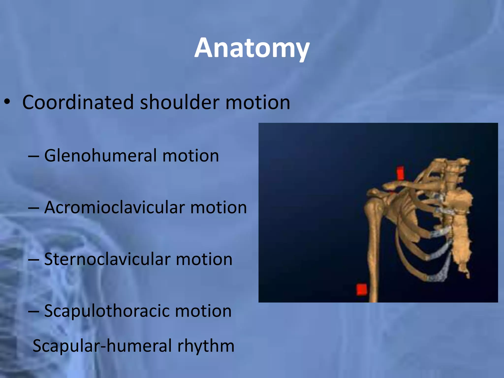
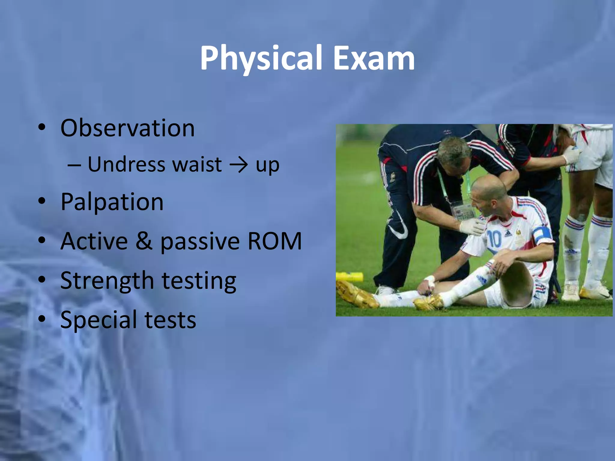
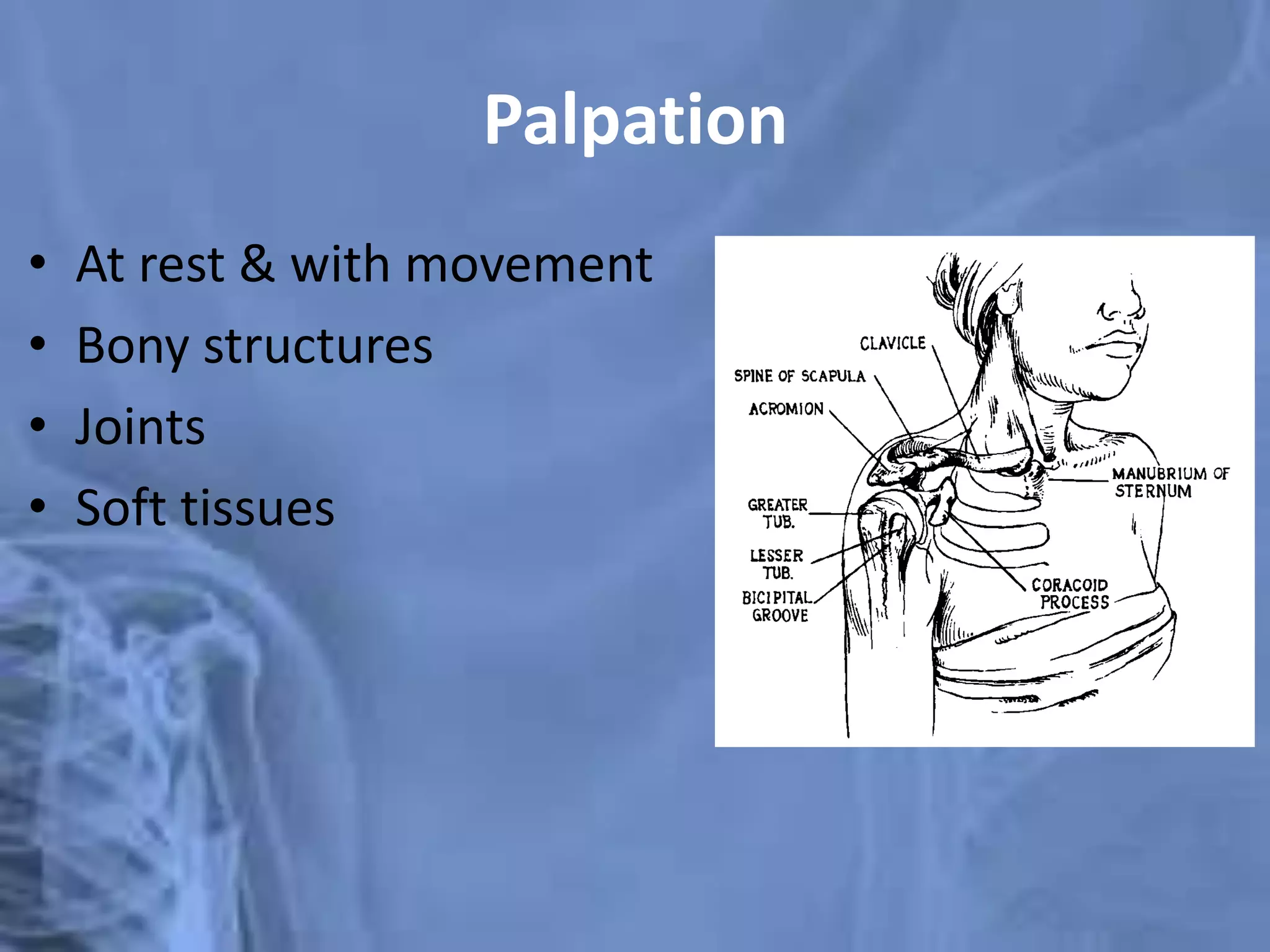
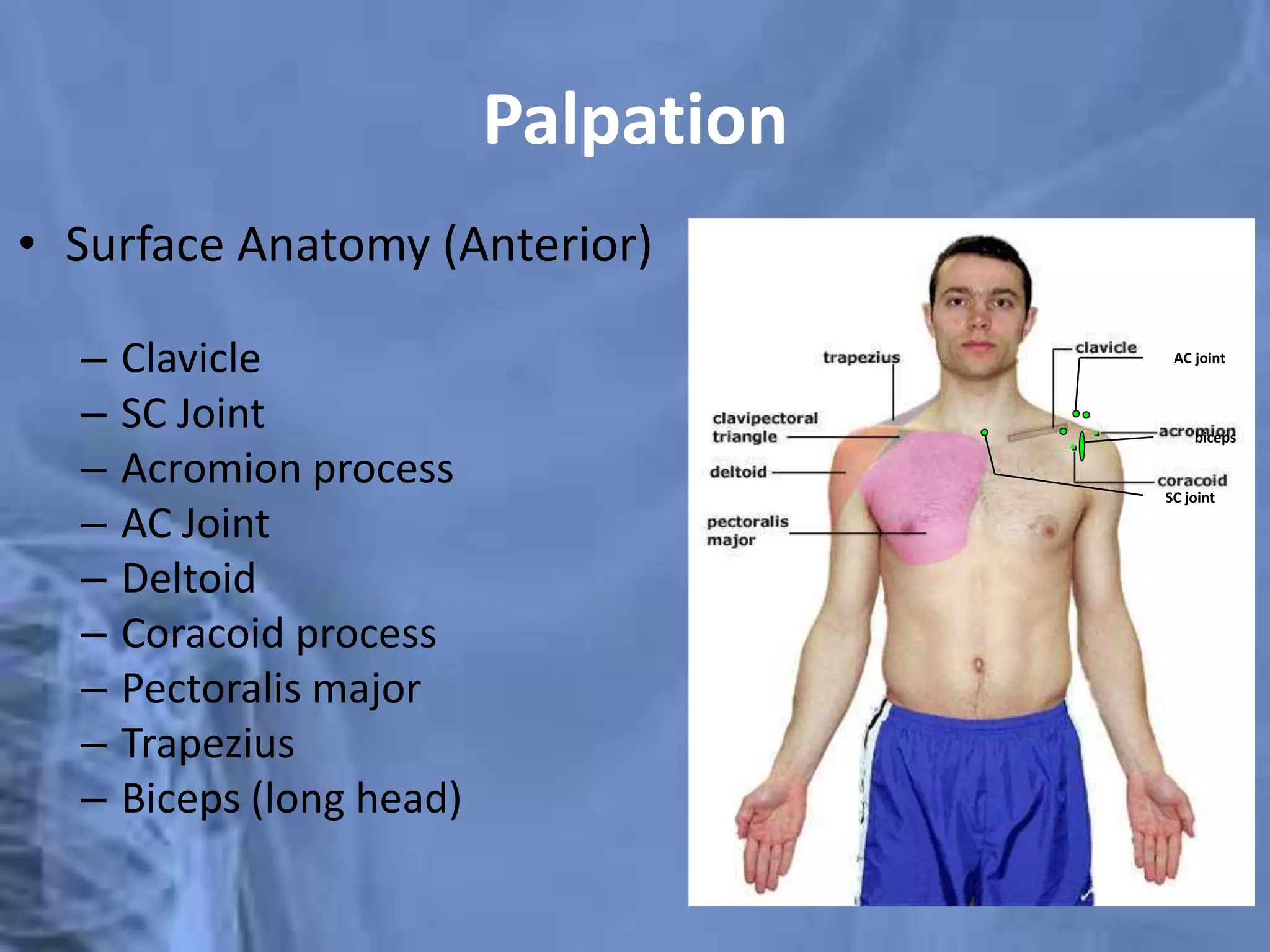
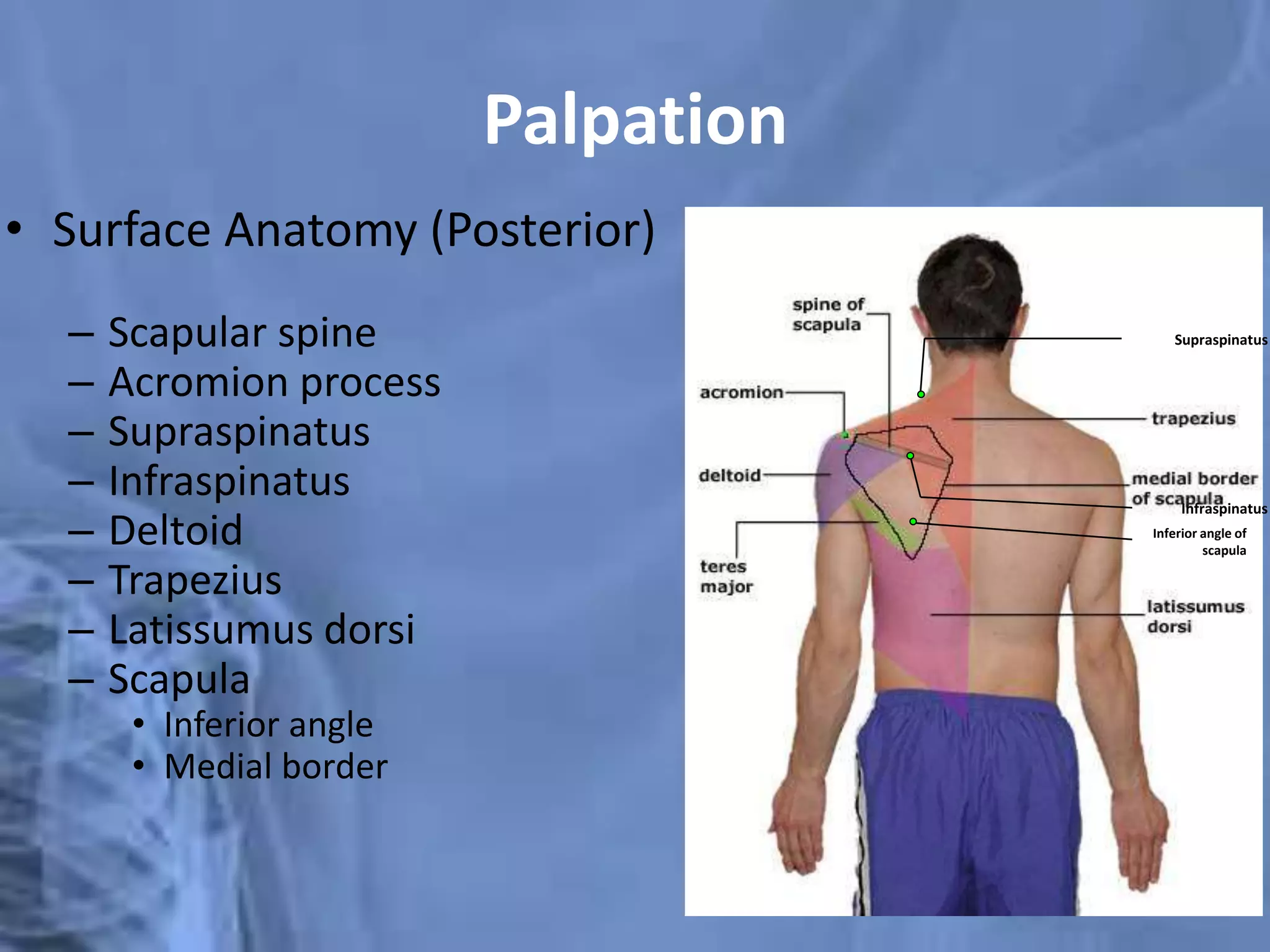
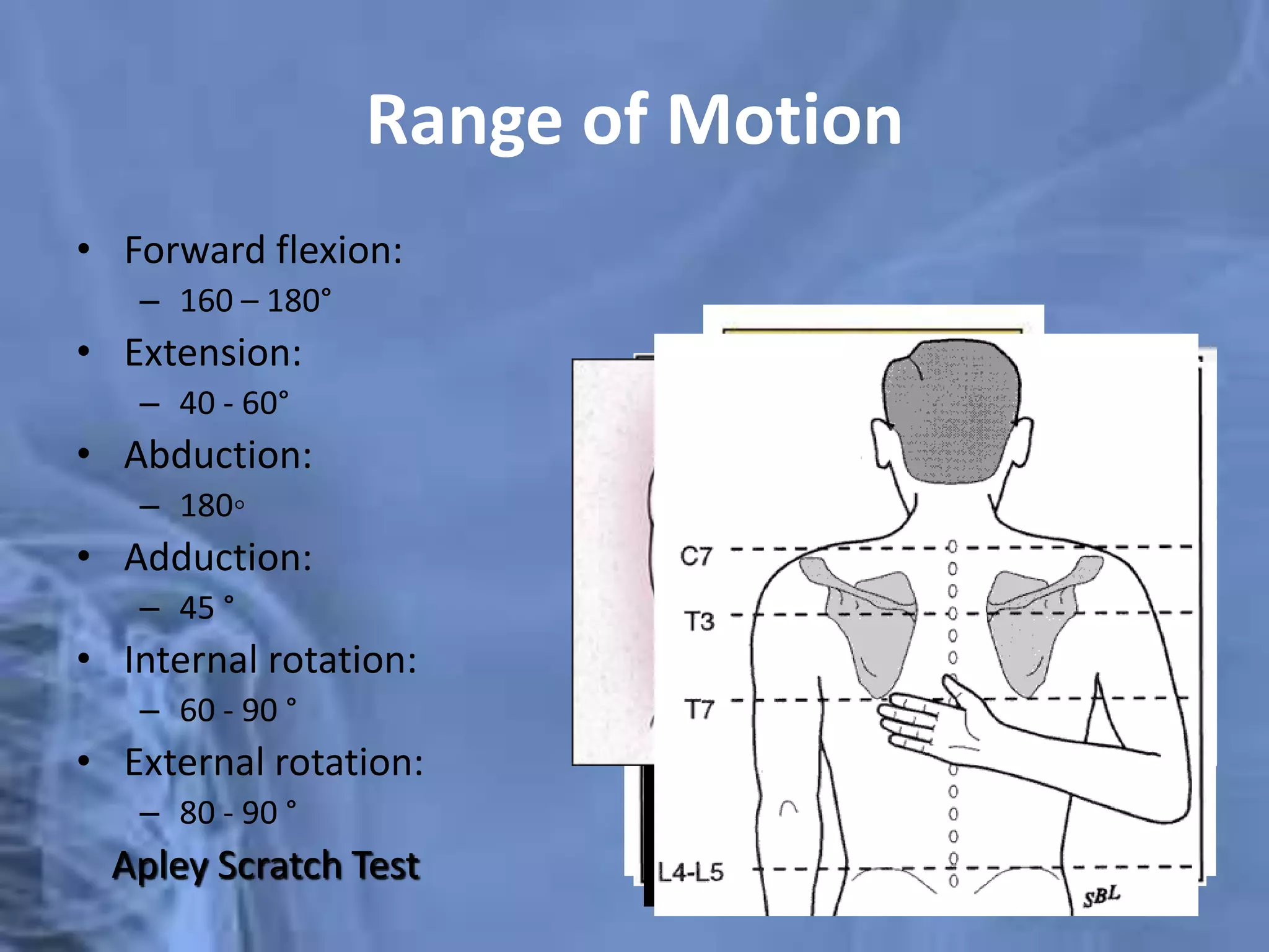
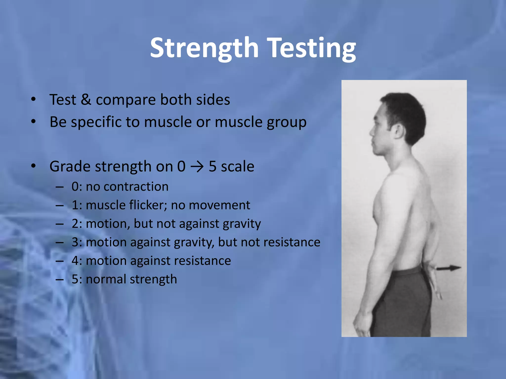
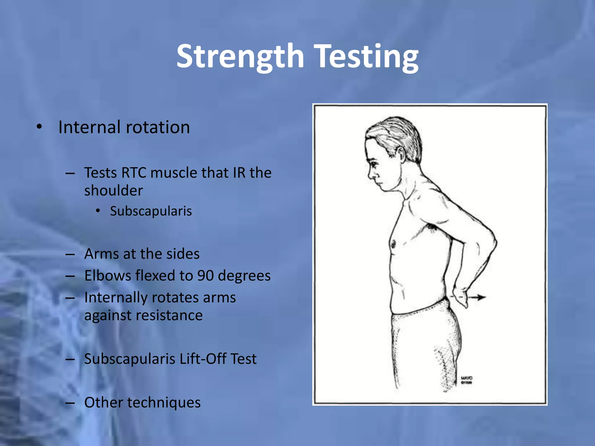
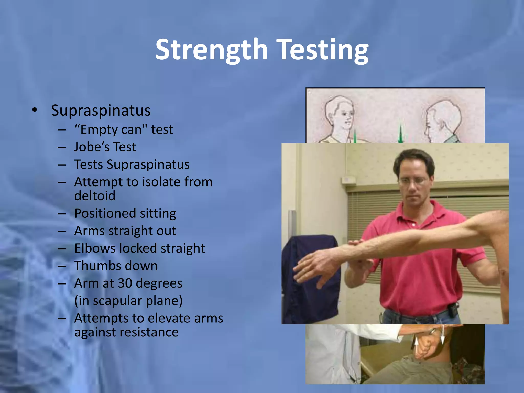
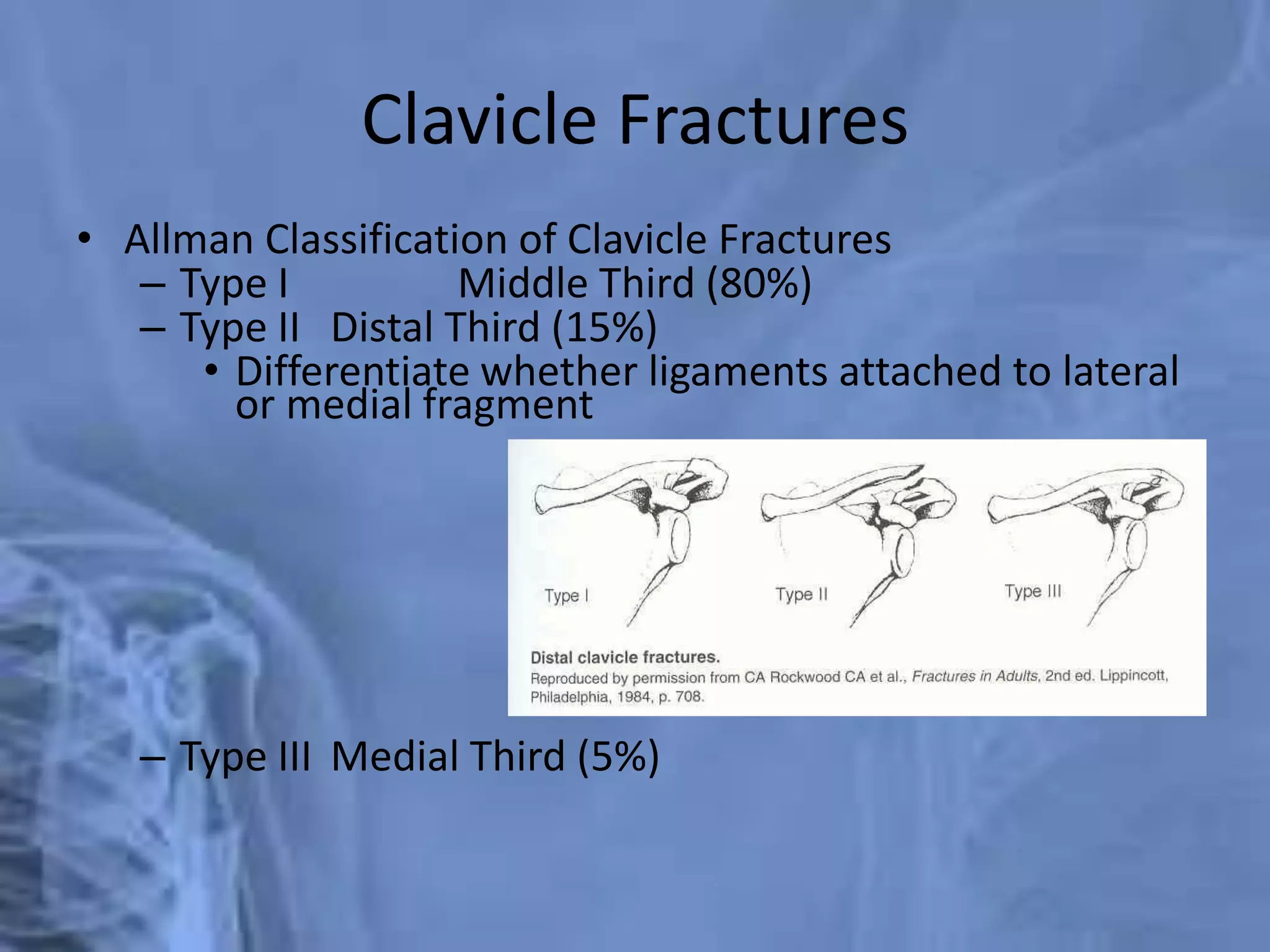
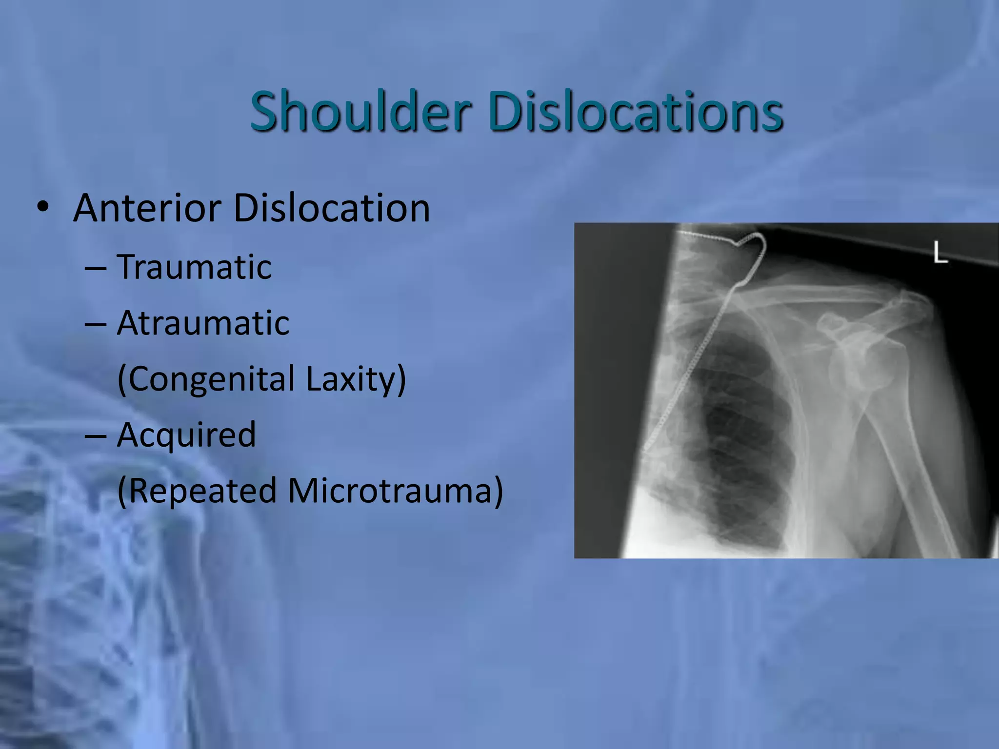
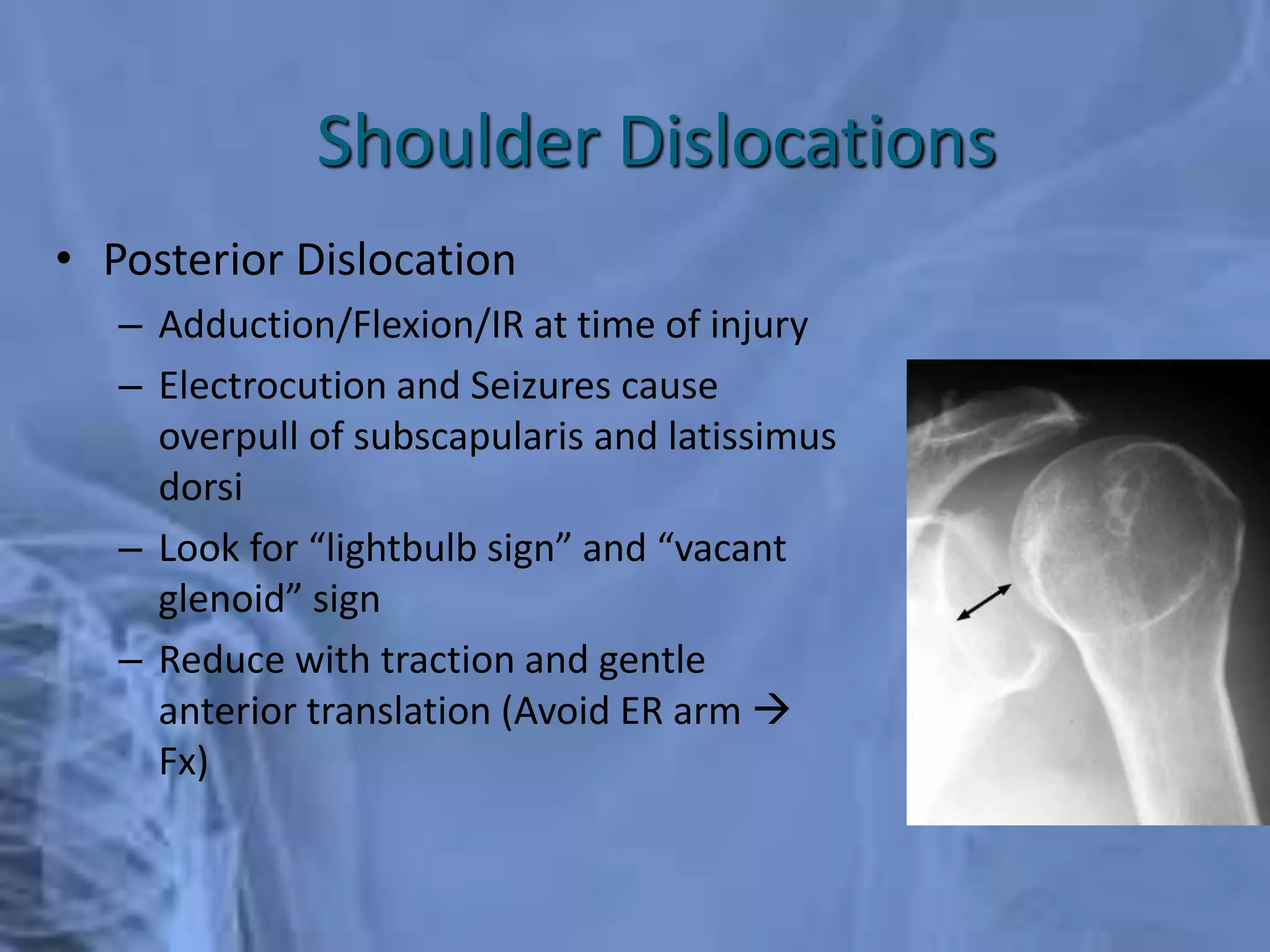
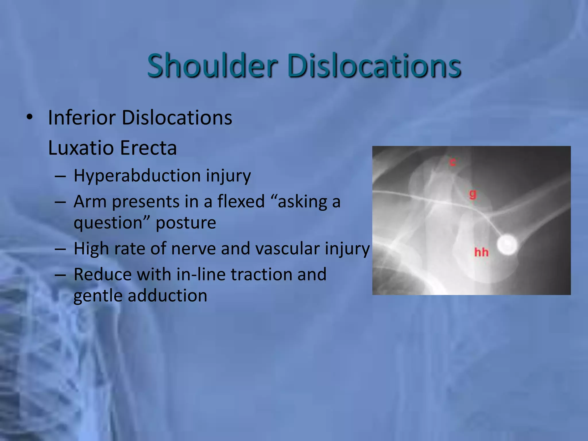
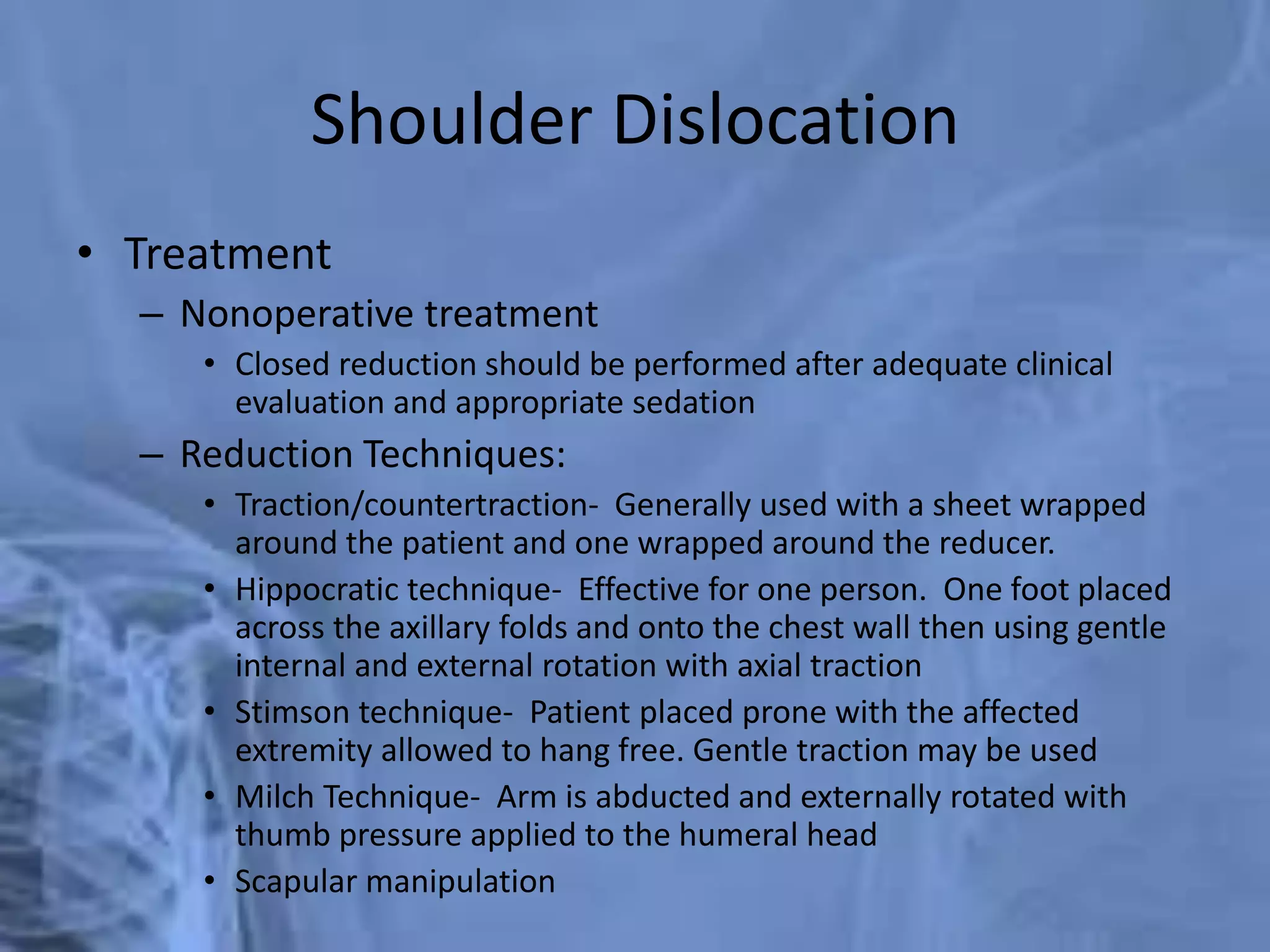
BY DR/ KHALED ALSAYANI
Shoulder JOINT
Brief Epidemiology
• Shoulder pain: a common complaint
in primary care
– 2nd only to knee pain for specialist
referrals
– Most common causes in adults (peak
ages 40-60)
• Subacromial impingement syndrome
• Rotator cuff problems
• Athletic injuries
– Shoulder: 8-13% of all athletic injuries
Anatomy
• 3 Bones
– Humerus
– Scapula
– Clavicle
• 3 Joints
– Glenohumeral
– Acromioclavicular
– Sternoclavicular
• 1 “Articulation”
– Scapulothoracic
Anatomy
• Humerus
– Head *
– Greater tubercle*
– Lesser tubercle*
– Intertubercular (bicipital) groove
– Deltoid tuberosity
• Scapula
– Angles
• Superior
• Inferior
• Lateral (Head)
Anatomy
• Scapula
– Glenoid
– Acromion
– Coracoid
– Subscapular fossa
– Scapular spine
– Supraspinatus fossa
– Infraspinatus fossa
Anatomy
• Glenohumeral joint
– “Ball and socket” vs
“Golf ball and tee”
– Very mobile
– Price: instability
– 45% of all dislocations
– Joint stability depends
on multiple factors
Anatomy
• Glenohumeral joint
– Passive stability
• Joint conformity
• Glenoid labrum (50%)
• Joint capsule
• Ligaments
• Bony restraints
Anatomy
• Muscles
– Deltoid
– Trapezius *
– Rhomboids *
– Levator scapulae *
– Rotator cuff
– Teres major
– Biceps
– Pectoralis muscles *
– Serratus anterior *
* Scapular stabilizers
Anatomy
• Rotator Cuff Muscles
– S – Supraspinatus
– I – Infraspinatus
– t - Teres minor
– S- Supscapularis
Anatomy
• Bursae
– Subacromial
(Subdeltoid)
– Subscapular
Anatomy
• Neurologic
– Nerve roots
– Brachial plexus
– Peripheral nerves
Anatomy
• Coordinated shoulder motion
– Glenohumeral motion
– Acromioclavicular motion
– Sternoclavicular motion
– Scapulothoracic motion
Scapular-humeral rhythm
Differential Diagnosis
• Impingement syndrome
– Subacromial bursitis
– Rotator cuff tendinopathy
– Rotator cuff tear
– Biceps tendinopathy
• Adhesive capsulitis
• SC joint arthritis, sprain
• AC joint arthritis, sprain
• Glenohumeral joint OA
• Instablity
– GH dislocation
– GH subluxation
– Labral tear (e.g. Bankart, SLAP, etc.)
• Clavicle fracture
• Proximal humerus fracture
• Scapular fracture
• Other arthritic disease
– Rheumatoid, Gout, SLE
– Septic, Lyme, etc.
• Avascular necrosis
• Neoplastic disease
• Thoracic outlet syndrome
• CRPS
• Myofascial pain
• Referred pain
– Cervical radiculopathy
– Cardiac
– Aortic aneurysm
– Abdominal / Diaphragm
– Other GI
Clinical History
• Characterize pain
• Location of pain
• Night pain
• Weakness
• Deformity
• Instability
• Locking / Clicking / Clunking
• Sport / Occupation
• Previous treatments
• Alleviating / Exacerbating
• Acute vs. Chronic
• Traumatic vs. Overuse
• History of prior injury
Clinical History
• Mechanism of Injury
Physical Exam
• Observation
– Undress waist → up
• Palpation
• Active & passive ROM
• Strength testing
• Special tests
Physical Exam – Observation /
Inspection
• Front & Back
• Height of shoulder &
scapulae
• Asymmetry
• Obvious deformity
• Ecchymosis
• Muscle atrophy
– Supraspinatus
– Infraspinatus
– Deltoid
Palpation
• At rest & with movement
• Bony structures
• Joints
• Soft tissues
Palpation
• Surface Anatomy (Anterior)
– Clavicle
– SC Joint
– Acromion process
– AC Joint
– Deltoid
– Coracoid process
– Pectoralis major
– Trapezius
– Biceps (long head)
AC joint
SC joint
biceps
Palpation
• Surface Anatomy (Posterior)
– Scapular spine
– Acromion process
– Supraspinatus
– Infraspinatus
– Deltoid
– Trapezius
– Latissumus dorsi
– Scapula
• Inferior angle
• Medial border
Supraspinatus
Infraspinatus
Inferior angle of
scapula
Range of Motion
• Forward flexion:
– 160 – 180°
• Extension:
– 40 - 60°
• Abduction:
– 180◦
• Adduction:
– 45 °
• Internal rotation:
– 60 - 90 °
• External rotation:
– 80 - 90 °
Apley Scratch Test
Range of Motion
• Scapular dyskinesis (Scapulothoracic
dysfuntion)
– Compare scapular motion through
ROM on both sides
– Wall push-ups
– Symmetrical
– Smooth
– No or minimal winging
Strength Testing
• Test & compare both sides
• Be specific to muscle or muscle group
• Grade strength on 0 → 5 scale
– 0: no contraction
– 1: muscle flicker; no movement
– 2: motion, but not against gravity
– 3: motion against gravity, but not resistance
– 4: motion against resistance
– 5: normal strength
Strength Testing
• External rotation
– Tests RTC muscles that ER the
shoulder
• Infraspinatus
• Teres minor
– Arms at the sides
– Elbows flexed to 90 degrees
– Externally rotates arms
against resistance
Strength Testing
• Internal rotation
– Tests RTC muscle that IR the
shoulder
• Subscapularis
– Arms at the sides
– Elbows flexed to 90 degrees
– Internally rotates arms
against resistance
– Subscapularis Lift-Off Test
– Other techniques
Strength Testing
• Supraspinatus
– “Empty can" test
– Jobe’s Test
– Tests Supraspinatus
– Attempt to isolate from
deltoid
– Positioned sitting
– Arms straight out
– Elbows locked straight
– Thumbs down
– Arm at 30 degrees
(in scapular plane)
– Attempts to elevate arms
against resistance
Fractures
Clavicle Fractures
Clavicle Fractures
• Mechanism
– Fall onto shoulder (87%)
– Direct blow (7%)
– Fall onto outstretched hand
(6%)
• Trimodal distribution
0
10
20
30
40
50
60
70
80
Group I
(13yrs)
Group 2
(47yrs)
Group 3
(59yrs)
Percent
The clavicle is the last
ossification center to
complete (sternal end)
at about 22-25yo.
Clavicle Fractures
• Clinical Evaluation
– Inspect and palpate for deformity/abnormal motion
– Thorough distal neurovascular exam
– Auscultate the chest for the possibility of lung injury or
pneumothorax
• Radiographic Exam
– AP chest radiographs.
– Clavicular 45deg A/P oblique X-rays
– Traction pictures may be used as well
Clavicle Fractures
• Allman Classification of Clavicle Fractures
– Type I Middle Third (80%)
– Type II Distal Third (15%)
• Differentiate whether ligaments attached to lateral
or medial fragment
– Type III Medial Third (5%)
Clavicle Fracture
• Closed Treatment
– Sling immobilization for usually 3-4 weeks with early ROM
encouraged
• Operative intervention
– Fractures with neurovascular injury
– Fractures with severe associated chest injuries
– Open fractures
– Group II, type II fractures
– Cosmetic reasons, uncontrolled deformity
– Nonunion
• Associated Injuries
– Brachial Plexus Injuries
• Contusions most common, penetrating (rare)
– Vascular Injury
– Rib Fractures
– Scapula Fractures
– Pneumothorax
Clavicle Fractures
Shoulder Dislocations
• Epidemiology
– Anterior: Most common
– Posterior: Uncommon, 10%, Think Electrocutions &
Seizures
– Inferior (Luxatio Erecta): Rare, hyperabduction injury
Shoulder Dislocations
• Clinical Evaluation
– Examine axillary nerve (deltoid function, not sensation
over lateral shoulder)
– Examine M/C nerve (biceps function and anterolateral
forearm sensation)
• Radiographic Evaluation
– True AP shoulder
– Axillary Lateral
– Scapular Y
– Stryker Notch View (Bony Bankart)
Shoulder Dislocations
• Anterior Dislocation Recurrence Rate
– Age 20: 80-92%
– Age 30: 60%
– > Age 40: 10-15%
• Look for Concomitant Injuries
– Bony: Bankart, Hill-Sachs Lesion, Glenoid Fracture, Greater
Tuberosity Fracture
– Soft Tissue: Subscapularis Tear, RCT (older pts with
dislocation)
– Vascular: Axillary artery injury (older pts with
atherosclerosis)
– Nerve: Axillary nerve neuropraxia
• Anterior Dislocation
– Traumatic
– Atraumatic
(Congenital Laxity)
– Acquired
(Repeated Microtrauma)
Shoulder Dislocations
• Posterior Dislocation
– Adduction/Flexion/IR at time of injury
– Electrocution and Seizures cause
overpull of subscapularis and latissimus
dorsi
– Look for “lightbulb sign” and “vacant
glenoid” sign
– Reduce with traction and gentle
anterior translation (Avoid ER arm 
Fx)
Shoulder Dislocations
• Inferior Dislocations
Luxatio Erecta
– Hyperabduction injury
– Arm presents in a flexed “asking a
question” posture
– High rate of nerve and vascular injury
– Reduce with in-line traction and
gentle adduction
Shoulder Dislocations
Shoulder Dislocation
• Treatment
– Nonoperative treatment
• Closed reduction should be performed after adequate clinical
evaluation and appropriate sedation
– Reduction Techniques:
• Traction/countertraction- Generally used with a sheet wrapped
around the patient and one wrapped around the reducer.
• Hippocratic technique- Effective for one person. One foot placed
across the axillary folds and onto the chest wall then using gentle
internal and external rotation with axial traction
• Stimson technique- Patient placed prone with the affected
extremity allowed to hang free. Gentle traction may be used
• Milch Technique- Arm is abducted and externally rotated with
thumb pressure applied to the humeral head
• Scapular manipulation
Shoulder Dislocations
• Postreduction
– Post reduction films are a must to confirm the position of
the humeral head
– Pain control
– Immobilization for 7-10 days then begin progressive ROM
• Operative Indications
– Irreducible shoulder (soft tissue interposition)
– Displaced greater tuberosity fractures
– Glenoid rim fractures bigger than 5 mm
– Elective repair for younger patients
Proximal Humerus Fractures
• Epidemiology
– Most common fracture of the humerus
– Higher incidence in the elderly, thought to be related to
osteoporosis
– Females 2:1 greater incidence than males
• Mechanism of Injury
– Most commonly a fall onto an outstretched arm from
standing height
– Younger patient typically present after high energy trauma
such as motor vehicle accident MVA
Proximal Humerus Fractures
• Clinical Evaluation
– Patients typically present with arm held close to chest by
contralateral hand. Pain and crepitus detected on
palpation
– Careful NV exam is essential, particularly with regards to
the axillary nerve. Test sensation over the deltoid. Deltoid
atony does not necessarily confirm an axillary nerve injury
Proximal Humerus Fractures
• Neer Classification
– Four parts
• Greater and lesser
tuberosities,
• Humeral shaft
• Humeral head
Proximal Humerus Fractures
• Treatment
– Minimally displaced fractures- Sling immobilization, early motion
– Two-part fractures-
• Anatomic neck fractures likely require Open reduction and internal
fixation ORIF. High incidence of osteonecrosis
– Three-part fractures
• Due to disruption of opposing muscle forces, these are unstable so closed
treatment is difficult. Displacement requires ORIF.
– Four-part fractures
• In general for displacement or unstable injuries ORIF in the young and
hemiarthroplasty in the elderly and those with severe comminution. High
rate of Avascular necrosis AVN (13-34%)
Humeral Shaft Fractures
• Mechanism of Injury
– Direct trauma is the most common especially MVA
– Indirect trauma such as fall on an outstretched hand
– Fracture pattern depends on stress applied
• Compressive- proximal or distal humerus
• Bending- transverse fracture of the shaft
• Torsional- spiral fracture of the shaft
• Torsion and bending- oblique fracture usually associated with a
butterfly fragment
Humeral Shaft Fractures
• Clinical evaluation
– Thorough history and
physical
– Patients typically present
with pain, swelling, and
deformity of the upper arm
– Careful NV exam important
as the radial nerve is in close
proximity to the humerus
and can be injured
Humeral Shaft Fractures
• Radiographic evaluation
– AP and lateral views of the humerus
– Traction radiographs may be indicated for hard to
classify secondary to severe displacement or a lot
of comminution
Humeral Shaft Fractures
• Conservative Treatment
– Goal of treatment is to establish
union with acceptable alignment
– >90% of humeral shaft fractures heal
with nonsurgical management
Humeral Shaft Fractures
• Treatment
– Operative Treatment
• Indications for operative treatment include inadequate
reduction, nonunion, open fractures, segmental fractures,
associated vascular or nerve injuries
• Most commonly treated with plates and screws but also IM
nails
SHOULDER INSTABILITY
Patient Evaluation
• Physical Exam
– Strength
– Muscle atrophy and scapular
winging
– Motor and sensory exam
– ROM assessment
– Soft-tissue laxity
• Beighton Score
– Special tests
• Sulcus Sign
• Load and Shift
• Apprehension Test
– Relocation
• Jerk Test
Shoulder Instability Imaging
• Radiographs
• CT
– Best to assess bone stock
– 3D reconstructions
• MRI
– With or without arthrogram
– Evaluate the soft-tissues
Shoulder Instability
• Traumatic vs. Atraumatic
• Direction of Instability
• Age
• Single episode vs.
Recurrent
• Arm dominance
• Activities/Sports
– Contact vs. Non-contact,
overhead
• Co-Morbidities
Anterior Instability
• Anterior Instability
– >90% of Dislocations
– Abducted and externally rotated arm
• Recurrent Dislocation
– Under 20 years old: Wide variation between
studies (<25% to >90%)
– Less likely as age increases
– Males higher risk than females at all ages
– Higher risks in contact sports
• Rotator Cuff Tears
– After 40 yrs old – 40%
• Initial Management
– Reduction, immobilization
Anterior Instability
• Bankart Lesion and
avulsion of IGHL
from glenoid
– >90%
– Includes bony
component at
times
• Humeral Avulsion
of Glenohumeral
Ligament (HAGL)
– <10%
Treatment
• Immobilization in internal vs. external
rotation
– No consensus, no apparent benefit in ER
• In season dislocations:
– AJSM 2004 study (Buss et al) showed
87% return to sport that season
• Averaged 1.4 incidents of recurrent
dislocation
• >50% had surgery the following off-
season
Treatment
• Risks with non-operative
management in a first time
dislocator, especially young male
in contact sports
– Recurrent instability
– Damage to joint, glenoid over time
necessitating open procedure
– Arthropathy
Surgical Treatment
• Open vs. Arthroscopic procedures
– Level 1 study showed no difference (AJSM 2006)
– Systematic review (Sports Health 2011)
• No significant difference in redislocation rate, return to activity, or
functional outcomes
• Better ROM in arthroscopic group
• High level contact athletes, some argue open is preferred method
• Bony deficiencies
– Glenoid- restore bone stock
• Latarjet Procedure
– Humerus (Hill-Sachs deformity)- fill in space
• Remplissage
Arthroscopic Management (Bankart
Lesion)
Arthroscopic Management (Bony
Bankart)
Open Management
Latarjet Procedure
Posterior Instability
• 4% of all glenohumeral dislocations
• High energy, seizure, electric shock
• Can see from repetitive use/stress
– e.g. Football linemen
• 50-80% missed at initial presentation
• Often present with internally rotated,
adducted upper extremity
• Radiographs – Axillary view essential
• Posterior Subluxation
– Apprehension sign: posteriorly directed
force with the adducted arm flexed 90°
and internally rotated 90°
Treatment
• Reduction if needed
– Traction, external rotation,
abduction
• Immobilization 4-6 weeks in neutral
rotation
• Surgical stabilization for
– Chronic instability/dislocations
– Articular fractures >20%
– Large glenoid rim fractures
– Displaced lesser tuberosity fracture
• Humeral head replacement
– Chronic dislocations > 3-6 months
– Fracture of humeral head >50%
Arthroscopic Management
Open Management
McLaughlin Procedure
Multidirectional Instability (MDI)
• Can be from overuse (microtrauma)
– Swimmers, gymnasts, volleyball,
throwers
• Associated with connective-tissue disorders
– Marfan’s, Ehler-Danlos
• Will possess patulous inferior capsule and
deficient rotator interval
• Primary treatment is always non-operative
– Dynamic strengthening
• Surgical treatment reserved for patients
who failed prolonged conservative
management
– Focus on the capsule
FROZEN SHOULDER (Syn:
Periarthritis, Adhesive Capsulitis)
FROZEN SHOULDER
• It is defined as a clinical syndrome
characterized by painful restriction of both
active and passive shoulder movements due
to causes within the shoulder joint or remote
(other parts of the body)
History
• Dupley first described it in 1872 and called it
as humeroscapular periarthritis. In 1934,
Codman coined the term Frozen shoulder, and
in 1945, Neviaser gave the name adhesive
capsulitis.
Epidemiology of Frozen Shoulder
• Incidence in general population is 2 percent.
• Incidence in diabetics is 10-35 percent.
• More common in females than males.
• Mean age is 40-60 years.
• Bilateral 12 percent.
Causes
• Primary: Here the exact cause is not known
and it could be idiopathic.
• Secondary: According to Lumberg, the
secondary causes could be: – Shoulder causes:
Problems directly related to shoulder joint which
can give rise to frozen shoulder are tendonitis of
rotator cuff, bicipital tendinitis, fractures and
dislocations around the shoulder, etc.
• – Nonshoulder causes: Problems not related
to shoulder joint like diabetes, cardiovascular
diseases with referred pain to the shoulder,
which keeps the joint immobile, reflex
sympathetic dystrophy, frozen hand shoulder
syndrome, a complication of Colles’ fracture,
can all lead to frozen shoulder. The reason
could be prolonged immobilization of the
shoulder joint due to referred pain, etc.
Pathology
• • During abduction, and repeated overhead
activities of the shoulder, long head of biceps
and rotator cuff undergo repeated strain. This
results in inflammation, fibrosis and
consequent thickening of the shoulder
capsule, which results in loss of movements If
the movements are continued, then the
fibrosis gradually breaks, movements return
but never come back to normal
• Prolonged activity causes small scapular and
biceps muscles to waste faster, load on joint
increases and degenerative changes sets in.
Capsule is fibrosed and shoulder movements
are decreased.
Clinical Features
A patient with frozen shoulder clinically presents as
follows:
• Decreased range of both active and passive shoulder
movements.
• The patient demonstrates a capsular pattern of
movement restrictions
• Pain is noted at the end stage of stretch.
• Accessory joint play is reduced.
• Resistive tests are generally pain free in the available
range of motion.
• Patient is unable to do routine daily activities
Clinical Stages
There are three classical stages in frozen shoulder,
according to Reeves:
• Stage I (stage of pain): Patient complains of acute
pain, decreased movements, external rotation
greatest followed by loss of abduction and then
forward flexion. Internal rotation is least affected.
This stage lasts for 10-36 weeks.
Note: Pain in frozen shoulder does not radiate
below the elbow
• Stage II (stage of stiffness): In this stage, pain
gradually decreases and the patient complains
of stiff shoulder. Slight movements are
present. This lasts for 4-12 months.
• Stage III (stage of recovery): Patient will have
no pain and movements would have
recovered but will never be regained to
normal. It lasts for 6 months to 2 years
Radiology
• X-ray of the shoulder is
usually normal; but in a
few cases, ‘sclerosis’
may be seen on the
outer edge of greater
tuberosity (Golding’s
sign)
Treatment
• Stage I: In this stage, long acting once a day
NSAIDs are usually preferred as this condition
usually runs a long course (10-36 weeks).
Intra-articular steroids may help to provide
transient relief of pain only.
• Stage II: In this stage, since the pain will have
reduced considerably, exercises both active
and passive are gradually begun followed by
physiotherapy, ultrasound, heat and shoulder
wheel exercises. The role of manipulation of
the shoulder is controversial but can be
attempted under general anesthesia in this
stage.
• Stage III: In this stage, active and passive
exercises, physiotherapy consisting of short
wave diathermy, ultrasound, etc. are
continued.

Shoulder anatomy & examination-2.pptx

  • 1.
    BY DR/ KHALEDALSAYANI Shoulder JOINT
  • 2.
    Brief Epidemiology • Shoulderpain: a common complaint in primary care – 2nd only to knee pain for specialist referrals – Most common causes in adults (peak ages 40-60) • Subacromial impingement syndrome • Rotator cuff problems • Athletic injuries – Shoulder: 8-13% of all athletic injuries
  • 3.
    Anatomy • 3 Bones –Humerus – Scapula – Clavicle • 3 Joints – Glenohumeral – Acromioclavicular – Sternoclavicular • 1 “Articulation” – Scapulothoracic
  • 4.
    Anatomy • Humerus – Head* – Greater tubercle* – Lesser tubercle* – Intertubercular (bicipital) groove – Deltoid tuberosity • Scapula – Angles • Superior • Inferior • Lateral (Head)
  • 5.
    Anatomy • Scapula – Glenoid –Acromion – Coracoid – Subscapular fossa – Scapular spine – Supraspinatus fossa – Infraspinatus fossa
  • 6.
    Anatomy • Glenohumeral joint –“Ball and socket” vs “Golf ball and tee” – Very mobile – Price: instability – 45% of all dislocations – Joint stability depends on multiple factors
  • 7.
    Anatomy • Glenohumeral joint –Passive stability • Joint conformity • Glenoid labrum (50%) • Joint capsule • Ligaments • Bony restraints
  • 8.
    Anatomy • Muscles – Deltoid –Trapezius * – Rhomboids * – Levator scapulae * – Rotator cuff – Teres major – Biceps – Pectoralis muscles * – Serratus anterior * * Scapular stabilizers
  • 9.
    Anatomy • Rotator CuffMuscles – S – Supraspinatus – I – Infraspinatus – t - Teres minor – S- Supscapularis
  • 10.
  • 11.
    Anatomy • Neurologic – Nerveroots – Brachial plexus – Peripheral nerves
  • 12.
    Anatomy • Coordinated shouldermotion – Glenohumeral motion – Acromioclavicular motion – Sternoclavicular motion – Scapulothoracic motion Scapular-humeral rhythm
  • 13.
    Differential Diagnosis • Impingementsyndrome – Subacromial bursitis – Rotator cuff tendinopathy – Rotator cuff tear – Biceps tendinopathy • Adhesive capsulitis • SC joint arthritis, sprain • AC joint arthritis, sprain • Glenohumeral joint OA • Instablity – GH dislocation – GH subluxation – Labral tear (e.g. Bankart, SLAP, etc.) • Clavicle fracture • Proximal humerus fracture • Scapular fracture • Other arthritic disease – Rheumatoid, Gout, SLE – Septic, Lyme, etc. • Avascular necrosis • Neoplastic disease • Thoracic outlet syndrome • CRPS • Myofascial pain • Referred pain – Cervical radiculopathy – Cardiac – Aortic aneurysm – Abdominal / Diaphragm – Other GI
  • 14.
    Clinical History • Characterizepain • Location of pain • Night pain • Weakness • Deformity • Instability • Locking / Clicking / Clunking • Sport / Occupation • Previous treatments • Alleviating / Exacerbating • Acute vs. Chronic • Traumatic vs. Overuse • History of prior injury
  • 15.
  • 16.
    Physical Exam • Observation –Undress waist → up • Palpation • Active & passive ROM • Strength testing • Special tests
  • 17.
    Physical Exam –Observation / Inspection • Front & Back • Height of shoulder & scapulae • Asymmetry • Obvious deformity • Ecchymosis • Muscle atrophy – Supraspinatus – Infraspinatus – Deltoid
  • 18.
    Palpation • At rest& with movement • Bony structures • Joints • Soft tissues
  • 19.
    Palpation • Surface Anatomy(Anterior) – Clavicle – SC Joint – Acromion process – AC Joint – Deltoid – Coracoid process – Pectoralis major – Trapezius – Biceps (long head) AC joint SC joint biceps
  • 20.
    Palpation • Surface Anatomy(Posterior) – Scapular spine – Acromion process – Supraspinatus – Infraspinatus – Deltoid – Trapezius – Latissumus dorsi – Scapula • Inferior angle • Medial border Supraspinatus Infraspinatus Inferior angle of scapula
  • 21.
    Range of Motion •Forward flexion: – 160 – 180° • Extension: – 40 - 60° • Abduction: – 180◦ • Adduction: – 45 ° • Internal rotation: – 60 - 90 ° • External rotation: – 80 - 90 ° Apley Scratch Test
  • 22.
    Range of Motion •Scapular dyskinesis (Scapulothoracic dysfuntion) – Compare scapular motion through ROM on both sides – Wall push-ups – Symmetrical – Smooth – No or minimal winging
  • 23.
    Strength Testing • Test& compare both sides • Be specific to muscle or muscle group • Grade strength on 0 → 5 scale – 0: no contraction – 1: muscle flicker; no movement – 2: motion, but not against gravity – 3: motion against gravity, but not resistance – 4: motion against resistance – 5: normal strength
  • 24.
    Strength Testing • Externalrotation – Tests RTC muscles that ER the shoulder • Infraspinatus • Teres minor – Arms at the sides – Elbows flexed to 90 degrees – Externally rotates arms against resistance
  • 25.
    Strength Testing • Internalrotation – Tests RTC muscle that IR the shoulder • Subscapularis – Arms at the sides – Elbows flexed to 90 degrees – Internally rotates arms against resistance – Subscapularis Lift-Off Test – Other techniques
  • 26.
    Strength Testing • Supraspinatus –“Empty can" test – Jobe’s Test – Tests Supraspinatus – Attempt to isolate from deltoid – Positioned sitting – Arms straight out – Elbows locked straight – Thumbs down – Arm at 30 degrees (in scapular plane) – Attempts to elevate arms against resistance
  • 27.
  • 28.
  • 29.
    Clavicle Fractures • Mechanism –Fall onto shoulder (87%) – Direct blow (7%) – Fall onto outstretched hand (6%) • Trimodal distribution 0 10 20 30 40 50 60 70 80 Group I (13yrs) Group 2 (47yrs) Group 3 (59yrs) Percent The clavicle is the last ossification center to complete (sternal end) at about 22-25yo.
  • 30.
    Clavicle Fractures • ClinicalEvaluation – Inspect and palpate for deformity/abnormal motion – Thorough distal neurovascular exam – Auscultate the chest for the possibility of lung injury or pneumothorax • Radiographic Exam – AP chest radiographs. – Clavicular 45deg A/P oblique X-rays – Traction pictures may be used as well
  • 31.
    Clavicle Fractures • AllmanClassification of Clavicle Fractures – Type I Middle Third (80%) – Type II Distal Third (15%) • Differentiate whether ligaments attached to lateral or medial fragment – Type III Medial Third (5%)
  • 32.
    Clavicle Fracture • ClosedTreatment – Sling immobilization for usually 3-4 weeks with early ROM encouraged • Operative intervention – Fractures with neurovascular injury – Fractures with severe associated chest injuries – Open fractures – Group II, type II fractures – Cosmetic reasons, uncontrolled deformity – Nonunion
  • 33.
    • Associated Injuries –Brachial Plexus Injuries • Contusions most common, penetrating (rare) – Vascular Injury – Rib Fractures – Scapula Fractures – Pneumothorax Clavicle Fractures
  • 34.
  • 35.
    Shoulder Dislocations • Epidemiology –Anterior: Most common – Posterior: Uncommon, 10%, Think Electrocutions & Seizures – Inferior (Luxatio Erecta): Rare, hyperabduction injury
  • 36.
    Shoulder Dislocations • ClinicalEvaluation – Examine axillary nerve (deltoid function, not sensation over lateral shoulder) – Examine M/C nerve (biceps function and anterolateral forearm sensation) • Radiographic Evaluation – True AP shoulder – Axillary Lateral – Scapular Y – Stryker Notch View (Bony Bankart)
  • 37.
    Shoulder Dislocations • AnteriorDislocation Recurrence Rate – Age 20: 80-92% – Age 30: 60% – > Age 40: 10-15% • Look for Concomitant Injuries – Bony: Bankart, Hill-Sachs Lesion, Glenoid Fracture, Greater Tuberosity Fracture – Soft Tissue: Subscapularis Tear, RCT (older pts with dislocation) – Vascular: Axillary artery injury (older pts with atherosclerosis) – Nerve: Axillary nerve neuropraxia
  • 38.
    • Anterior Dislocation –Traumatic – Atraumatic (Congenital Laxity) – Acquired (Repeated Microtrauma) Shoulder Dislocations
  • 39.
    • Posterior Dislocation –Adduction/Flexion/IR at time of injury – Electrocution and Seizures cause overpull of subscapularis and latissimus dorsi – Look for “lightbulb sign” and “vacant glenoid” sign – Reduce with traction and gentle anterior translation (Avoid ER arm  Fx) Shoulder Dislocations
  • 40.
    • Inferior Dislocations LuxatioErecta – Hyperabduction injury – Arm presents in a flexed “asking a question” posture – High rate of nerve and vascular injury – Reduce with in-line traction and gentle adduction Shoulder Dislocations
  • 41.
    Shoulder Dislocation • Treatment –Nonoperative treatment • Closed reduction should be performed after adequate clinical evaluation and appropriate sedation – Reduction Techniques: • Traction/countertraction- Generally used with a sheet wrapped around the patient and one wrapped around the reducer. • Hippocratic technique- Effective for one person. One foot placed across the axillary folds and onto the chest wall then using gentle internal and external rotation with axial traction • Stimson technique- Patient placed prone with the affected extremity allowed to hang free. Gentle traction may be used • Milch Technique- Arm is abducted and externally rotated with thumb pressure applied to the humeral head • Scapular manipulation
  • 43.
    Shoulder Dislocations • Postreduction –Post reduction films are a must to confirm the position of the humeral head – Pain control – Immobilization for 7-10 days then begin progressive ROM • Operative Indications – Irreducible shoulder (soft tissue interposition) – Displaced greater tuberosity fractures – Glenoid rim fractures bigger than 5 mm – Elective repair for younger patients
  • 44.
  • 46.
    Proximal Humerus Fractures •Epidemiology – Most common fracture of the humerus – Higher incidence in the elderly, thought to be related to osteoporosis – Females 2:1 greater incidence than males • Mechanism of Injury – Most commonly a fall onto an outstretched arm from standing height – Younger patient typically present after high energy trauma such as motor vehicle accident MVA
  • 47.
    Proximal Humerus Fractures •Clinical Evaluation – Patients typically present with arm held close to chest by contralateral hand. Pain and crepitus detected on palpation – Careful NV exam is essential, particularly with regards to the axillary nerve. Test sensation over the deltoid. Deltoid atony does not necessarily confirm an axillary nerve injury
  • 48.
    Proximal Humerus Fractures •Neer Classification – Four parts • Greater and lesser tuberosities, • Humeral shaft • Humeral head
  • 49.
    Proximal Humerus Fractures •Treatment – Minimally displaced fractures- Sling immobilization, early motion – Two-part fractures- • Anatomic neck fractures likely require Open reduction and internal fixation ORIF. High incidence of osteonecrosis – Three-part fractures • Due to disruption of opposing muscle forces, these are unstable so closed treatment is difficult. Displacement requires ORIF. – Four-part fractures • In general for displacement or unstable injuries ORIF in the young and hemiarthroplasty in the elderly and those with severe comminution. High rate of Avascular necrosis AVN (13-34%)
  • 50.
  • 51.
    Humeral Shaft Fractures •Mechanism of Injury – Direct trauma is the most common especially MVA – Indirect trauma such as fall on an outstretched hand – Fracture pattern depends on stress applied • Compressive- proximal or distal humerus • Bending- transverse fracture of the shaft • Torsional- spiral fracture of the shaft • Torsion and bending- oblique fracture usually associated with a butterfly fragment
  • 52.
    Humeral Shaft Fractures •Clinical evaluation – Thorough history and physical – Patients typically present with pain, swelling, and deformity of the upper arm – Careful NV exam important as the radial nerve is in close proximity to the humerus and can be injured
  • 53.
    Humeral Shaft Fractures •Radiographic evaluation – AP and lateral views of the humerus – Traction radiographs may be indicated for hard to classify secondary to severe displacement or a lot of comminution
  • 54.
    Humeral Shaft Fractures •Conservative Treatment – Goal of treatment is to establish union with acceptable alignment – >90% of humeral shaft fractures heal with nonsurgical management
  • 55.
    Humeral Shaft Fractures •Treatment – Operative Treatment • Indications for operative treatment include inadequate reduction, nonunion, open fractures, segmental fractures, associated vascular or nerve injuries • Most commonly treated with plates and screws but also IM nails
  • 56.
  • 57.
    Patient Evaluation • PhysicalExam – Strength – Muscle atrophy and scapular winging – Motor and sensory exam – ROM assessment – Soft-tissue laxity • Beighton Score – Special tests • Sulcus Sign • Load and Shift • Apprehension Test – Relocation • Jerk Test
  • 58.
    Shoulder Instability Imaging •Radiographs • CT – Best to assess bone stock – 3D reconstructions • MRI – With or without arthrogram – Evaluate the soft-tissues
  • 59.
    Shoulder Instability • Traumaticvs. Atraumatic • Direction of Instability • Age • Single episode vs. Recurrent • Arm dominance • Activities/Sports – Contact vs. Non-contact, overhead • Co-Morbidities
  • 60.
    Anterior Instability • AnteriorInstability – >90% of Dislocations – Abducted and externally rotated arm • Recurrent Dislocation – Under 20 years old: Wide variation between studies (<25% to >90%) – Less likely as age increases – Males higher risk than females at all ages – Higher risks in contact sports • Rotator Cuff Tears – After 40 yrs old – 40% • Initial Management – Reduction, immobilization
  • 61.
    Anterior Instability • BankartLesion and avulsion of IGHL from glenoid – >90% – Includes bony component at times • Humeral Avulsion of Glenohumeral Ligament (HAGL) – <10%
  • 62.
    Treatment • Immobilization ininternal vs. external rotation – No consensus, no apparent benefit in ER • In season dislocations: – AJSM 2004 study (Buss et al) showed 87% return to sport that season • Averaged 1.4 incidents of recurrent dislocation • >50% had surgery the following off- season
  • 63.
    Treatment • Risks withnon-operative management in a first time dislocator, especially young male in contact sports – Recurrent instability – Damage to joint, glenoid over time necessitating open procedure – Arthropathy
  • 64.
    Surgical Treatment • Openvs. Arthroscopic procedures – Level 1 study showed no difference (AJSM 2006) – Systematic review (Sports Health 2011) • No significant difference in redislocation rate, return to activity, or functional outcomes • Better ROM in arthroscopic group • High level contact athletes, some argue open is preferred method • Bony deficiencies – Glenoid- restore bone stock • Latarjet Procedure – Humerus (Hill-Sachs deformity)- fill in space • Remplissage
  • 65.
  • 66.
  • 67.
  • 68.
    Posterior Instability • 4%of all glenohumeral dislocations • High energy, seizure, electric shock • Can see from repetitive use/stress – e.g. Football linemen • 50-80% missed at initial presentation • Often present with internally rotated, adducted upper extremity • Radiographs – Axillary view essential • Posterior Subluxation – Apprehension sign: posteriorly directed force with the adducted arm flexed 90° and internally rotated 90°
  • 69.
    Treatment • Reduction ifneeded – Traction, external rotation, abduction • Immobilization 4-6 weeks in neutral rotation • Surgical stabilization for – Chronic instability/dislocations – Articular fractures >20% – Large glenoid rim fractures – Displaced lesser tuberosity fracture • Humeral head replacement – Chronic dislocations > 3-6 months – Fracture of humeral head >50%
  • 70.
  • 71.
  • 72.
    Multidirectional Instability (MDI) •Can be from overuse (microtrauma) – Swimmers, gymnasts, volleyball, throwers • Associated with connective-tissue disorders – Marfan’s, Ehler-Danlos • Will possess patulous inferior capsule and deficient rotator interval • Primary treatment is always non-operative – Dynamic strengthening • Surgical treatment reserved for patients who failed prolonged conservative management – Focus on the capsule
  • 73.
  • 74.
    FROZEN SHOULDER • Itis defined as a clinical syndrome characterized by painful restriction of both active and passive shoulder movements due to causes within the shoulder joint or remote (other parts of the body)
  • 75.
    History • Dupley firstdescribed it in 1872 and called it as humeroscapular periarthritis. In 1934, Codman coined the term Frozen shoulder, and in 1945, Neviaser gave the name adhesive capsulitis.
  • 76.
    Epidemiology of FrozenShoulder • Incidence in general population is 2 percent. • Incidence in diabetics is 10-35 percent. • More common in females than males. • Mean age is 40-60 years. • Bilateral 12 percent.
  • 77.
    Causes • Primary: Herethe exact cause is not known and it could be idiopathic. • Secondary: According to Lumberg, the secondary causes could be: – Shoulder causes: Problems directly related to shoulder joint which can give rise to frozen shoulder are tendonitis of rotator cuff, bicipital tendinitis, fractures and dislocations around the shoulder, etc.
  • 78.
    • – Nonshouldercauses: Problems not related to shoulder joint like diabetes, cardiovascular diseases with referred pain to the shoulder, which keeps the joint immobile, reflex sympathetic dystrophy, frozen hand shoulder syndrome, a complication of Colles’ fracture, can all lead to frozen shoulder. The reason could be prolonged immobilization of the shoulder joint due to referred pain, etc.
  • 79.
    Pathology • • Duringabduction, and repeated overhead activities of the shoulder, long head of biceps and rotator cuff undergo repeated strain. This results in inflammation, fibrosis and consequent thickening of the shoulder capsule, which results in loss of movements If the movements are continued, then the fibrosis gradually breaks, movements return but never come back to normal
  • 80.
    • Prolonged activitycauses small scapular and biceps muscles to waste faster, load on joint increases and degenerative changes sets in. Capsule is fibrosed and shoulder movements are decreased.
  • 82.
    Clinical Features A patientwith frozen shoulder clinically presents as follows: • Decreased range of both active and passive shoulder movements. • The patient demonstrates a capsular pattern of movement restrictions • Pain is noted at the end stage of stretch. • Accessory joint play is reduced. • Resistive tests are generally pain free in the available range of motion. • Patient is unable to do routine daily activities
  • 83.
    Clinical Stages There arethree classical stages in frozen shoulder, according to Reeves: • Stage I (stage of pain): Patient complains of acute pain, decreased movements, external rotation greatest followed by loss of abduction and then forward flexion. Internal rotation is least affected. This stage lasts for 10-36 weeks. Note: Pain in frozen shoulder does not radiate below the elbow
  • 85.
    • Stage II(stage of stiffness): In this stage, pain gradually decreases and the patient complains of stiff shoulder. Slight movements are present. This lasts for 4-12 months. • Stage III (stage of recovery): Patient will have no pain and movements would have recovered but will never be regained to normal. It lasts for 6 months to 2 years
  • 86.
    Radiology • X-ray ofthe shoulder is usually normal; but in a few cases, ‘sclerosis’ may be seen on the outer edge of greater tuberosity (Golding’s sign)
  • 87.
    Treatment • Stage I:In this stage, long acting once a day NSAIDs are usually preferred as this condition usually runs a long course (10-36 weeks). Intra-articular steroids may help to provide transient relief of pain only.
  • 88.
    • Stage II:In this stage, since the pain will have reduced considerably, exercises both active and passive are gradually begun followed by physiotherapy, ultrasound, heat and shoulder wheel exercises. The role of manipulation of the shoulder is controversial but can be attempted under general anesthesia in this stage.
  • 89.
    • Stage III:In this stage, active and passive exercises, physiotherapy consisting of short wave diathermy, ultrasound, etc. are continued.