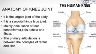
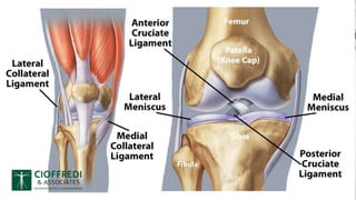
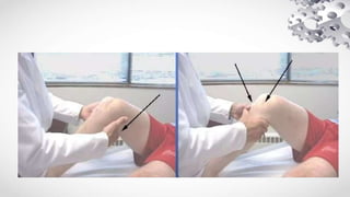
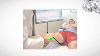
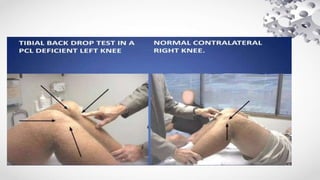
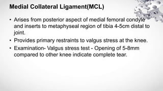
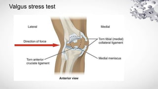
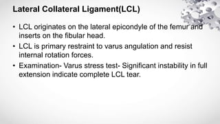
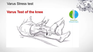
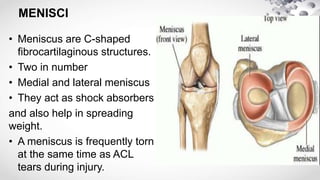
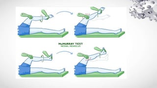
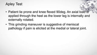
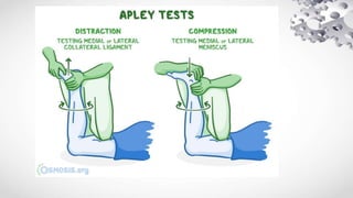
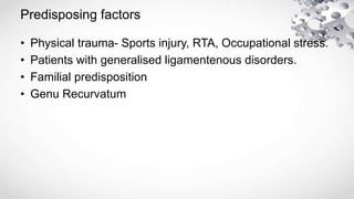
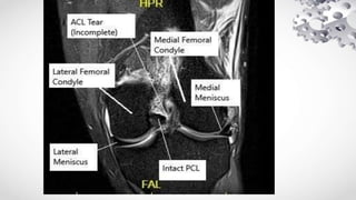
2. Internal derangements of knee (IDK)
• It is a term used to cover the diseases involving disruption
of the normal functioning of the ligaments or menscii of
the knee joints.
• The commonest derangement is Medial collateral
ligament injury followed by medial meniscus and ACL.
3. ANATOMY OF KNEE JOINT
• It is the largest joint of the body.
• It is a synovial hinge type joint.
• Mainly articulation of four
bones:femur,tibia,patella and
fibula.
• The primary articulation is
between the condyles of femur
and tibia.
4. LIGAMENTS
Two major ligaments (inside knee joint)
-Anterior and posterior cruciate ligament
Two other ligament (outside knee joint)
-Medial and lateral collateral ligament
-They act to stabilise knee sideways motion.
5.
6. Anterior cruciate ligament(ACL)
• Origin at the medial wall of the lateral femoral condyle and
inserts into the middle of the intercondylar area.
• It have four main functions
-Restrains anterior translation of tibia.
-Prevent hyperextension of knee.
-Acts as secondary stabiliser to valgus stress test.
-Controls rotation of tibia on femur.
7. Examination of ACL
Anterior Drawer Test
• With the knee flexed to 90deg, verification of relaxation of
hamstrings.With foot stabilised and in neutral rotation, a
firm gentle grip on proximal tibia is achieved.
• An anterior force is applied to proximal tibia with gentle to
and fro motion to assess for increased translation as
compared to C/l knee. %mm is the upper limit of anterior
tibial displacement normally.
8.
9. Lachmans test
• One hand secures and stabilises the distal femur while
the other hand grasps the proximal tibia.
• A gentle anterior translation force is applied to proximal
tibia.
• There is significant translation of tibia on the femur in an
ACL deficient knee.
10.
11. • Patient rotated 20deg from supine towards the affected side. with
slight distal traction on the leg, a valgus and internal rotation force
is applied to the extended knee.
• With maintainence of force noted above, the knee is flexed past
30deg.
• Pivot shift in an ACL deficient knee, in the initial stages of knee
flexion, the tibia will be anterolaterally subluxed on the distal
femur with application of valgus and internal rotation at the knee.
• With further flexion of knee (past 30deg) the illiotibial band goes
from an extendor to flexor of knee and tibial anterolateral
subluxation reduces bak in place.
Pivot Shift test
12.
13. Posterior cruciate Ligament (PCL)
-Originates from the anterolateral aspect of the medial
femoral condyle within the notch and inserts along the
posterior aspect of the tibial plateau, approximately 1 cm
distal to the joint line.
-Functions
Provide restraint against hyperextension.
Against posteior displacement of the tibia in flexed knee
14. Examination
• Posterior Drawer Test
-With knee flexed to approx 90deg,foot in neutral rotation
and stabilised, a firm but gentle posterior translation force is
applied to proximal tibia.
-Application of posterior translation force results in posterior
subluxation of tibia on the femur in patient with PCL
deficient knee.
15.
16. Tibial Back drop Test
• In this test, we compare the prominences of proximal tibia
to the femoral condykles with the knee flexed to 80deg.
• In a PCL deficient knee, the knee will be posteriorly
subluxed due to gravity.
17.
18.
19. Medial Collateral Ligament(MCL)
• Arises from posterior aspect of medial femoral condyle
and inserts to metaphyseal region of tibia 4-5cm distal to
joint.
• Provides primary restraints to valgus stress at the knee.
• Examination- Valgus stress test - Opening of 5-8mm
compared to other knee indicate complete tear.
21. Lateral Collateral Ligament(LCL)
• LCL originates on the lateral epicondyle of the femur and
inserts on the fibular head.
• LCL is primary restraint to varus angulation and resist
internal rotation forces.
• Examination- Varus stress test- Significant instability in full
extension indicate complete LCL tear.
23. MENISCI
• Meniscus are C-shaped
fibrocartilaginous structures.
• Two in number
• Medial and lateral meniscus
• They act as shock absorbers
and also help in spreading
weight.
• A meniscus is frequently torn
at the same time as ACL
tears during injury.
24. Examination
• Joint Line tenderness- It is an accurate clinical sign
occuring in 75-85% of patients
• Effusion- present in approx 50% cases.
• Range of motion- Restricted by pain and swelling.
25. Mc Murray test
This maneuver usually elicits pain or a reproducible click in the
presence of a meniscal tear
The Medial meniscus is evaluated by extending the fully flexed
knee with foot/tibia internally rotated while a varus stress is
applied.
The Lateral meniscus is evaluated by extending the knee from
the fully flexed position, with the foot/tibia externally rotated
while a valgus stress is applied to the knee.
26.
27. Apley Test
• Patient lie prone and knee flexed 90deg. An axial load is
applied through the heel as the lower leg is internally and
externally rotated.
• This grinding maneuver is suggestive of meniscal
pathology if pain is elicited at the medial or lateral joint.
30. Mode of injury
• MCL injury - Forced valgus stress.
• LCL injury - Forced varus stress.
• ACL- Forced Valgus in fully extended knee.
• PCL- High velocity Trauma with posterior dislocation of
tibia in a flexed knee.
• Meniscus tear- Rotational stresses in flexed knees.
31. Imaging Modalities
• Xray- To rule out Bony injuries.
• MRI
• Arthroscopy- It is diagnostic as well as theraputic.
32.
33. Treatment Protocols
• Choice of Treatment depends on 3 major factors:
-Age
-Functional disability
-Functional requirement
• Treatment varies according to the ligament/cartilage
injured.
- It can be Conservative (Non Operative) and Operative.
34. NON OPERATIVE RX
Methods
• Progressive physical therapy and rehabilitation
• Educating the patient how to prevent knee instability.
• Use of Hinged knee brace.
Indications
• Isolated tears wtih no symptoms
• When growth plates are still open(children)
• People who do light work.
35. OPERATIVE RX
Indications
• Patients with knee instability, pain or swelling.
• Progressive premature degenerative changes in patient
with unstable knee
Methods
• Repair
• Reconstruction
• Debridement and removal