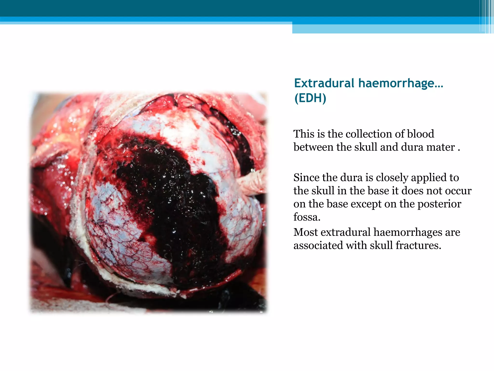
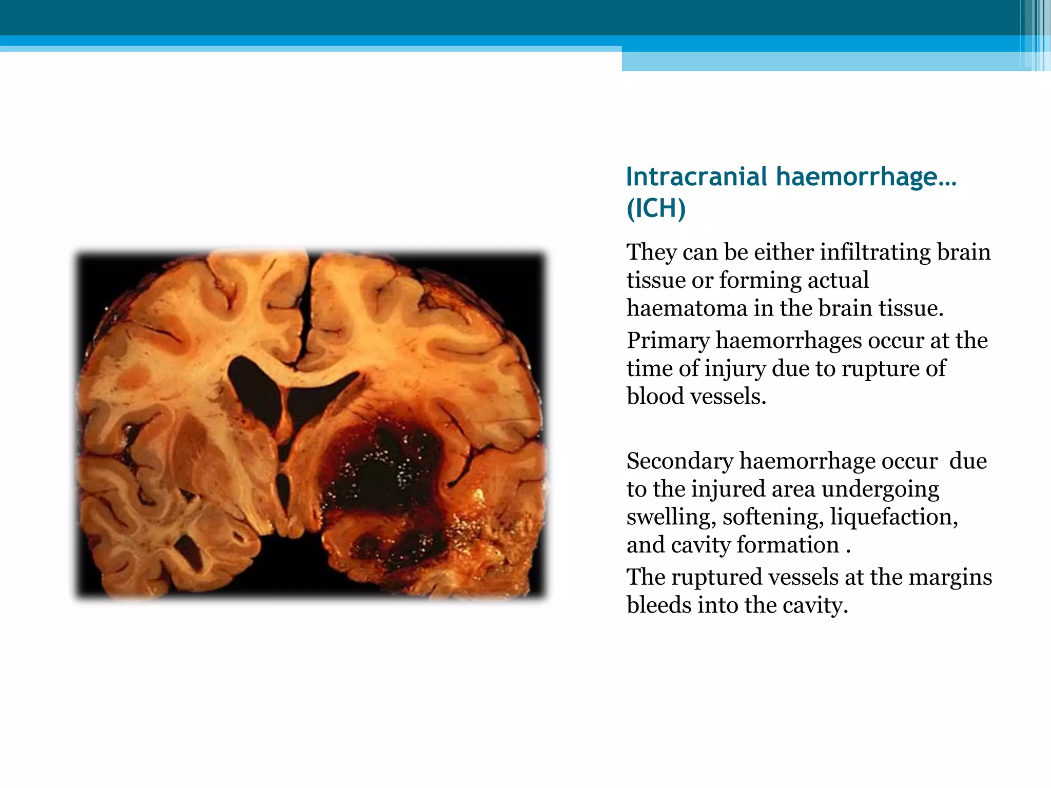
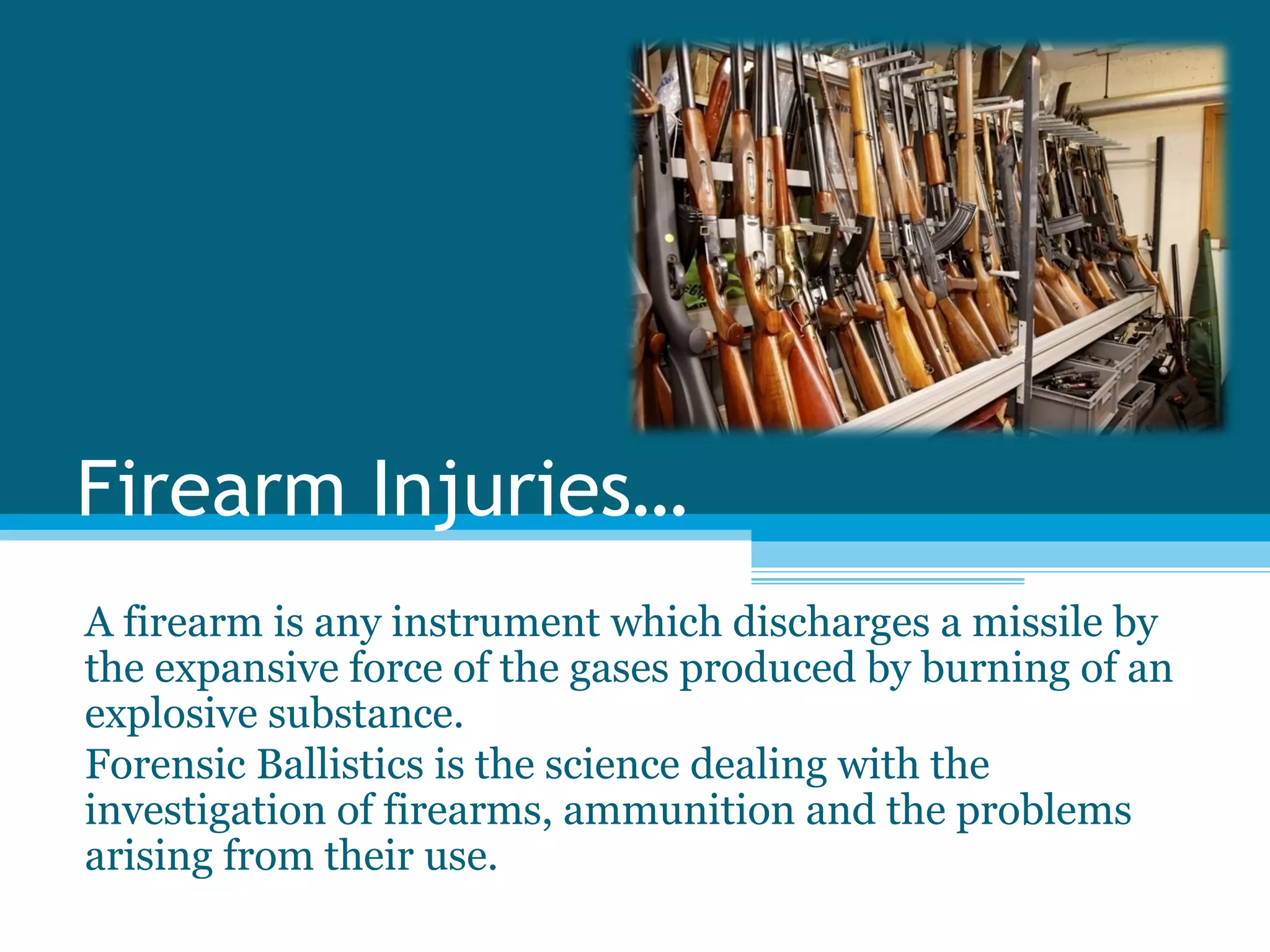
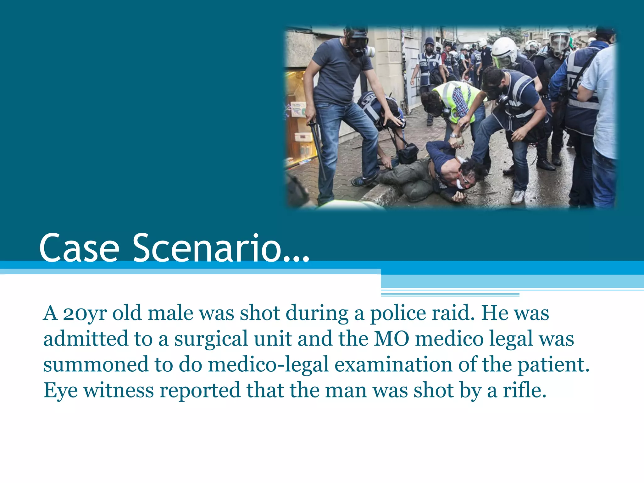
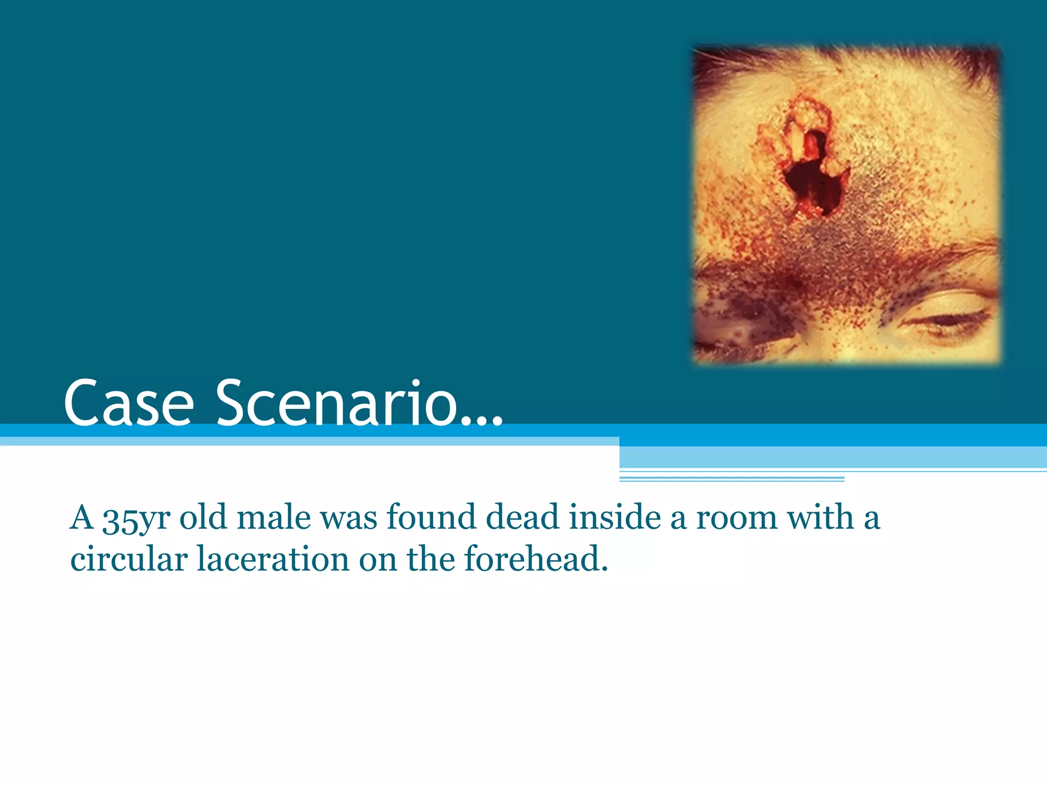
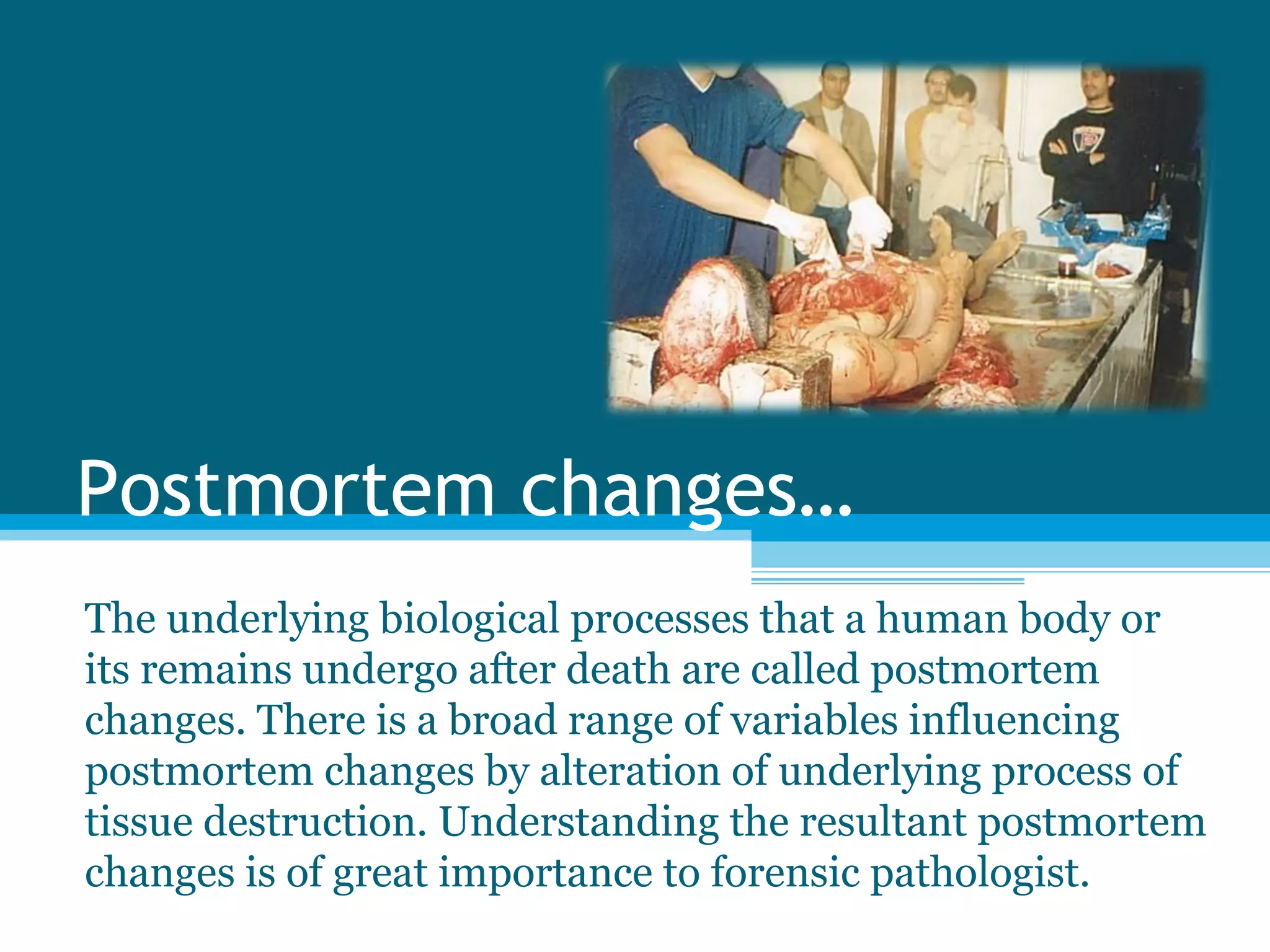
Fixed Learning Session - 3 
Intracranial injuries… 
Firearm & Blast injuries… 
Postmortem changes…
Objectives… 
You should utilize this FLS to achieve the following; 
 Intracranial injuries 
Identify the different features and causation of injuries. 
• Fire arm 
Identify the injuries caused by different types of fire arm. 
Identification of entry and exit wound 
Identify circumstances of firearm injury 
• Postmortem changes 
Identify the different types of postmortem changes at 
different time intervals.
Intracranial Injuries… 
The brain, its membranes and blood vessels are said to be 
the most fragile of all the organs of the body. That is why 
they are encased in a rigid bony box called the skull. 
Intracranial injuries include injury to the above soft tissues.
Objectives… 
You should utilize this FLS to achieve the following; 
 Intracranial injuries 
Identify the different features and causation of 
injuries.
Case Scenario… 
A 25 yr old pedestrian was knocked down by a car and 
thrown onto the road. She was admitted to the ward in 
unconscious state.
1. List the possible types of 
intracranial haemorrhages 
that may be observed in a 
patient with head injury.
1. List the possible types of 
intracranial haemorrhages 
that may be observed in a 
patient with head injury. 
1. Extradural haemorrhage 
(EDH) 
2. Subdural haemorrhage (SDH) 
3. Subarrachnoid haemorrhage 
(SAH) 
4. Intracerebral haemorrhage 
(ICH)
2. Identify the lesion seen in 
this photograph.
2. Identify the lesion seen in 
this photograph. 
Diffuse subarachnoid haemorrhage 
(SAH) over the cerebral 
hemispheres 
This subarachnoid haemorrhage 
has resulted from rupturing of 
bridging vessels during rotation of 
the brain.
3. What are the other 
possible causes for SAH?
4. What are the other 
possible causes for SAH? 
Extension of traumatic 
intracerebral haemorrhage 
Rupture of Berry aneurysm 
Rupture of the AV malformation 
Rupture/laceration of vertebral 
artery
Case Scenario… 
A patient who had a severe headache was admitted to the 
medical ward in an unconscious state. He was a 
hypertensive patient. Following abnormalities were 
detected during the autopsy.
1. Identify the abnormality 
pointed by the arrow.
1. Identify the abnormality 
pointed by the arrow. 
Ruptured berry aneurysm
2. What is the type of 
intracranial haemorrhage you 
would expect to see in this 
person?
2. What is the type of 
intracranial haemorrhage you 
would expect to see in this 
person? 
Basal subarchnoid haemorrhage
3. What is the most likely 
mechanism of death?
3. What is the most likely 
mechanism of death? 
Basal subarachnoid haemorrhage 
causes vide spread vascular spasm 
which may have an effect on vital 
centers of the brain stem causing 
sudden collapse and death.
Case Scenario… 
A 40yr old man was assaulted with a wooden pole over the 
right side of the head. On examination there was a 
laceration over the right temporo-parietal region. The Xray 
of the skull showed an underlying fracture. Despite the 
medical intervention the patient succumbed to the injuries.
1. Identify the intracranial 
haemorrhage shown by this 
photograph.
1. Identify the intracranial 
haemorrhage shown by this 
photograph. 
Extra dural haemorrhage
2. Name the responsible 
blood vessel causing above 
haemorrhage.
2. Name the responsible 
blood vessel causing above 
haemorrhage. 
Middle meningeal artery
3. Describe the clinical 
picture of this person.
3. Describe the clinical 
picture of this person. 
Lose of conciseness due to concussion 
Followed by recovery and “lucid 
interval” 
Gradual decoration of the level of 
consciousness
Fact file… 
Intracranial Haemorrhages…
Extradural haemorrhage… 
(EDH) 
This is the collection of blood 
between the skull and dura mater . 
Since the dura is closely applied to 
the skull in the base it does not occur 
on the base except on the posterior 
fossa. 
Most extradural haemorrhages are 
associated with skull fractures.
Extradural haemorrhage… 
(EDH) 
The classical clinical sign of 
extradural haemorrhage includes 
‘lucid interval’. 
A doctor may be accused of medical 
negligence if a patient is discharged 
from the hospital after the recovery of 
the concussion and died at home 
from complications of extradural 
haemorrhage.
Subdural haemorrhage… 
(SDH) 
This is collection of blood in the 
sudural space and it is much more 
common than extradural 
haemorrhage. 
It can be classified in to acute and 
chronic and commoner in both 
extremes of life.
Subdural haemorrhage… 
(SDH) 
In young it is infamously associated 
with fatal child abuse. 
Acute SDH is a common result of any 
significant head injury. Majority of 
SDH are associated with skull 
fractures.
Subarachnoid haemorrhage… 
(SAH) 
This is collection of blood in the 
subarachnoid space. 
The blood in the subarachnoid space 
tend to mix with CSF. Therefore 
unlike other haemorrhage less likely 
to clot.
Subarachnoid haemorrhage… 
(SAH) 
SAH can also occur as a result of 
natural causes such as rupture of 
berry aneurysm and arterior-venous 
malformations. 
Death from SAH may be 
instantaneous, particularly when 
there is haemorrhage around the 
brainstem.
Intracranial haemorrhage… 
(ICH) 
They can be either infiltrating brain 
tissue or forming actual 
haematoma in the brain tissue. 
Primary haemorrhages occur at the 
time of injury due to rupture of 
blood vessels. 
Secondary haemorrhage occur due 
to the injured area undergoing 
swelling, softening, liquefaction, 
and cavity formation . 
The ruptured vessels at the margins 
bleeds into the cavity.
Firearm Injuries… 
A firearm is any instrument which discharges a missile by 
the expansive force of the gases produced by burning of an 
explosive substance. 
Forensic Ballistics is the science dealing with the 
investigation of firearms, ammunition and the problems 
arising from their use.
Objectives… 
You should utilize this FLS to achieve the following; 
• Fire arm 
Identify the injuries caused by different types of fire 
arm 
Identification of entry and exit wound 
Identify circumstances of firearm injury
Fact file… 
There are 2 types of firearms…
2 types of firearms… 
Rifle Smooth bore
Case Scenario… 
A 50yr old male who was later revealed to be a leader of a 
local drug dealing gang was found dead at a junction of a 
road. There were multiple firearm injuries on the body of 
the victim. The victim was found to be shot from 
intermediate range.
1. How would you identify the type of firearm by examining the injuries ? 
A 
B
1. How would you identify the type of firearm by examining the injuries? 
Rifle (A) Smooth bore (B) 
Edge of the 
wound 
Circular hole with inverted 
margins 
Scalloped edges (rat hole) 
Satellite wounds Absent Present 
Wad mark Absent Present as a 
laceration/contusion below 
the main wound but not 
visible in this case 
Abrasion collar Present Absent 
Grease ring Present Absent
Case Scenario… 
A 20yr old male was shot during a police raid. He was 
admitted to a surgical unit and the MO medico legal was 
summoned to do medico-legal examination of the patient. 
Eye witness reported that the man was shot by a rifle.
1. What are the features that would help the MO medico-legal to 
differentiate an entry wound from an exit wound? 
A B
1. What are the features that would help the MO medico-legal to 
differentiate an entry wound from an exit wound? 
Entry wound (A) Exit wound (B) 
Shape Circular hole with inverted 
margins 
Stellate and everted 
Abrasion collar Present Absent 
Burning 
Blackening 
Tattooing 
May be present depending on 
the range of fire but not 
present in this case 
absent
Case Scenario… 
A 35yr old male was found dead inside a room with a 
circular laceration on the forehead.
1. Which of the following features will indicate this case as a firearm suicide? 
1. Room is locked from inside. 
1. Weapon is absent from the scene. 
1. Presence of more than one fatal injury. 
1. Elective site. 
1. Entry wound is compatible with the handedness. 
1. Injuries present in inaccessible area.
1. Which of the following features will indicate this case as a firearm suicide? 
1. Room is locked from inside. (T) 
1. Weapon is absent from the scene. (F) 
1. Presence of more than one fatal injury. (F) 
1. Elective site. (T) 
1. Entry wound is compatible with the handedness. (T) 
1. Injuries present in inaccessible area. (F)
Postmortem changes… 
The underlying biological processes that a human body or 
its remains undergo after death are called postmortem 
changes. There is a broad range of variables influencing 
postmortem changes by alteration of underlying process of 
tissue destruction. Understanding the resultant postmortem 
changes is of great importance to forensic pathologist.
Objectives… 
You should utilize this FLS to achieve the following; 
• Postmortem changes 
Identify the different types of postmortem changes at 
different time intervals.
Early postmortem changes… 
Include; 
•Rigor mortis 
•Hypostasis 
•Dilation of pupils 
•Dropping of intraocular pressure 
•Loss of skin elasticity
Case Scenario… 
The body of a 27yr old adult male was found inside a motor 
car with a running engine. Following picture was taken 
during the autopsy.
1. What could be the likely 
cause of death?
1. What could be the likely 
cause of death? 
CO poisoning
2. How would you confirm 
your diagnosis?
2. How would you confirm 
your diagnosis? 
Venous blood sample for CO level from 
femoral vein
Fact file… 
The colour of hypostasis may indicate the cause of death…
Different colours of hypostasis…
Case Scenario… 
A body of a 20 year old male was recovered from a river. 
Following photograph was taken during the autopsy.
1. Identify the postmortem 
change seen in this 
photograph.
1. Identify the postmortem 
change seen in this 
photograph. 
Cadaveric spasm 
Cadaveric/Postmortem spasm is a 
form of muscular stiffening that 
occurs at the time of death 
persisting into the period of rigor 
mortis. It is usually associated 
with violent deaths with intense 
emotion.
2. What is the medico-legal 
significance of this?
2. What is the medico-legal 
significance of this? 
It provides a clue that the 
individual was alive at the time of 
entering into the water.
Case Scenario… 
A 70 year old man who was living alone was found dead by 
the neighbors.
1. Identify the postmortem changes.
1. 1. Identify the postmortem 
changes 
Skin blisters 
Skin slippage 
Bloating of the body 
Colour change of the skin
1. Depending on the 
postmortem changes roughly 
estimate the time since 
death.
1. Depending on the 
postmortem changes roughly 
estimate the time since 
death. 
Time since death 48-72 hours
Late postmortem changes… 
Include; 
•Mummification 
•Adipocere formation 
•Skeletalization
Case Scenario… 
A body of a six (6) months old infant was exhumed for 
autopsy following allegations by relatives.
1. Identify the postmortem 
change seen in this 
photograph.
1. Identify the postmortem 
change seen in this 
photograph. 
Adipocere formation 
This is a rare phenomenon. 
The fat coverage of the 
body hydrolyse and 
hydrogenases. 
This forms a greyish white, 
greasy, friable substance 
with cheesy amonical smell. 
The time of onset is highly 
variable and is between 1- 
6/12.
2. What is the environmental 
condition that is favourable 
for this .
2. What is the environmental 
condition that is favourable 
for this . 
Warm and moist environment
2. List the important medico-legal 
significances of this 
change.
2. List the important medico-legal 
significances of this 
change. 
• Condition of disposal 
• Preserve external features 
• Identification of injuries 
• Time since death : Minimum 3 -6 
wks
Case Scenario… 
A body of an adult male was recovered from a desert like 
place. He went missing from home four (4) weeks ago.
1. Identify the postmortem 
change seen in this 
photograph.
1. Identify the postmortem 
change seen in this 
photograph. 
Mummification 
Needs hot dry and windy condition. 
Commonly seen in deserts. 
Tissues will dry and shrivelled 
preventing putrefaction. This 
results in stretching the skin. The 
condition may co –exists with 
adipocere.
2. List the important medico-legal 
significances of this 
change.
2. List the important medico-legal 
significances of this 
change. 
• Condition of disposal 
• Identification 
• Identification of injuries 
• Time since death - 2 - 4 wks
Identify the given 
postmortem artifact and click 
on the correct answer. 
Rat bites 
Ant bites 
Fish bites 
Predator attacks
Correct! 
Click here for the 
explanation.
Wrong Answer. 
Click here for the 
explanation.
Postmortem ant bites… 
Ants can be present at all stages of 
decomposition as they are typically 
observed shortly after death or during 
the early postmortem period. 
The feeding action of ants can cause 
many irregular scalloped areas of 
superficial skin loss, and small punctuate 
and scratch-type lesions with well 
defined margins. 
Usually ant injuries are orange-pink to 
yellow in color and diffusely scattered 
over the skin surface. 
These injuries can be easily 
misinterpreted as antemortem abrasions 
but the absence of inflammatory 
reactions helps to differentiate them 
from antemortem injuries.
Identify the given 
postmortem artifact and click 
on the correct answer. 
Rat bites 
Ant bites 
Fish bites 
Predator attacks
Correct! 
Click here for the 
explanation.
Wrong Answer. 
Click here for the 
explanation.
Predator attacks… 
There are multiple parallel claw 
marks with no vital reactions 
probably inflicted by a water 
monitor.
Now can you? 
 Identify the different features and causation of injuries. 
• Identify the injuries caused by different types of fire arm. 
• Identify the different types of postmortem changes at 
different time intervals.
Head injuries in forensic medicine

Head injuries in forensic medicine

  • 1.
    Fixed Learning Session- 3 Intracranial injuries… Firearm & Blast injuries… Postmortem changes…
  • 2.
    Objectives… You shouldutilize this FLS to achieve the following;  Intracranial injuries Identify the different features and causation of injuries. • Fire arm Identify the injuries caused by different types of fire arm. Identification of entry and exit wound Identify circumstances of firearm injury • Postmortem changes Identify the different types of postmortem changes at different time intervals.
  • 3.
    Intracranial Injuries… Thebrain, its membranes and blood vessels are said to be the most fragile of all the organs of the body. That is why they are encased in a rigid bony box called the skull. Intracranial injuries include injury to the above soft tissues.
  • 4.
    Objectives… You shouldutilize this FLS to achieve the following;  Intracranial injuries Identify the different features and causation of injuries.
  • 5.
    Case Scenario… A25 yr old pedestrian was knocked down by a car and thrown onto the road. She was admitted to the ward in unconscious state.
  • 6.
    1. List thepossible types of intracranial haemorrhages that may be observed in a patient with head injury.
  • 7.
    1. List thepossible types of intracranial haemorrhages that may be observed in a patient with head injury. 1. Extradural haemorrhage (EDH) 2. Subdural haemorrhage (SDH) 3. Subarrachnoid haemorrhage (SAH) 4. Intracerebral haemorrhage (ICH)
  • 8.
    2. Identify thelesion seen in this photograph.
  • 9.
    2. Identify thelesion seen in this photograph. Diffuse subarachnoid haemorrhage (SAH) over the cerebral hemispheres This subarachnoid haemorrhage has resulted from rupturing of bridging vessels during rotation of the brain.
  • 10.
    3. What arethe other possible causes for SAH?
  • 11.
    4. What arethe other possible causes for SAH? Extension of traumatic intracerebral haemorrhage Rupture of Berry aneurysm Rupture of the AV malformation Rupture/laceration of vertebral artery
  • 12.
    Case Scenario… Apatient who had a severe headache was admitted to the medical ward in an unconscious state. He was a hypertensive patient. Following abnormalities were detected during the autopsy.
  • 13.
    1. Identify theabnormality pointed by the arrow.
  • 14.
    1. Identify theabnormality pointed by the arrow. Ruptured berry aneurysm
  • 15.
    2. What isthe type of intracranial haemorrhage you would expect to see in this person?
  • 16.
    2. What isthe type of intracranial haemorrhage you would expect to see in this person? Basal subarchnoid haemorrhage
  • 17.
    3. What isthe most likely mechanism of death?
  • 18.
    3. What isthe most likely mechanism of death? Basal subarachnoid haemorrhage causes vide spread vascular spasm which may have an effect on vital centers of the brain stem causing sudden collapse and death.
  • 19.
    Case Scenario… A40yr old man was assaulted with a wooden pole over the right side of the head. On examination there was a laceration over the right temporo-parietal region. The Xray of the skull showed an underlying fracture. Despite the medical intervention the patient succumbed to the injuries.
  • 20.
    1. Identify theintracranial haemorrhage shown by this photograph.
  • 21.
    1. Identify theintracranial haemorrhage shown by this photograph. Extra dural haemorrhage
  • 22.
    2. Name theresponsible blood vessel causing above haemorrhage.
  • 23.
    2. Name theresponsible blood vessel causing above haemorrhage. Middle meningeal artery
  • 24.
    3. Describe theclinical picture of this person.
  • 25.
    3. Describe theclinical picture of this person. Lose of conciseness due to concussion Followed by recovery and “lucid interval” Gradual decoration of the level of consciousness
  • 26.
  • 27.
    Extradural haemorrhage… (EDH) This is the collection of blood between the skull and dura mater . Since the dura is closely applied to the skull in the base it does not occur on the base except on the posterior fossa. Most extradural haemorrhages are associated with skull fractures.
  • 28.
    Extradural haemorrhage… (EDH) The classical clinical sign of extradural haemorrhage includes ‘lucid interval’. A doctor may be accused of medical negligence if a patient is discharged from the hospital after the recovery of the concussion and died at home from complications of extradural haemorrhage.
  • 29.
    Subdural haemorrhage… (SDH) This is collection of blood in the sudural space and it is much more common than extradural haemorrhage. It can be classified in to acute and chronic and commoner in both extremes of life.
  • 30.
    Subdural haemorrhage… (SDH) In young it is infamously associated with fatal child abuse. Acute SDH is a common result of any significant head injury. Majority of SDH are associated with skull fractures.
  • 31.
    Subarachnoid haemorrhage… (SAH) This is collection of blood in the subarachnoid space. The blood in the subarachnoid space tend to mix with CSF. Therefore unlike other haemorrhage less likely to clot.
  • 32.
    Subarachnoid haemorrhage… (SAH) SAH can also occur as a result of natural causes such as rupture of berry aneurysm and arterior-venous malformations. Death from SAH may be instantaneous, particularly when there is haemorrhage around the brainstem.
  • 33.
    Intracranial haemorrhage… (ICH) They can be either infiltrating brain tissue or forming actual haematoma in the brain tissue. Primary haemorrhages occur at the time of injury due to rupture of blood vessels. Secondary haemorrhage occur due to the injured area undergoing swelling, softening, liquefaction, and cavity formation . The ruptured vessels at the margins bleeds into the cavity.
  • 34.
    Firearm Injuries… Afirearm is any instrument which discharges a missile by the expansive force of the gases produced by burning of an explosive substance. Forensic Ballistics is the science dealing with the investigation of firearms, ammunition and the problems arising from their use.
  • 35.
    Objectives… You shouldutilize this FLS to achieve the following; • Fire arm Identify the injuries caused by different types of fire arm Identification of entry and exit wound Identify circumstances of firearm injury
  • 36.
    Fact file… Thereare 2 types of firearms…
  • 37.
    2 types offirearms… Rifle Smooth bore
  • 38.
    Case Scenario… A50yr old male who was later revealed to be a leader of a local drug dealing gang was found dead at a junction of a road. There were multiple firearm injuries on the body of the victim. The victim was found to be shot from intermediate range.
  • 39.
    1. How wouldyou identify the type of firearm by examining the injuries ? A B
  • 40.
    1. How wouldyou identify the type of firearm by examining the injuries? Rifle (A) Smooth bore (B) Edge of the wound Circular hole with inverted margins Scalloped edges (rat hole) Satellite wounds Absent Present Wad mark Absent Present as a laceration/contusion below the main wound but not visible in this case Abrasion collar Present Absent Grease ring Present Absent
  • 41.
    Case Scenario… A20yr old male was shot during a police raid. He was admitted to a surgical unit and the MO medico legal was summoned to do medico-legal examination of the patient. Eye witness reported that the man was shot by a rifle.
  • 42.
    1. What arethe features that would help the MO medico-legal to differentiate an entry wound from an exit wound? A B
  • 43.
    1. What arethe features that would help the MO medico-legal to differentiate an entry wound from an exit wound? Entry wound (A) Exit wound (B) Shape Circular hole with inverted margins Stellate and everted Abrasion collar Present Absent Burning Blackening Tattooing May be present depending on the range of fire but not present in this case absent
  • 44.
    Case Scenario… A35yr old male was found dead inside a room with a circular laceration on the forehead.
  • 45.
    1. Which ofthe following features will indicate this case as a firearm suicide? 1. Room is locked from inside. 1. Weapon is absent from the scene. 1. Presence of more than one fatal injury. 1. Elective site. 1. Entry wound is compatible with the handedness. 1. Injuries present in inaccessible area.
  • 46.
    1. Which ofthe following features will indicate this case as a firearm suicide? 1. Room is locked from inside. (T) 1. Weapon is absent from the scene. (F) 1. Presence of more than one fatal injury. (F) 1. Elective site. (T) 1. Entry wound is compatible with the handedness. (T) 1. Injuries present in inaccessible area. (F)
  • 47.
    Postmortem changes… Theunderlying biological processes that a human body or its remains undergo after death are called postmortem changes. There is a broad range of variables influencing postmortem changes by alteration of underlying process of tissue destruction. Understanding the resultant postmortem changes is of great importance to forensic pathologist.
  • 48.
    Objectives… You shouldutilize this FLS to achieve the following; • Postmortem changes Identify the different types of postmortem changes at different time intervals.
  • 49.
    Early postmortem changes… Include; •Rigor mortis •Hypostasis •Dilation of pupils •Dropping of intraocular pressure •Loss of skin elasticity
  • 50.
    Case Scenario… Thebody of a 27yr old adult male was found inside a motor car with a running engine. Following picture was taken during the autopsy.
  • 51.
    1. What couldbe the likely cause of death?
  • 52.
    1. What couldbe the likely cause of death? CO poisoning
  • 53.
    2. How wouldyou confirm your diagnosis?
  • 54.
    2. How wouldyou confirm your diagnosis? Venous blood sample for CO level from femoral vein
  • 55.
    Fact file… Thecolour of hypostasis may indicate the cause of death…
  • 56.
    Different colours ofhypostasis…
  • 57.
    Case Scenario… Abody of a 20 year old male was recovered from a river. Following photograph was taken during the autopsy.
  • 58.
    1. Identify thepostmortem change seen in this photograph.
  • 59.
    1. Identify thepostmortem change seen in this photograph. Cadaveric spasm Cadaveric/Postmortem spasm is a form of muscular stiffening that occurs at the time of death persisting into the period of rigor mortis. It is usually associated with violent deaths with intense emotion.
  • 60.
    2. What isthe medico-legal significance of this?
  • 61.
    2. What isthe medico-legal significance of this? It provides a clue that the individual was alive at the time of entering into the water.
  • 62.
    Case Scenario… A70 year old man who was living alone was found dead by the neighbors.
  • 63.
    1. Identify thepostmortem changes.
  • 64.
    1. 1. Identifythe postmortem changes Skin blisters Skin slippage Bloating of the body Colour change of the skin
  • 65.
    1. Depending onthe postmortem changes roughly estimate the time since death.
  • 66.
    1. Depending onthe postmortem changes roughly estimate the time since death. Time since death 48-72 hours
  • 67.
    Late postmortem changes… Include; •Mummification •Adipocere formation •Skeletalization
  • 68.
    Case Scenario… Abody of a six (6) months old infant was exhumed for autopsy following allegations by relatives.
  • 69.
    1. Identify thepostmortem change seen in this photograph.
  • 70.
    1. Identify thepostmortem change seen in this photograph. Adipocere formation This is a rare phenomenon. The fat coverage of the body hydrolyse and hydrogenases. This forms a greyish white, greasy, friable substance with cheesy amonical smell. The time of onset is highly variable and is between 1- 6/12.
  • 71.
    2. What isthe environmental condition that is favourable for this .
  • 72.
    2. What isthe environmental condition that is favourable for this . Warm and moist environment
  • 73.
    2. List theimportant medico-legal significances of this change.
  • 74.
    2. List theimportant medico-legal significances of this change. • Condition of disposal • Preserve external features • Identification of injuries • Time since death : Minimum 3 -6 wks
  • 75.
    Case Scenario… Abody of an adult male was recovered from a desert like place. He went missing from home four (4) weeks ago.
  • 76.
    1. Identify thepostmortem change seen in this photograph.
  • 77.
    1. Identify thepostmortem change seen in this photograph. Mummification Needs hot dry and windy condition. Commonly seen in deserts. Tissues will dry and shrivelled preventing putrefaction. This results in stretching the skin. The condition may co –exists with adipocere.
  • 78.
    2. List theimportant medico-legal significances of this change.
  • 79.
    2. List theimportant medico-legal significances of this change. • Condition of disposal • Identification • Identification of injuries • Time since death - 2 - 4 wks
  • 80.
    Identify the given postmortem artifact and click on the correct answer. Rat bites Ant bites Fish bites Predator attacks
  • 81.
    Correct! Click herefor the explanation.
  • 82.
    Wrong Answer. Clickhere for the explanation.
  • 83.
    Postmortem ant bites… Ants can be present at all stages of decomposition as they are typically observed shortly after death or during the early postmortem period. The feeding action of ants can cause many irregular scalloped areas of superficial skin loss, and small punctuate and scratch-type lesions with well defined margins. Usually ant injuries are orange-pink to yellow in color and diffusely scattered over the skin surface. These injuries can be easily misinterpreted as antemortem abrasions but the absence of inflammatory reactions helps to differentiate them from antemortem injuries.
  • 84.
    Identify the given postmortem artifact and click on the correct answer. Rat bites Ant bites Fish bites Predator attacks
  • 85.
    Correct! Click herefor the explanation.
  • 86.
    Wrong Answer. Clickhere for the explanation.
  • 87.
    Predator attacks… Thereare multiple parallel claw marks with no vital reactions probably inflicted by a water monitor.
  • 88.
    Now can you?  Identify the different features and causation of injuries. • Identify the injuries caused by different types of fire arm. • Identify the different types of postmortem changes at different time intervals.