

Recommended
More Related Content
What's hot
What's hot (20)
Similar to Selaginella: features, morphology ,anatomy and reproduction.
Similar to Selaginella: features, morphology ,anatomy and reproduction. (20)
More from Cherry
More from Cherry (20)
Recently uploaded
Recently uploaded (20)
Selaginella: features, morphology ,anatomy and reproduction.
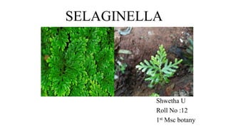
- 1. SELAGINELLA Shwetha U Roll No :12 1st Msc botany
- 2. SELAGINELLA Systematic position class : Lycopodiopsida order : Selaginellales family : Selaginellaceae genus : Selaginella
- 3. SELAGINELLA spike moss or club moss Habitat • About 700 species of selaginella are distributed in the tropical and temperature areas. • Most of the selaginella species grow in damp shady areas . • Some species are xerophytes. e.g : S. lepidophylla, S. rupestris • Some are epiphytic eg: s.oregana • Common species S. rupestris S. megaphylla S. kraussiana S. bryopteris
- 4. MORPHOLOGY OF SELAGINELLA • The plant body is sporophyte and differentiated into: Root Stem Leaves Ligules Rhizophore
- 5. ROOT • The root is adventitious grow from the tip of rhizophore or directly from the stem. • Root are arise endogenously and branched dichotomously. • Aerial root contain cap.(in epiphyte) • Root hair is present.
- 6. STEM • Stem is green, herbaceous, dichotomously branched , erect or prostrate with erect branches. • Stem is covered with leaves. • They are also pseudomonopodia ( false growth from one point). • The shoot apex consists of a single apical cell in most cases.
- 7. LEAVES • Leaves are microphyllous with a single unbranched midrib. • On the upper surface a small, membranous, tongue like outgrowth called ligule is present. • The leaves are dorsiventral.
- 8. • Two types of leaf condition: 1. Homoeophyllum or (isophyllous) 2. Heterophyllum or (anisophyllous) Homoeophyllum Heterophyllum Leaves in a plant are all one kind such condition is called is isophylly and the leaves are called isophyllous. Leaves of plants are two kinds: 1. Small , 2. Large such condition is called anisophylly and the leaves are called anisophyllous
- 9. LIGULE • On the upper surface a small, membranous, tongue like or leaf like outgrowth called ligule present on the adaxial side of the leaf. • A mature ligule has a prominent basal portion called the glossopodium. • Definite function of the ligule is unknown.
- 10. RHIZOPHORE • It is a colourless, leafless, unbranched and cylindrical structure. • This structure arises from the prostrate axis at the point of dichotomy and elongates downward. • free end of rhizophore touches the soil it develops a tuft of adventitious roots . • Root cap is absent . It develops adventitious root at its tip.
- 11. ANATOMY OF SELAGINELLA Stem anatomy Leaf anatomy Ligule anatomy Root anatomy Rhizophore anatomy
- 12. STEM ANATOMY • Epidermis is without hair and stomata. It is surrounded by a cuticle. • Epidermis is followed by a well defined cortex region. It is parenchymatous or is differentiated into outer sclerenchymatous and inner parenchymatous regions. • Stele is generally protostelic, where xylem is surrounded by phloem cells. The number of steles varies in different species. • Central portion is separated from cortex by a cavity having air spaces • Endodermis :the cortex and central tissue is connected by radially elongated cells called trabeculae. • Stele is surrounded by a pericycle that is single layered. Pith is generally absent.
- 14. LEAF ANATOMY • Epidermis single layered on the lower and upper side stoma is present. • Stomata present on the upper surface .Epidermis contain chloroplast. • Mesophyll is uniformly formed either all of spongy or all palisade like elongated cells with air spaces • Simple vascular bundle at the center.
- 15. LIGULE ANATOMY • The ligule arise from several short rows of superficial cells. • Fully developed ligule consists of a distinct and hemispherical basal region where cells are large and thin walled and contain vacuolated cytoplasam this region is glossopodium. • Glossopodium surrounded by a sheath called glossopodium sheath.
- 16. ROOT ANATOMY • Outermost layer is epidermis ( large single layer ), covered by cuticle. • Root hairs are present and arise from some epidermal cells. • Lower region of the epidermis, wide zone of cortex is present 1. Outer hypodermis ( have sclerenchyma cells) 2. Inner parenchyma cells. • Endodermis layer is present ( inconspicuous ) • Single layered pericycle is present just below the endodermis. • Xylem is surrounded by phloem
- 18. RHIZOPHORE ANATOMY • Outermost layer is epidermis. It is of thick walled and single layer cells. • Root hairs are absent. • Just below the epidermis there is cortex region 1. Hypodermis ( thick walled) few layered. 2. Thin walled parenchymatous region • After cortex region endodermis layer around the pericycle. • Thin walled pericycle is present around the vascular tissue. • Stele is protostele ( xylem is in center and phloem surrounds the xylem)
- 20. REPRODUCTION 1.Vegetative Reproduction i. Fragmentation ii. Tubers iii. Resting Buds 2. Sexual reproduction / gametophyte
- 21. VEGETATIVE REPRODUCTION • Fragmentation Under humid conditions in S. rupestris,branches of the stem develop some roots . These branches later disjoin from the parent plant and develop into separate individual plants. • Tubers These appear towards the end of the growing season. The tubers may be aerial, developing at the tips of underground branches (e.g., S. chrysocaulos) (e.g., S. chrysorrhizos). Under favourable conditions tubers germinate into a new plant.
- 22. • Resting Buds The resting buds develop at the tip of some aerial branches. They pass the unfavourable conditions and develop rhizophore in the favourable conditions.
- 23. Sexual reproduction by spores • Selaginella is a sporophytic plant and reproduce sexually. • Selaginella is a heterosporous i.e., produce two different types of spores— megaspores and microspores. • These spores are produced in megasporangia and microsporangia, respectively which, in turn, are produced on fertile leaves known as megasporophylls and microsporophylls respectively. • Usually both these structures are grouped together to form a compact structure known as strobilus which is usually a terminal structure.
- 24. STROBILUS: It is a reproductive structure formed by the aggregation of ligulate sporophylls at the apex of the branches of stem. The length of the strobilus varies from 1/4 inch to 2-3 inches in different species. In some species as for e.g., S cuspidata, S. patula etc. the growth of the stem continues beyond the strobilus and such condition is called selago condition
- 26. SPORANGIA • Mature sporangia is a stalked structure consist short stalk and capsule. • Stalk is multicellular and multiseriate , capsule is 2 layered wall called jacket. • Two types of sporangia: 1. Megasporangia (large and pale) 2. Microsporangia (small , slightly elongated )
- 27. Development of sporangia • Sporangium development is of the eusporangiate type. Development of megasporangia and microsporangia similar up to formation of spore mother cell. • Sporangia initial present in axil of the leaf. It divides to form outer jacket initial and inner archesporial initials. • The archesporial initial divides to form mass of sporogenous tissue. outer most layer of sporogenous tissue forms tapetum. • Jacket initial by further division rise to a jacket layer . • All sporogenous cells in the microsporangium become spore mother cells. SMC separate from each other and they under go meiosis to form microspores and several SMC produce megasporangium. • But only one SMC is functional rest are disintegrate. The spore mother cell divide meiotically to produce 4 megaspore. • The development of megaspore started before their shedding from the sporangia.
- 29. Gametophytic generation Spores and gametophyte • Spore is the first cell of gametophytic generation and they are haploid and uninucleate. • Selaginella is heterosporous they produce two types spores : 1. Microspores it produce microsporangium: Tetrahedral , two layered outer exine and inner intine. Exine thick and intine thin 2. Megaspores it produce megasporangium: Tetrahedral with triradiate ridge and spore coat is thick it may be 2or3 layered. If it is 2 layered outer exine and inner intine. If it is 3 layered outer exine ,middle mesospore and inner endospore. exine layer is thick and inner intine thin.
- 30. GERMINATION • Spore germination takes place by segmentation . • Initial stage of germination is spores remain in sporangia. • Thus, spores are shed as multicellular gametophyte. • Microspores germinate to form : (Male) microgametophyte (Female) megagametophyte
- 31. Germination of microspore and development of microgametophyte • Microspore is first divides unequally . • Resulting form small prothalli cell and large antheridial initial. • Prothalli cell does not under go division simply become vegetative part of prothallus. • Antheridial cell undergo division until 12 cell formed.4 of them form a central core and other are arranged around them. • The central cells called primary androgonial cell and marginal cell called jacket cells. • In this stage microgametophyte consist 13 cells namely 4 androgonial cell,1 prothallial cell,8 jacket cell.
- 32. Germination of megaspore and development of megagametophyte • Megaspore nucleus divides many times forming a coenocytic mass of numerous free nuclei. • Now wall formation begins between the nuclei of the apical part and a cushion like cellular mass is formed in the part. • Below this tissue the gametophyte is still in the free nuclear coenocytic stage. • Most of the superficial cells in the apical cushion become archegonial initial. • Few archegonial initial appear in the apical region
- 33. FERTILIZATION • Water is necessary to carry out the process of fertilization. • The swimming antherozoids reach the egg through the neck of archegonium and the nucleus of antherozoid fuses with the egg nucleus thus forming a zygotic nucleus. • The fertilized egg secretes a wall around it forming a diploid structure known as zygote or oospore (2x). • Thus the gametophytic generation ends and the initial stage of sporophytic generation is formed.
- 34. Embryo Development (Young Sporophyte): • Oospore or zygote is the initial stage of sporophytic generation. • oospore first divides by a transverse division into an upper suspensor initial (epibasal) and a lower embryo initial (hypobasal) . • The suspensor initial further divides in all directions forming a multicellular suspensor . • The embryo initial divides by 2 vertical divisions at right angle to each other thus forming 4 cells . • One of these 4 cells divides by an oblique wall forming a shoot initial. • Now the cells except the shoot initial divide sporophyte transversely forming 2 tiers of 4 cells each.