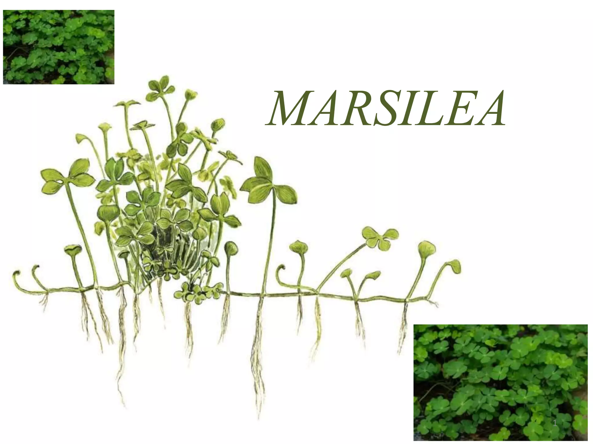
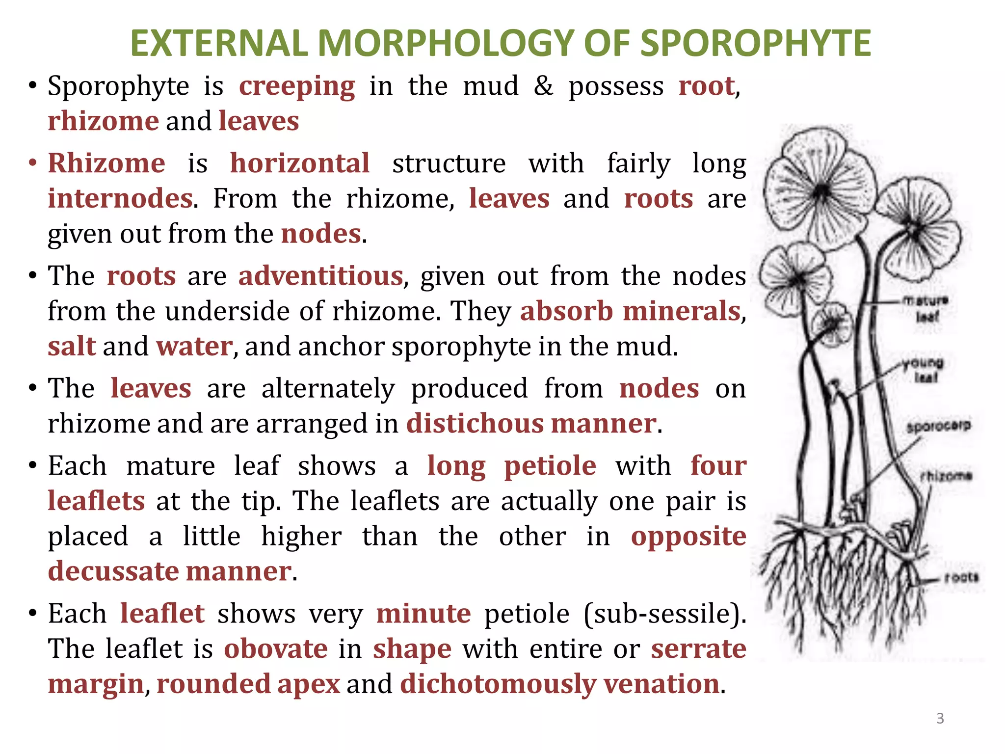
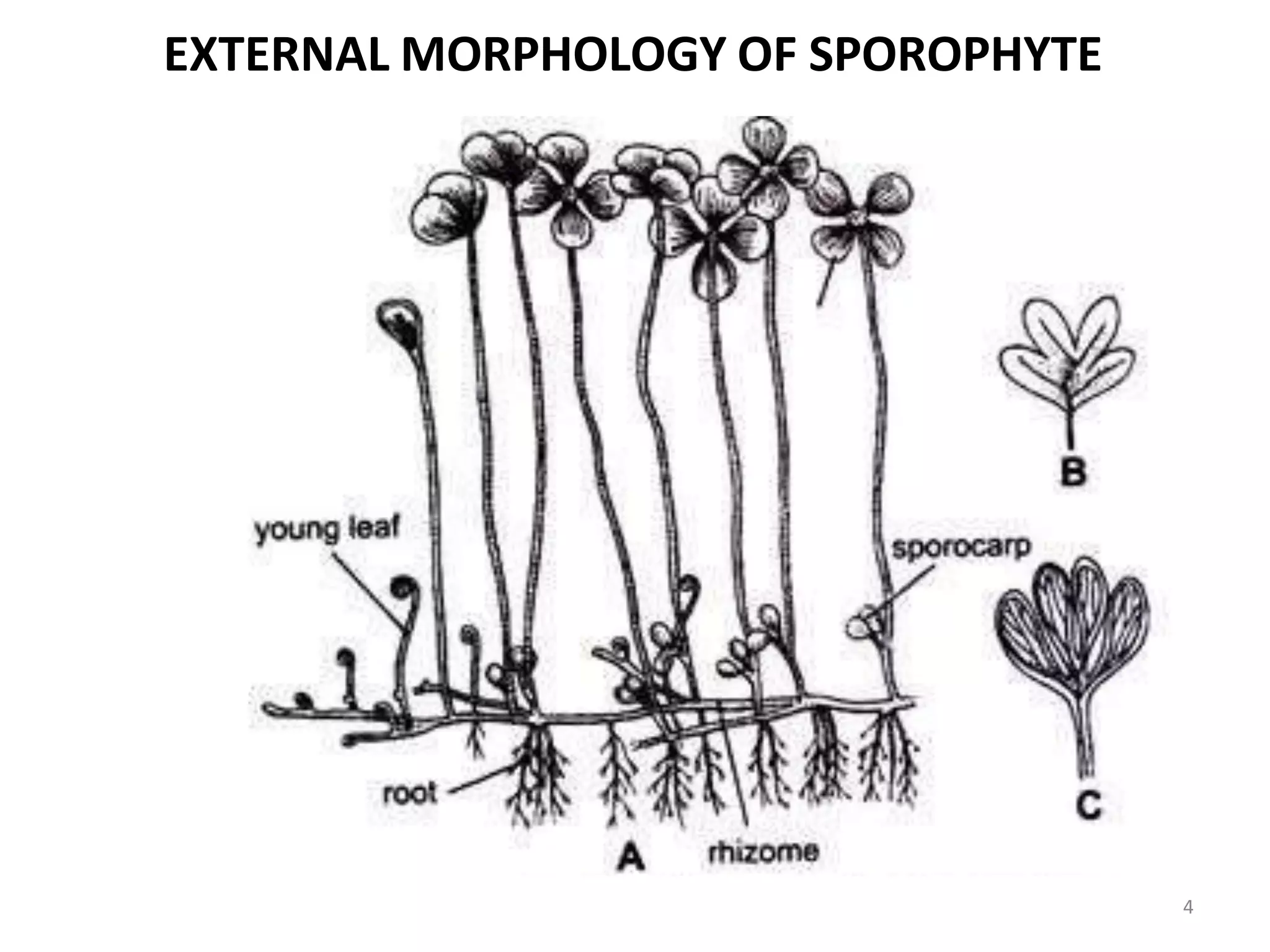
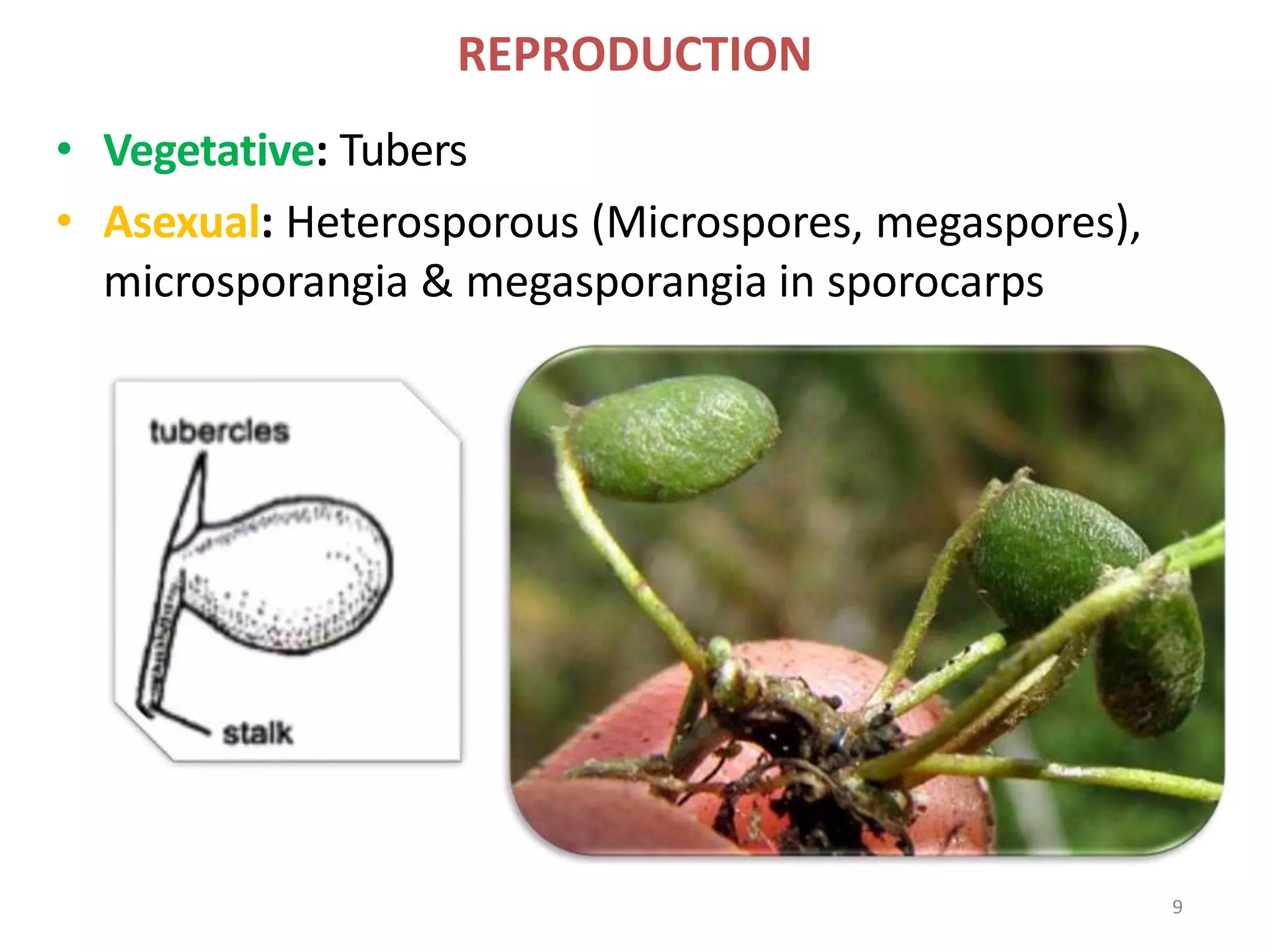
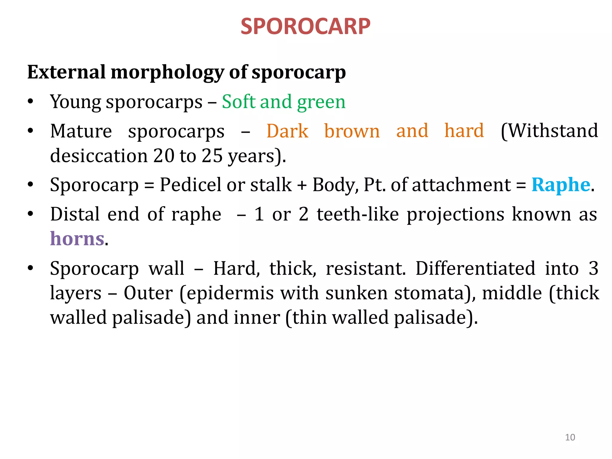
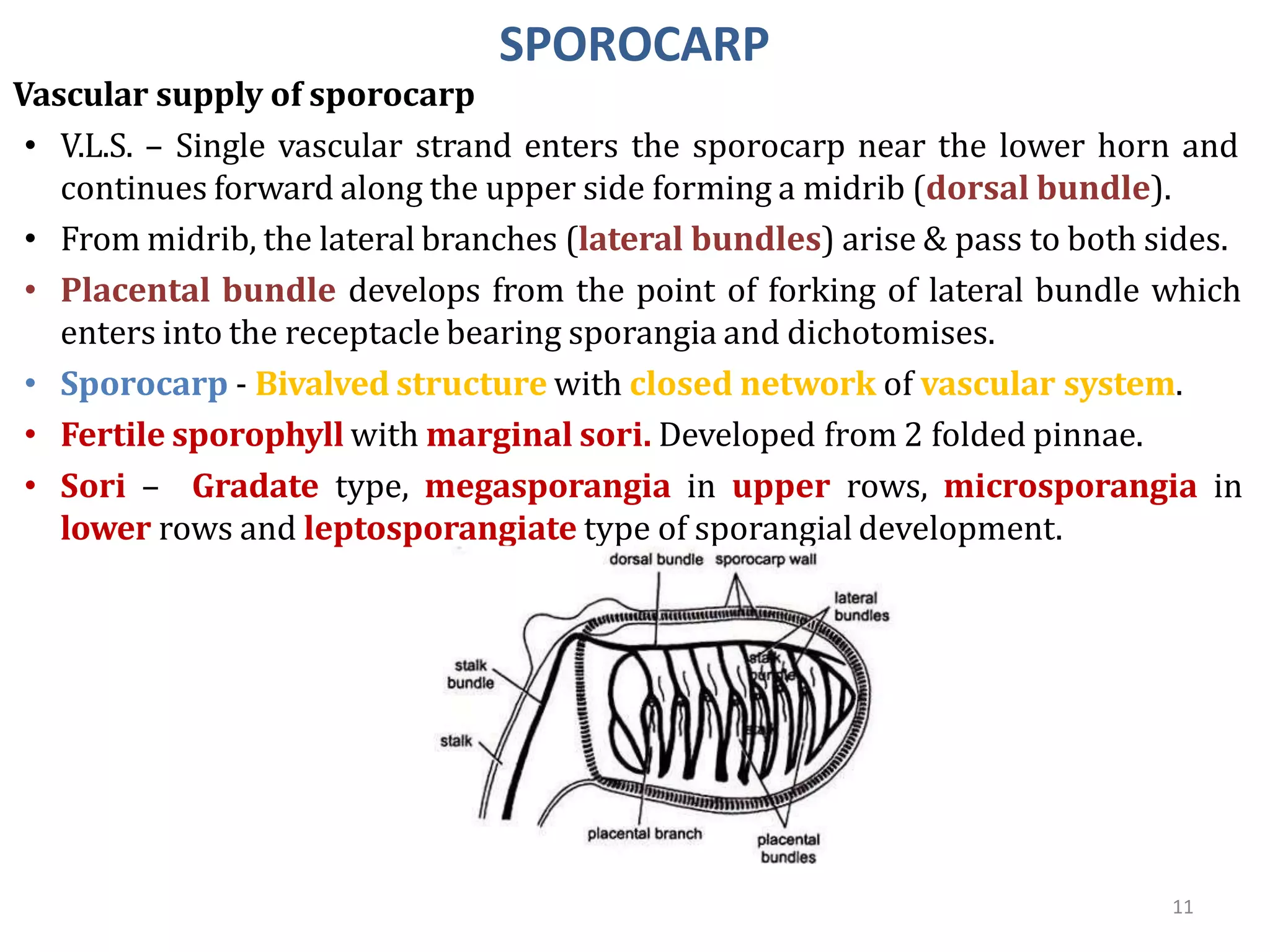
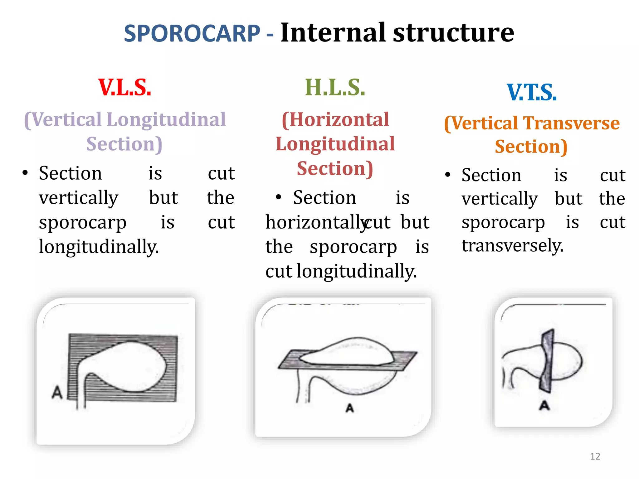
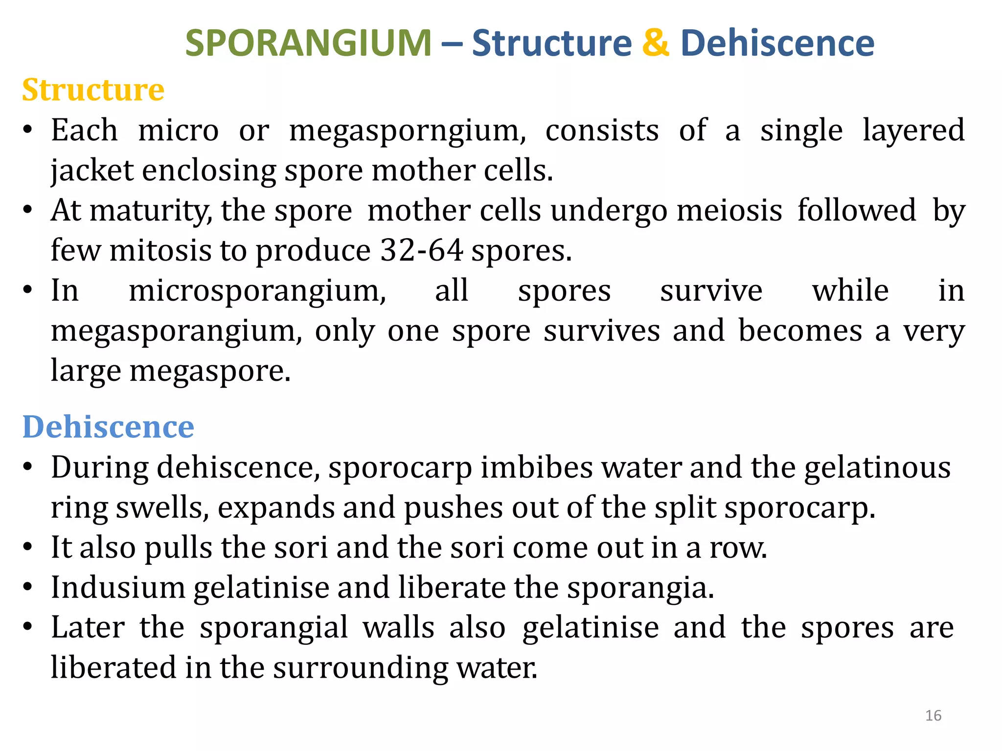
MARSILEA
1
Classification of marsilea
Division :pterophyta
Class: leptosporangiopsida
Order:Marisileales
Family:Marsieaceae
Genus:Marsilea
EXTERNAL MORPHOLOGY OF SPOROPHYTE
• Sporophyte is creeping in the mud & possess root,
rhizome and leaves
• Rhizome is horizontal structure with fairly long
internodes. From the rhizome, leaves and roots are
given out from the nodes.
• The roots are adventitious, given out from the nodes
from the underside of rhizome. They absorb minerals,
salt and water, and anchor sporophyte in the mud.
• The leaves are alternately produced from nodes on
rhizome and are arranged in distichous manner.
• Each mature leaf shows a long petiole with four
leaflets at the tip. The leaflets are actually one pair is
placed a little higher than the other in opposite
decussate manner.
• Each leaflet shows very minute petiole (sub-sessile).
The leaflet is obovate in shape with entire or serrate
margin, rounded apex and dichotomously venation.
3
EXTERNAL MORPHOLOGY OF SPOROPHYTE
4
INTERNAL MORPHOLOGY OF SPOROPHYTE
exarch xylem. 5
T.S. OF ROOT:
• Epidermis
• Cortex: Outer
aeparenchymatous
cortex, middle
parenchymatous
cortex & inner
cortex
sclerenchymatous.
• Stele:
Endodermis,
pericycle, radial
vascular bundles
with diarch &
INTERNAL MORPHOLOGY OF SPOROPHYTE
T.S. OF RHIZOME:
• Epidermis
• Cortex:
parenchymatous
with a ring
Outer
cortex
of air
chambers
(aerenchymatous), inner
parenchymatous cortex
with sclerenchymatous
patch.
• Stele: Amphiphloic
centre and can
parenchymatous
submerged plants)
siphonostele.
• Pith: Present in the
be
(in
or
sclerenchymatous
(terrestrial plants)
5
INTERNAL MORPHOLOGY OF SPOROPHYTE
T.S. OF PETIOLE:
• Epidermis
• Cortex: Outer cortex
parenchymatous,
Middle cortex
aerenchymatous
(septate), inner cortex
parenchymatous.
• Stele: Protostele
(plectostelic). Xylem
consists of two plates
with metaxylem in the
centre & protoxylem
towards the periphery.
7
INTERNAL MORPHOLOGY OF SPOROPHYTE
8
T.S. OF LEAFLET:
• Upper epidermis
• Mesophyll: Isobilateral (in the submerged species),
dorsiventral with aerenchyama (in terrestrial species)
• Vascular bundles: Concentric vascular bundles arranged
in parallel series.
• Lower epidermis
REPRODUCTION
• Vegetative: Tubers
• Asexual: Heterosporous (Microspores, megaspores),
microsporangia & megasporangia in sporocarps
9
10
SPOROCARP
External morphology of sporocarp
• Young sporocarps – Soft and green
• Mature sporocarps – Dark brown
desiccation 20 to 25 years).
and hard (Withstand
• Sporocarp = Pedicel or stalk + Body, Pt. of attachment = Raphe.
• Distal end of raphe – 1 or 2 teeth-like projections known as
horns.
• Sporocarp wall – Hard, thick, resistant. Differentiated into 3
layers – Outer (epidermis with sunken stomata), middle (thick
walled palisade) and inner (thin walled palisade).
SPOROCARP
Vascular supply of sporocarp
• V.L.S. – Single vascular strand enters the sporocarp near the lower horn and
continues forward along the upper side forming a midrib (dorsal bundle).
• From midrib, the lateral branches (lateral bundles) arise & pass to both sides.
• Placental bundle develops from the point of forking of lateral bundle which
enters into the receptacle bearing sporangia and dichotomises.
• Sporocarp - Bivalved structure with closed network of vascular system.
• Fertile sporophyll with marginal sori. Developed from 2 folded pinnae.
• Sori – Gradate type, megasporangia in upper rows, microsporangia in
lower rows and leptosporangiate type of sporangial development.
11
SPOROCARP - Internal structure
V.L.S.
(Vertical Longitudinal
Section)
• Section
vertically
sporocarp
is cut
but the
is cut
longitudinally.
V.T.S.
(Vertical Transverse
Section)
• Section is cut
vertically but the
sporocarp is cut
transversely.
H.L.S.
(Horizontal
Longitudinal
Section)
horizontally
the sporocarp
• Section is
cut but
is
cut longitudinally.
12
SPOROCARP - Internal structure
Vertical longitudinal
section (V.L.S.)
• Many sori arranged in
vertical rows.
• Either megasporangia or
microsporangia are
visible.
• Each sorus is
surrounded by an
indusium.
• The development of sori
is of gradate type.
• The gelatinous mucilage
ring is more prominent
in dorsal side.
13
SPOROCARP - Internal structure
and ventral sides. 14
Horizontal Longitudinal
Section (H.L.S.)
• Each sorus
transversely
• Sorus is an
is cut
elongated
structure, covered by a
delicate indusium.
• Sori are of gradate type,
in basipetal pattern.
• Sorus consists of a row
of megasporangia at top
and two rows of
microsporangia on
either sides.
• Mucilage ring is present
in two masses on dorsal
SPOROCARP - Internal structure
Vertical transverse section (V.T.S.)
• Two sori opposite to each other.
• Each sorus shows many megasporangia in the middle while 1-2
microsporangia at the ends.
• The mucilage ring is present only on the dorsal side.
15
16
SPORANGIUM – Structure & Dehiscence
Structure
• Each micro or megasporngium, consists of a single layered
jacket enclosing spore mother cells.
• At maturity, the spore mother cells undergo meiosis followed by
few mitosis to produce 32-64 spores.
• In microsporangium, all spores survive while in
megasporangium, only one spore survives and becomes a very
large megaspore.
Dehiscence
• During dehiscence, sporocarp imbibes water and the gelatinous
ring swells, expands and pushes out of the split sporocarp.
• It also pulls the sori and the sori come out in a row.
• Indusium gelatinise and liberate the sporangia.
• Later the sporangial walls also gelatinise and the spores are
liberated in the surrounding water.
17
GAMETOPHYTE – Male
Microspore and male gametophyte
• Microspore – Yellowish, spherical haploid with a triradiate ridge &
consists of uninucleate cytoplasm is surrounded by spore wall.
• Spore wall – 2 layered – endosporium & exosporium.
• Spore absorbs water and increase considerably in size.
• Nucleus divides to form a small prothalial cell & large apical cell.
• Apical cell divides to form two antheridial initials.
• Antheridial initial – few jacket cells externally & one spermatogenous
cell internally.
• Spermatogenous cell forms 16 androcytes which represent one
antheridium.
• At this stage, prothalial cell and jacket disintegrate and two groups of
androcytes remain free but within the microspore.
• Each androcyte now develops into multiciliated, coiled spermatozoid
with vesicle at one end.
• Thus, development of male gametophyte is endosporic.
GAMETOPHYTE – Male
18
19
GAMETOPHYTE – Female
Megaspore and female gametophyte
• Mature megaspore is elliptical with a short papilla at one end.
• Spore wall – 2 layered – endosporium & exosporium.
• Papilla is surrounded only by endosporium.
• Nucleus is located in the apical papilla and is surrounded by a dense cytoplasm.
• The rest of the spore is filled with watery cytoplasm & food.
• Megaspore germinates to give rise to female gametophyte. The development is
endosporic.
• Apical nucleus is divided into unequal nuclei. One nucleus remains in the dense
cytoplasm while the larger one migrates to watery cytoplasm.
• Transverse wall is formed at the base of the papilla separating the upper small
cell and lower larger cell called prothalial cell.
• Prothalial cell do not divide further and acts as nutritive cell.
• Upper cell soon develops an archegonium with a short neck and venter. The
neck has single neck canal cell surrounded by jacket made up of two tiers of four
cells each.
• Venter contains a venter canal cell. At maturity, megaspore absorbs water, the tip
of the megaspore splits in tri-radiate fissure and the archegonium is exposed.
• Female gametophyte is surrounded by a gelatinous mass & a funnel shaped
opening at the top.
GAMETOPHYTE – Fertilization
20
• During fertilization, spermatozoids or male gametes are liberated
from ruptured male gametophyte and are attracted by the chemical
substances present in the gelatinous mass.
• Movement of spermatozoids under the influence of chemical
substances is called chemotaxis.
• Spermatozoids swarm around and enter the gelatinous matrix of the
female gametophyte. Only one spermatozoid enters the open neck
and fertilizes the egg to form diploid zygote.
SPOROPHYTE – Development
• Zygote develops into embryo. Embryo shows cotyledons and roots
developed from upper half of the embryo while stem and foot
developed from lower half of embryo.
• The cotyledon comes out from calyptra. The rhizoids are developed
at first on the root. Very soon the embryo settles in the mud and

Marsilea.pptx

  • 1.
  • 2.
    Classification of marsilea Division:pterophyta Class: leptosporangiopsida Order:Marisileales Family:Marsieaceae Genus:Marsilea
  • 3.
    EXTERNAL MORPHOLOGY OFSPOROPHYTE • Sporophyte is creeping in the mud & possess root, rhizome and leaves • Rhizome is horizontal structure with fairly long internodes. From the rhizome, leaves and roots are given out from the nodes. • The roots are adventitious, given out from the nodes from the underside of rhizome. They absorb minerals, salt and water, and anchor sporophyte in the mud. • The leaves are alternately produced from nodes on rhizome and are arranged in distichous manner. • Each mature leaf shows a long petiole with four leaflets at the tip. The leaflets are actually one pair is placed a little higher than the other in opposite decussate manner. • Each leaflet shows very minute petiole (sub-sessile). The leaflet is obovate in shape with entire or serrate margin, rounded apex and dichotomously venation. 3
  • 4.
  • 5.
    INTERNAL MORPHOLOGY OFSPOROPHYTE exarch xylem. 5 T.S. OF ROOT: • Epidermis • Cortex: Outer aeparenchymatous cortex, middle parenchymatous cortex & inner cortex sclerenchymatous. • Stele: Endodermis, pericycle, radial vascular bundles with diarch &
  • 6.
    INTERNAL MORPHOLOGY OFSPOROPHYTE T.S. OF RHIZOME: • Epidermis • Cortex: parenchymatous with a ring Outer cortex of air chambers (aerenchymatous), inner parenchymatous cortex with sclerenchymatous patch. • Stele: Amphiphloic centre and can parenchymatous submerged plants) siphonostele. • Pith: Present in the be (in or sclerenchymatous (terrestrial plants) 5
  • 7.
    INTERNAL MORPHOLOGY OFSPOROPHYTE T.S. OF PETIOLE: • Epidermis • Cortex: Outer cortex parenchymatous, Middle cortex aerenchymatous (septate), inner cortex parenchymatous. • Stele: Protostele (plectostelic). Xylem consists of two plates with metaxylem in the centre & protoxylem towards the periphery. 7
  • 8.
    INTERNAL MORPHOLOGY OFSPOROPHYTE 8 T.S. OF LEAFLET: • Upper epidermis • Mesophyll: Isobilateral (in the submerged species), dorsiventral with aerenchyama (in terrestrial species) • Vascular bundles: Concentric vascular bundles arranged in parallel series. • Lower epidermis
  • 9.
    REPRODUCTION • Vegetative: Tubers •Asexual: Heterosporous (Microspores, megaspores), microsporangia & megasporangia in sporocarps 9
  • 10.
    10 SPOROCARP External morphology ofsporocarp • Young sporocarps – Soft and green • Mature sporocarps – Dark brown desiccation 20 to 25 years). and hard (Withstand • Sporocarp = Pedicel or stalk + Body, Pt. of attachment = Raphe. • Distal end of raphe – 1 or 2 teeth-like projections known as horns. • Sporocarp wall – Hard, thick, resistant. Differentiated into 3 layers – Outer (epidermis with sunken stomata), middle (thick walled palisade) and inner (thin walled palisade).
  • 11.
    SPOROCARP Vascular supply ofsporocarp • V.L.S. – Single vascular strand enters the sporocarp near the lower horn and continues forward along the upper side forming a midrib (dorsal bundle). • From midrib, the lateral branches (lateral bundles) arise & pass to both sides. • Placental bundle develops from the point of forking of lateral bundle which enters into the receptacle bearing sporangia and dichotomises. • Sporocarp - Bivalved structure with closed network of vascular system. • Fertile sporophyll with marginal sori. Developed from 2 folded pinnae. • Sori – Gradate type, megasporangia in upper rows, microsporangia in lower rows and leptosporangiate type of sporangial development. 11
  • 12.
    SPOROCARP - Internalstructure V.L.S. (Vertical Longitudinal Section) • Section vertically sporocarp is cut but the is cut longitudinally. V.T.S. (Vertical Transverse Section) • Section is cut vertically but the sporocarp is cut transversely. H.L.S. (Horizontal Longitudinal Section) horizontally the sporocarp • Section is cut but is cut longitudinally. 12
  • 13.
    SPOROCARP - Internalstructure Vertical longitudinal section (V.L.S.) • Many sori arranged in vertical rows. • Either megasporangia or microsporangia are visible. • Each sorus is surrounded by an indusium. • The development of sori is of gradate type. • The gelatinous mucilage ring is more prominent in dorsal side. 13
  • 14.
    SPOROCARP - Internalstructure and ventral sides. 14 Horizontal Longitudinal Section (H.L.S.) • Each sorus transversely • Sorus is an is cut elongated structure, covered by a delicate indusium. • Sori are of gradate type, in basipetal pattern. • Sorus consists of a row of megasporangia at top and two rows of microsporangia on either sides. • Mucilage ring is present in two masses on dorsal
  • 15.
    SPOROCARP - Internalstructure Vertical transverse section (V.T.S.) • Two sori opposite to each other. • Each sorus shows many megasporangia in the middle while 1-2 microsporangia at the ends. • The mucilage ring is present only on the dorsal side. 15
  • 16.
    16 SPORANGIUM – Structure& Dehiscence Structure • Each micro or megasporngium, consists of a single layered jacket enclosing spore mother cells. • At maturity, the spore mother cells undergo meiosis followed by few mitosis to produce 32-64 spores. • In microsporangium, all spores survive while in megasporangium, only one spore survives and becomes a very large megaspore. Dehiscence • During dehiscence, sporocarp imbibes water and the gelatinous ring swells, expands and pushes out of the split sporocarp. • It also pulls the sori and the sori come out in a row. • Indusium gelatinise and liberate the sporangia. • Later the sporangial walls also gelatinise and the spores are liberated in the surrounding water.
  • 17.
    17 GAMETOPHYTE – Male Microsporeand male gametophyte • Microspore – Yellowish, spherical haploid with a triradiate ridge & consists of uninucleate cytoplasm is surrounded by spore wall. • Spore wall – 2 layered – endosporium & exosporium. • Spore absorbs water and increase considerably in size. • Nucleus divides to form a small prothalial cell & large apical cell. • Apical cell divides to form two antheridial initials. • Antheridial initial – few jacket cells externally & one spermatogenous cell internally. • Spermatogenous cell forms 16 androcytes which represent one antheridium. • At this stage, prothalial cell and jacket disintegrate and two groups of androcytes remain free but within the microspore. • Each androcyte now develops into multiciliated, coiled spermatozoid with vesicle at one end. • Thus, development of male gametophyte is endosporic.
  • 18.
  • 19.
    19 GAMETOPHYTE – Female Megasporeand female gametophyte • Mature megaspore is elliptical with a short papilla at one end. • Spore wall – 2 layered – endosporium & exosporium. • Papilla is surrounded only by endosporium. • Nucleus is located in the apical papilla and is surrounded by a dense cytoplasm. • The rest of the spore is filled with watery cytoplasm & food. • Megaspore germinates to give rise to female gametophyte. The development is endosporic. • Apical nucleus is divided into unequal nuclei. One nucleus remains in the dense cytoplasm while the larger one migrates to watery cytoplasm. • Transverse wall is formed at the base of the papilla separating the upper small cell and lower larger cell called prothalial cell. • Prothalial cell do not divide further and acts as nutritive cell. • Upper cell soon develops an archegonium with a short neck and venter. The neck has single neck canal cell surrounded by jacket made up of two tiers of four cells each. • Venter contains a venter canal cell. At maturity, megaspore absorbs water, the tip of the megaspore splits in tri-radiate fissure and the archegonium is exposed. • Female gametophyte is surrounded by a gelatinous mass & a funnel shaped opening at the top.
  • 20.
    GAMETOPHYTE – Fertilization 20 •During fertilization, spermatozoids or male gametes are liberated from ruptured male gametophyte and are attracted by the chemical substances present in the gelatinous mass. • Movement of spermatozoids under the influence of chemical substances is called chemotaxis. • Spermatozoids swarm around and enter the gelatinous matrix of the female gametophyte. Only one spermatozoid enters the open neck and fertilizes the egg to form diploid zygote. SPOROPHYTE – Development • Zygote develops into embryo. Embryo shows cotyledons and roots developed from upper half of the embryo while stem and foot developed from lower half of embryo. • The cotyledon comes out from calyptra. The rhizoids are developed at first on the root. Very soon the embryo settles in the mud and