
Recommended
More Related Content
What's hot
What's hot (20)
Similar to PSILOTUM : structure, morphology, anatomy, reproduction , life cycle etc.
Similar to PSILOTUM : structure, morphology, anatomy, reproduction , life cycle etc. (20)
More from Silpa Selvaraj
More from Silpa Selvaraj (14)
Recently uploaded
Recently uploaded (20)
PSILOTUM : structure, morphology, anatomy, reproduction , life cycle etc.
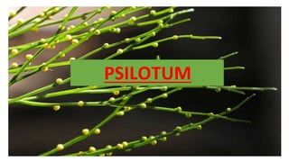
- 1. PSILOTUM
- 2. SYSTEMATIC POSITION • Class: Psilotopsida • Order: Psilotales • Family: Psilotaceae • Genus: Psilotum
- 3. DISTRIBUTION • Commonly known as Whisk fern. • Represented by only two species P.nudum and P.flaccidum. • P.nudum is distributed throughout the tropics and subtropics. It is found in Florida,Hawaii,New Zealand etc and in India it is found in Assam,Bengal,Kulu etc. • P.nudum is an erect,slender,shrubby plant found in the crevices of rocks but sometimes it occur as epiphyte also. Attains to a height of 25cm. It is cultivated in green houses and Botanical gardens. • P.flaccidum is found in Mexico, Jamaica and some pacific islands.It is a rare species and a pendulous epiphyte which can grow up to 90cm in length. • Psilotum species occur as epiphytes generally on trees,on the trunks of coconut trees or at the vase of trees. P.flaccidum differs from P.nudum in possessing flattened stem.
- 5. • Plant body is sporophytic and consists of subterranean rhizome and an erect aerial shoot. RHIZOME • Rhizome is cylindrical , irregularly, extensively and dichotomously branched,prostrate. • It is brown in colour and lack roots. • Rhizoids are present instead of roots which helps in absorption and anchorage. • Often associated with the rhizome,there will be an intracellular mycorrhizal fungus whose hyphae enter through the rhizoids and reach the cortex. • Sometimes the tips of immature rhizoids give rise to gemmae which helps in vegetative propagation. • The branches of rhizome, which are near the surface and also exposed to light ,ultimately turn into aerial shoots.
- 6. AERIAL SHOOT • Aerial shoots are slender,green,upright, ridged and dichotomously branched. • In terrestrial forms, they grow upright and in epiphytes, the hangs down from the host plant. • The basal part of aerial shoot is usually smooth and cylindrical. But their distal part is longitudinally ribbed in P.nudum and flat in P.flaccidum. • The aerial shoots of P.nudum are short (15- 20cm) while those of P.flaccidum are long (upto 90cm) . • In the absence of true leaves, green aerial shoots function as photosynthetic organs.
- 7. APPENDAGES • Aerial shoots are devoid of true leaves. They bear small,scale like appendages which are devoid of vascular traces and stomata. • In P.nudum ,they are arranged spirally or maybe irregularly distributed. • In P.flaccidum they are arranged in subopposite pairs. • The appendages are absent on rhizome and at the extreme basal part of the aerial shoots. • In the terminal part of the shoots,fertile appendages can be seen. In the axils of these fertile appendages, trilobite sporangia called synangia can be seen.
- 8. ANATOMY
- 9. I. AERIAL SHOOT Transverse section of the aerial shoot is irregular in outline due to the presence of ridges and grooves. It consist of three parts namely Epidermis, Cortex and Stele. 1.EPIDERMIS • well developed single layered epidermis. • Cells are elongated and protected by thick cuticle. • Sunken stomata present. It shows the xerophytic feature. • The stomata are without subsidiary cells ,a situation comparable to gymnosperms.
- 10. 2. CORTEX A well developed cortex is present which is differentiated into three zones. OUTER CORTEX: • Made up of 2 to 5 layers of thin walled,elongated, chlorophyll containing cells. • Intercellular spaces present. • This zone is photosynthetic. MIDDLE CORTEX • Made up of 4 to 5 layers of closely packed,thick walled, sclerenchymatous cells. • Provides mechanical support. INNER CORTEX • Broadest zone. • Consist of thin walled parenchymatous cells with no intercellular spaces. • Cells rich in starch reserves. Endodermis: Well developed with casparian strips.
- 11. 3. STELE • In the rhizome,it is a protostele with central core of xylem completely surrounded by phloem.The xylem core is not starshaped or lobed.There is no pith. • In the transit zone between rhizome and aerial shoot,the xylem becomes lobed and as many as ten lobes can be counted.The protoxylem occupies the tips of the lobes.The phloem occurs in between the lobes and forms irregular patches. Here the stele is actinostelic protostele. There is no pith. • In the middle of aerial shoot,the number of xylem lobes are reduced to 5 or 7 and pith appears in the centre. Here it changes from protostele to siphonostele.
- 12. • In all these cases the xylem is exarch and consist of only tracheids. • The protoxylem has spiral tracheids whereas the metaxylem has scalariform or pitted tracheids. • The phloem consist of sieve tubes and phloem parenchyma.The sieve tubes are vertically elongated with slight lignification and have sieve plates.
- 13. C.S
- 14. III. RHIZOME The anatomy of rhizome is somewhat similar to aerial shoot. very small rhizomes of less than 1mm diameter is without any vascular tissues and is uniformly parenchymatous. The stele gets differentiated in some larger rhizomes. The following structures can be observed in rhizomes with a diameter of more than 2mm. ★ Epidermis:single layered,uninterrupted,made up of thin walled cells. ★ Cortex:well developed and differentiated into three regions. ★ Outer cortex:contain many hyphae of endophytic mycorrhizal fungus. ★ Middle cortex:consist of parenchymatous cells storing starch. ★ Inner cortex:two to four layered,cells are brown in colour due to deposition of phlobaphene which is compound formed as result of oxidation and condensation of tannins. ★ Endodermis: distinct with casparian strips. ★ Pericycle:single layered and parenchymatous. ★ Stele: protostele,xylem surrounded by phloem, xylem not differentiated into metaxylem and protoxylem.Pith absent.
- 15. III. APPENDAGES The anatomy of appendage is simple .It consist of Epidermis and Mesophyll. ❖ Epidermis: single layered,cutinized,stomata absent. ❖ Mesophyll: consist of chlorophyll containing parenchymatous cells • No differentiation into palisade and spongy tissues. • More intercellular spaces in Psilotum nudum than Psilotum flaccidum. In the absence of stomata and vascular traces,the leaves are not photosynthetic in function.
- 16. REPRODUCTION
- 17. The sporophytic plant reproduces vegetatively and also by spores. VEGETATIVE REPRODUCTION • Vegetative reproduction takesplace by means of ovoid,minute,one cell thick,multicellular outgrowths developing on the rhizome. These outgrowths are called gemmae.Gemmae cells contain starch. • They undergo germination and produce new sporophytes which in turn produce new gemmae and repeat the cycle. • Germination of sporophytic gemmae occur when they remain attached to the parent plant and also after falling to an ideal substratum. • Gemmae like structures also develop on the gametophyte also. They are structurally similar to those occuring on the sporophyte. They germinate and produce new prothalli to repeat the cycle. • Vegetative reproduction also takesplace by the death of the older parts of the rhizome. The younger parts of rhizome separate from the dead rhizome and grow as independent plants.
- 18. REPRODUCTION BY SPORES • Reproduction by spores occur when the sporophytic plant attains maturity. • Spores are produced within the trilobite sporangia called synangia which is formed by the fusion of 2 or more sporangia. • A synangium develop on a small fertile appendage, subtended by a forked bract. • They together constitute of sporangium complex.
- 19. STRUCTURE OF SYNANGIUM • Synangium is a trilocular (three chambered) spore bearing structure in Psilotum. • It is considered as a fusion product of three sporangia. • Location: axils of fertile appendages on the aerial shoot. • Wall is 3 to 4 layered. • Thick outer wall forms the epidermis. • Inner wall separates the three chambers. • Each locule is filled with large numbers of bean shaped spores and they are of homosporous type. • Synangium splits along the three longitudinal lines of dehiscence thereby releasing spores.
- 20. DEVELOPMENT OF SPORANGIUM • Development of sporangium is of eusporangiate type. • Early stages of the sporangial development indicate that each of the three sporangia of a group develops from a single epidermal cell of the sporangiophore. • This initial cell divides periclinally to form outer primary jacket cell and an inner primary archesporial cell. • The jacket cell undergoes repeated anticlinal and periclinal divisions to form 4 to 5 cells thick jacket layer. The outermost layer becomes the epidermis. • The primary archesporial cell divides repeatedly to form sporogenous cells which differentiate into sporogenous tissue. • Psilotum differ from most of the other pteridophytes because neither its outermost sporogenous cells nor innermost jacket cells develop into tapetum.
- 21. • Some of the cells of the sporogenous tissue divide and redivide and give rise to spore mother cells (SMC) while the remaining cells of the sporogenous tissue develop into a plasmodial mass of spores. It provides nourishment to the sporocytes at the same time. • Spore mother cells undergo meiosis to form large number of spore tetrads within the Sporangium.The spores formed in the tetrads are colorless and kidney shaped and they are homosporous. • The spores are dispersed along the line of dehiscence.
- 23. SPORES IN PSILOTUM • Spores are homosporus. • Mature spores are kidney shaped.(average 0.065-0.032mm) • The two curved ends of the mature spores are joined by a narrow slit. • On either side of the slit,thick,Smooth, border called lip is present. • Outer coat of the spore is exine and inner coat is called the intine.
- 24. DEVELOPMENT & STRUCTURE OF GAMETOPHYTE • The spore germination is a slow process which takes 3-4months . • The germinating spore absorbs water and swells.as a result, the turgor pressure inside increases, the exine opens along the median slit. All the inner contents, covered by the thin membrane of intine,project out in the form of a conical mound. • With the formation of transverse wall,a basal large spherical cell is separated from the upper extruded cell. The basal cell remains covered by the portions of the ruptured outer wall. • The upper cell divides by two intersecting oblique divisions to form and apical cell. • This apical cell leads to the formation of the prothallus
- 26. • This results in the formation of prothallus provided with rhizoids. Prothallus represents the gametophyte. • The prothallus is simple,irregularly dichotomising,cylindrical, subterraneous measuring 0.5 to 2mm in diameter and upto 18mm in length. It is pale yellow to dark brown in color and covered with hair like rhizoids. Over the entire surface of the prothallus, antheridia and archegonia are scattered without any definite arrangement. • The prothalli are penetrated by an endophytic fungus in their early stages of development, which provides nutrients to the prothallus. Thus the prothallus is saprophytic. It usually lacks vascular tissue.
- 28. SEX ORGANS The gametophyte is monoecious with male and female sex organs,they are scattered over the surface of the gametophyte. ANTHERIDIUM • It is the male sex organ. • In Psilotum it is projected out or emergent. • Mature antheridium is a semi spherical structure. • It is surrounded by a well defined jacket which is single layered and one cell thick.
- 29. • The jacket encloses a mass of spirally coiled and multiflagellate antherozoids which are unicellular, uninucleate and mutlciliate in Psilotum. The jacket contains only one opercular cell. • They are liberated through a passage formed by the disintegration of the opercular cell.
- 30. ARCHEGONIUM • It is the female sex organ. • It is usually embedded in the prothallus and consist of projecting neck . • The neck consist of generally six tiers of four cells each,2 neck canal nuclei and venter region encloses one venter canal cell and an egg cell. • 2 cover cells are present at the tip of the neck region.
- 31. FERTILIZATION • Fertilization takes place with the help of water. • Antherozoids are attracted chemotactically towards the archegonium. • A chemical substance comes out from the open archegonium, it functions as a sperm attractor, contains large amount of organic and inorganic compounds, especially malic acid and fumaric acid. • The neck canal nuclei and venter canal nuclei disintegrate to form a free passage for the entry of antherozoids. • During fertilization antherozoids swim down along this canal and fuses with the egg to form a diploid zygote.
- 32. DEVELOPMENT OF SPOROPHYTE • Zygote is the first cell of sporophytic generation. • It enlarges in size and completely fills the venter cavity. • It divides to form an outer epibasal cell and an inner hypobasal cell. • The epibasal cell undergo repeated division to form shoot and hypobasal cell form the foot. • This type of embryogeny in which the shoot forming cell is directed towards the neck of the archegonium is called exoscopic embryogeny.
- 33. • After the formation of epibasal and hypobasal cells, both of them undergo longitudinal division to form four cells. • Further divisions are irregular (Holloway,1939) • Some of the cells of the foot undergo transverse division to form finger like rows of cells. • These finger like structures penetrate into the gametophytic tissue. • With the help of some regular divisions of the epibasal cell, a three sided apical cell is formed. • A typical rhizome is soon formed which is infected by an endophytic mycorrhizal fungus. • Then the rhizome branches dichotomously. Some of these branches turn upward and develop to aerial shoots and thus a new sporophytic plant is formed.
- 34. LIFE CYCLE • The plant body of Psilotum is a diploid sporophyte. • Synangium is the trilobite sporangia which bears the spores. • Inside the Synangium, the diploid SMC undergo meiosis to form haploid spores. • They germinate to form the haploid prothallus(monoecious ). • Antheridium produces antherozoids and archegonium produces egg • Fertilization occurs and a diploid zygote is formed. • The zygote develop into embryo which later form the mature plant body ,diploid sporophyte and the cycle is repeated.
- 36. Thank you Submitted to , Dr. Liza Jacob Head of the department Department of Botany St Teresa’s college Ernakulam Submitted by, Silpa Selvaraj Roll no: 13 I Msc Botany St Teresa’s college Ernakulam