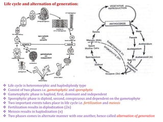
1. Anthoceros is a genus of hornworts that reproduces both sexually and asexually. The life cycle involves an alternation of generations between a dominant haploid gametophyte and a diploid sporophyte.
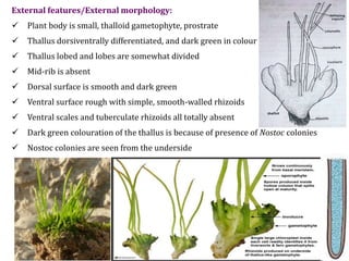
2. The gametophyte is a small, thalloid structure that produces male antheridia and female archegonia for sexual reproduction. Fertilization of an egg cell within the archegonium forms a zygote that develops into the sporophyte.
3. The sporophyte is an elongated structure that bears haploid spores through meiosis. Upon germination, the spores develop into new gametophyte plants, completing the


![Polymer [ बहुलक ] Chemistry Notes PDF - Irfanullah Mehar - JJ Sir Chemistry.pdf](https://cdn.slidesharecdn.com/ss_thumbnails/polymerchemistrynotespdf-irfanullahmehar-jjsirchemistry-260210172118-3f9b37f7-thumbnail.jpg?width=640&height=640&fit=bounds)


