SlideShare a Scribd company logo
1 of 28
Selaginella ( club moss or
spike moss)
Lycopsida
Taxonomic position
šŸ ¶ Division : Lycophyta
šŸ ¶ Class: ligulopsida
šŸ ¶ Order: Selaginellalies
šŸ ¶ Family: Selaginellaceae
šŸ ¶ Genus: Selaginella
Distribution and habitat
šŸ ¶ It is commonly called as club moss and spike
moss.
šŸ ¶ It has world wide distribution
šŸ ¶ Abundant in tropics and grows in ground and
shady places
šŸ ¶ Most common species is
šŸ ¶ Selaginella kraussiana
Vegetative morphology
šŸ ¶ The plant body is sporophyte and
it is differentiated in to
1. Root
2. Stem
3. Leaves
4. Ligules
5. rhizophores
Root
šŸ ¶ The root of young sporophyte is of
primary root while others are
adventitious
šŸ ¶ The adventitious roots are at the tips of
rhizophores
šŸ ¶ Aerial roots have developed caps, and
cutinized epidermal cells And enter soil.
Stem
šŸ ¶ Stem is green, dorsiventral and prostrate
with short erect branches
šŸ ¶ The branches are arranged dichotomously
šŸ ¶ They are also pseudomonopodia ( false
,growth from one point)
šŸ ¶ The shoot apex consists of a single apical
cell in most cases
Rhizophore
šŸ ¶ In some species , leafless and colorless branches arise
from the prostrate stem near point of branching.
šŸ ¶ These grow downwards and have group of
adventitious roots
šŸ ¶ They are called as rhizophores
šŸ ¶ Some scientist consider them branches and some
consider them as roots and still other consider it as
an organ for protection or other function.
šŸ ¶ But recently they are known as adventitious roots
that have dichotomous branches at tip.
Leaves
šŸ ¶ Microphylls are present. ( leaves
are small and single veined. They
are of 2 types
1. Isophyllous
2. Anisophyllous.
The anisophyllous leaves are in pairs.
They may be
šŸ ¶ Small: these are inserted on the
dorsal side of stem
šŸ ¶ Larger: these leaves are inserted
on the ventral side of stem
Ligules
šŸ ¶ Ligule: there is small outgrowth on adaxial
side ( upper side) of the leaf near base. It is
vestigial organ and provide water .
Anatomy
šŸ ¶ Stem anatomy
šŸ ¶ Steler system
šŸ ¶ Leaf anatomy
šŸ ¶ Root anatomy
Stem anatomy
šŸ ¶ Epidermis : thick epidermis , thin walled, rectangular
cells, covered with cuticle
šŸ ¶ cortex : many layered , outer 2-4 are thick walled
called as hypodermis
Below is thin walled parenchyma having chloroplast,
have small intercellular spaces.
Central portion is separated from cortex by a cavity
having air spaces
šŸ ¶ endodermis :the cortex and central tissue is
connected by radially elongated cells called
trabeculae
They contain casparian strips, trabuculae are modified
endodermal cells.
Steler system
šŸ ¶ Pericycle : there is single layer of pericycle formed of thin
walled cells, enclosing vascular tissue ( xylem and
phloem)
šŸ ¶ Phloem : there are phloem composed of sieve cells and
phloem parenchyma, companion cells are absent ,
phloem surrounds the xylem completely
šŸ ¶ Xylem : present in center , it consists of
1. Protoxylem : occupies two ends of meta xylem
2. Metaxylem: occupies the major portion of stele
These are composed of tracheid and parenchyma cells
Fibers are absent.
Rhizophore
šŸ ¶ Outermost layer is epidermis
šŸ ¶ It is of thick walled and single layer cells
šŸ ¶ Beneath the epidermis there is cortex
1. Hypodermis ( thick walled)
2. Thin walled parenchymatous region
3. Inner most layer is endodermis
šŸ ¶ Thin walled pericycle is present around the vascular
tissue
šŸ ¶ Stele is protostele ( xylem is in center and phloem
surrounds the xylem)
Leaf anatomy
šŸ ¶ The upper and lower epidermis are present
šŸ ¶ They are one celled thick and contain chloroplast
šŸ ¶ Stomata are present on upper and lower epidermis (
but majority have in lower side)
šŸ ¶ Below the epidermis there is mesophyll tissue having
thin walled parenchyma cells, these contain chloroplast
and have small and large air spaces
šŸ ¶ Vascular tissue is present in center
šŸ ¶ Phloem has few sieve cells and many parenchyma
šŸ ¶ Vascular bundle is surrounded by single layer forming
bundle sheath.
Ligule
Root anatomy
ļ‚“ by thin cuticle
ļ‚“ šŸ ¶ Root hairs are present and arise from epidermis
ļ‚“ šŸ ¶ Beneath the epidermis, wide zone of cortex is present
1. Outer hypodermis ( have sclerenchyma cells)
2. Endodermis ( inconspicuous )
ļ‚“ šŸ ¶ Single layered pericycle is present
ļ‚“ šŸ ¶ Protostele is next
ļ‚“ šŸ ¶ Xylem is surrounded by phloem
šŸ ¶ Outermost layer is epidermis ( single layer ), covered
Reproduction
šŸ ¶ Life cycle in Selaginella is characterized
by alternation of generation
šŸ ¶ Both spore producing and gamete
producing generations are independent .
šŸ ¶ Some species reproduce by vegetative
reproduction
The sporophyte : Vegetative and Asexual
reproduction
There are following methods for vegetative
reproduction
šŸ ¶ Adventitious branches
šŸ ¶ Tuber production during unfavorable
conditions
šŸ ¶ By production of resting buds at the ends of
aerial branches. ( these are surrounded by
leaves and can survive in adverse
conditions, upon reaching suitable
conditions they develop into new plants.
Asexual reproduction
šŸ ¶ These are hetrosporous
1. Microspore of smaller size
produced in microsporangia
2. Macrospores of larger size
produced in macrosporangia .
Sporangioferous spike
šŸ ¶ Sporangia are produced on the axils
of ligulated leaves called sporophylls
šŸ ¶ These sporophylls are organized to
form strobili at the ends of shoots
šŸ ¶ The sporophylls in strobilus is
arranged just like bracts in
angiosperm plants. Thatā€™s why it is
also called as sporangioferous spike
Structure of sporangia
šŸ ¶ Microsporangia : they are small, stalked, oval
and varying in shapes,
šŸ ¶ Mega sporangia : they are stalked and 4
lobes, larger in size and present at base of
strobilus , spores are of larger size.
1. Both consist of 2 layered sporangial wall
surrounding the tapetum and sporogenous
tissue.
2. Tapetum is developed from innermost layer
of sporangial wall.
3. Both differ in their size, location, and number
of spores
4. To release spore, both sporangia form vertical
cleft in wall .
Sexual reproduction ( gametophyte )
šŸ ¶ Microspores and macrospores
develop into male and female
gametophyte
šŸ ¶ Germination is precocoious (
within the walls of sporangia)
šŸ ¶ Male gametophyte is released
at 13 celled stage while female
gametophyte comes at various
stages , depending upon the
species.
Microspore ( male
gametophyte)
The microspores are very minute in
size and range in diameter from 0.015
to 0.05 mm. Soon after separation
from the tetrad they will be triradiate
but gradually assume a sub-spherical
shape. The spore wall is two- layered.
The outer exine (exospore) is very
thick and is sculptured. The inner
inline (endospore) is thin and
delicate. The spore consists of reserve
food material in the form of oil
globules and nitrogenous material.
Megaspores female
gametophyte)
The megaspores are much bigger in size
than the microspores and range in
diameter from 1.5-5 mm. When they are in
tetrad the spores have a triadiate shape but
become sub- spherical on separation. The
wall of the megaspore is very thick and
consists of a sculptured exine, a middle
mesospore and a thin intine.
The cytoplasm consists of reserve food in
the form of oil globules and nitrogenous
material. The amount of nitrogenous
material present is considerably less in
comparison with the microspore.
Chemical analysis of the stored food in
megaspores of Selaginella reveals that
they have 48% fats, 0.43% nitrogenous
matter and 1.26% mineral material.
selaginella species characters and distribution .pptx

More Related Content

Similar to selaginella species characters and distribution .pptx

Similar to selaginella species characters and distribution .pptx (20)

Funaria botany
Funaria botanyFunaria botany
Funaria botany
Ā 
MARSILEA notes in detail for II year Botany.ppt
MARSILEA  notes in detail for II year Botany.pptMARSILEA  notes in detail for II year Botany.ppt
MARSILEA notes in detail for II year Botany.ppt
Ā 
Life cycle of_funaria-jb
Life cycle of_funaria-jbLife cycle of_funaria-jb
Life cycle of_funaria-jb
Ā 
Life cycle of_funaria
Life cycle of_funariaLife cycle of_funaria
Life cycle of_funaria
Ā 
Type polytrichum
Type polytrichumType polytrichum
Type polytrichum
Ā 
Seminar on osmunda,dryopteris, 2
Seminar on osmunda,dryopteris, 2Seminar on osmunda,dryopteris, 2
Seminar on osmunda,dryopteris, 2
Ā 
Lycopodium
LycopodiumLycopodium
Lycopodium
Ā 
Structure, reproduction, life history and systematic position of Lycopodium
Structure, reproduction, life history and systematic position of LycopodiumStructure, reproduction, life history and systematic position of Lycopodium
Structure, reproduction, life history and systematic position of Lycopodium
Ā 
Psilotum
PsilotumPsilotum
Psilotum
Ā 
Lycopodium.pptx
Lycopodium.pptxLycopodium.pptx
Lycopodium.pptx
Ā 
Anthoceros
AnthocerosAnthoceros
Anthoceros
Ā 
MARSILIALES.pptx
MARSILIALES.pptxMARSILIALES.pptx
MARSILIALES.pptx
Ā 
Marchantia jb
Marchantia jbMarchantia jb
Marchantia jb
Ā 
Riccia
RicciaRiccia
Riccia
Ā 
Riccia Bryophytes presentation
Riccia             Bryophytes         presentationRiccia             Bryophytes         presentation
Riccia Bryophytes presentation
Ā 
Different type of plant cell in tissue system of plant
Different type of plant cell in tissue system of plantDifferent type of plant cell in tissue system of plant
Different type of plant cell in tissue system of plant
Ā 
Ginko
GinkoGinko
Ginko
Ā 
Structure of Anther
Structure of  AntherStructure of  Anther
Structure of Anther
Ā 
2. anthocerospp
2. anthocerospp2. anthocerospp
2. anthocerospp
Ā 
Life cycle of pinus
Life cycle of pinusLife cycle of pinus
Life cycle of pinus
Ā 

Recently uploaded

STERILITY TESTING OF PHARMACEUTICALS ppt by DR.C.P.PRINCE
STERILITY TESTING OF PHARMACEUTICALS ppt by DR.C.P.PRINCESTERILITY TESTING OF PHARMACEUTICALS ppt by DR.C.P.PRINCE
STERILITY TESTING OF PHARMACEUTICALS ppt by DR.C.P.PRINCEPRINCE C P
Ā 
Analytical Profile of Coleus Forskohlii | Forskolin .pdf
Analytical Profile of Coleus Forskohlii | Forskolin .pdfAnalytical Profile of Coleus Forskohlii | Forskolin .pdf
Analytical Profile of Coleus Forskohlii | Forskolin .pdfSwapnil Therkar
Ā 
Grafana in space: Monitoring Japan's SLIM moon lander in real time
Grafana in space: Monitoring Japan's SLIM moon lander  in real timeGrafana in space: Monitoring Japan's SLIM moon lander  in real time
Grafana in space: Monitoring Japan's SLIM moon lander in real timeSatoshi NAKAHIRA
Ā 
Work, Energy and Power for class 10 ICSE Physics
Work, Energy and Power for class 10 ICSE PhysicsWork, Energy and Power for class 10 ICSE Physics
Work, Energy and Power for class 10 ICSE Physicsvishikhakeshava1
Ā 
Artificial Intelligence In Microbiology by Dr. Prince C P
Artificial Intelligence In Microbiology by Dr. Prince C PArtificial Intelligence In Microbiology by Dr. Prince C P
Artificial Intelligence In Microbiology by Dr. Prince C PPRINCE C P
Ā 
TOPIC 8 Temperature and Heat.pdf physics
TOPIC 8 Temperature and Heat.pdf physicsTOPIC 8 Temperature and Heat.pdf physics
TOPIC 8 Temperature and Heat.pdf physicsssuserddc89b
Ā 
Dashanga agada a formulation of Agada tantra dealt in 3 Rd year bams agada tanta
Dashanga agada a formulation of Agada tantra dealt in 3 Rd year bams agada tantaDashanga agada a formulation of Agada tantra dealt in 3 Rd year bams agada tanta
Dashanga agada a formulation of Agada tantra dealt in 3 Rd year bams agada tantaPraksha3
Ā 
Analytical Profile of Coleus Forskohlii | Forskolin .pptx
Analytical Profile of Coleus Forskohlii | Forskolin .pptxAnalytical Profile of Coleus Forskohlii | Forskolin .pptx
Analytical Profile of Coleus Forskohlii | Forskolin .pptxSwapnil Therkar
Ā 
SOLUBLE PATTERN RECOGNITION RECEPTORS.pptx
SOLUBLE PATTERN RECOGNITION RECEPTORS.pptxSOLUBLE PATTERN RECOGNITION RECEPTORS.pptx
SOLUBLE PATTERN RECOGNITION RECEPTORS.pptxkessiyaTpeter
Ā 
Recombinant DNA technology( Transgenic plant and animal)
Recombinant DNA technology( Transgenic plant and animal)Recombinant DNA technology( Transgenic plant and animal)
Recombinant DNA technology( Transgenic plant and animal)DHURKADEVIBASKAR
Ā 
A relative description on Sonoporation.pdf
A relative description on Sonoporation.pdfA relative description on Sonoporation.pdf
A relative description on Sonoporation.pdfnehabiju2046
Ā 
Boyles law module in the grade 10 science
Boyles law module in the grade 10 scienceBoyles law module in the grade 10 science
Boyles law module in the grade 10 sciencefloriejanemacaya1
Ā 
Biopesticide (2).pptx .This slides helps to know the different types of biop...
Biopesticide (2).pptx  .This slides helps to know the different types of biop...Biopesticide (2).pptx  .This slides helps to know the different types of biop...
Biopesticide (2).pptx .This slides helps to know the different types of biop...RohitNehra6
Ā 
CALL ON āž„8923113531 šŸ”Call Girls Kesar Bagh Lucknow best Night Fun service šŸŖ”
CALL ON āž„8923113531 šŸ”Call Girls Kesar Bagh Lucknow best Night Fun service  šŸŖ”CALL ON āž„8923113531 šŸ”Call Girls Kesar Bagh Lucknow best Night Fun service  šŸŖ”
CALL ON āž„8923113531 šŸ”Call Girls Kesar Bagh Lucknow best Night Fun service šŸŖ”anilsa9823
Ā 
Isotopic evidence of long-lived volcanism on Io
Isotopic evidence of long-lived volcanism on IoIsotopic evidence of long-lived volcanism on Io
Isotopic evidence of long-lived volcanism on IoSĆ©rgio Sacani
Ā 
Call Girls in Munirka Delhi šŸ’ÆCall Us šŸ”9953322196šŸ” šŸ’ÆEscort.
Call Girls in Munirka Delhi šŸ’ÆCall Us šŸ”9953322196šŸ” šŸ’ÆEscort.Call Girls in Munirka Delhi šŸ’ÆCall Us šŸ”9953322196šŸ” šŸ’ÆEscort.
Call Girls in Munirka Delhi šŸ’ÆCall Us šŸ”9953322196šŸ” šŸ’ÆEscort.aasikanpl
Ā 
PossibleEoarcheanRecordsoftheGeomagneticFieldPreservedintheIsuaSupracrustalBe...
PossibleEoarcheanRecordsoftheGeomagneticFieldPreservedintheIsuaSupracrustalBe...PossibleEoarcheanRecordsoftheGeomagneticFieldPreservedintheIsuaSupracrustalBe...
PossibleEoarcheanRecordsoftheGeomagneticFieldPreservedintheIsuaSupracrustalBe...SĆ©rgio Sacani
Ā 
Recombination DNA Technology (Microinjection)
Recombination DNA Technology (Microinjection)Recombination DNA Technology (Microinjection)
Recombination DNA Technology (Microinjection)Jshifa
Ā 
Genomic DNA And Complementary DNA Libraries construction.
Genomic DNA And Complementary DNA Libraries construction.Genomic DNA And Complementary DNA Libraries construction.
Genomic DNA And Complementary DNA Libraries construction.k64182334
Ā 

Recently uploaded (20)

Engler and Prantl system of classification in plant taxonomy
Engler and Prantl system of classification in plant taxonomyEngler and Prantl system of classification in plant taxonomy
Engler and Prantl system of classification in plant taxonomy
Ā 
STERILITY TESTING OF PHARMACEUTICALS ppt by DR.C.P.PRINCE
STERILITY TESTING OF PHARMACEUTICALS ppt by DR.C.P.PRINCESTERILITY TESTING OF PHARMACEUTICALS ppt by DR.C.P.PRINCE
STERILITY TESTING OF PHARMACEUTICALS ppt by DR.C.P.PRINCE
Ā 
Analytical Profile of Coleus Forskohlii | Forskolin .pdf
Analytical Profile of Coleus Forskohlii | Forskolin .pdfAnalytical Profile of Coleus Forskohlii | Forskolin .pdf
Analytical Profile of Coleus Forskohlii | Forskolin .pdf
Ā 
Grafana in space: Monitoring Japan's SLIM moon lander in real time
Grafana in space: Monitoring Japan's SLIM moon lander  in real timeGrafana in space: Monitoring Japan's SLIM moon lander  in real time
Grafana in space: Monitoring Japan's SLIM moon lander in real time
Ā 
Work, Energy and Power for class 10 ICSE Physics
Work, Energy and Power for class 10 ICSE PhysicsWork, Energy and Power for class 10 ICSE Physics
Work, Energy and Power for class 10 ICSE Physics
Ā 
Artificial Intelligence In Microbiology by Dr. Prince C P
Artificial Intelligence In Microbiology by Dr. Prince C PArtificial Intelligence In Microbiology by Dr. Prince C P
Artificial Intelligence In Microbiology by Dr. Prince C P
Ā 
TOPIC 8 Temperature and Heat.pdf physics
TOPIC 8 Temperature and Heat.pdf physicsTOPIC 8 Temperature and Heat.pdf physics
TOPIC 8 Temperature and Heat.pdf physics
Ā 
Dashanga agada a formulation of Agada tantra dealt in 3 Rd year bams agada tanta
Dashanga agada a formulation of Agada tantra dealt in 3 Rd year bams agada tantaDashanga agada a formulation of Agada tantra dealt in 3 Rd year bams agada tanta
Dashanga agada a formulation of Agada tantra dealt in 3 Rd year bams agada tanta
Ā 
Analytical Profile of Coleus Forskohlii | Forskolin .pptx
Analytical Profile of Coleus Forskohlii | Forskolin .pptxAnalytical Profile of Coleus Forskohlii | Forskolin .pptx
Analytical Profile of Coleus Forskohlii | Forskolin .pptx
Ā 
SOLUBLE PATTERN RECOGNITION RECEPTORS.pptx
SOLUBLE PATTERN RECOGNITION RECEPTORS.pptxSOLUBLE PATTERN RECOGNITION RECEPTORS.pptx
SOLUBLE PATTERN RECOGNITION RECEPTORS.pptx
Ā 
Recombinant DNA technology( Transgenic plant and animal)
Recombinant DNA technology( Transgenic plant and animal)Recombinant DNA technology( Transgenic plant and animal)
Recombinant DNA technology( Transgenic plant and animal)
Ā 
A relative description on Sonoporation.pdf
A relative description on Sonoporation.pdfA relative description on Sonoporation.pdf
A relative description on Sonoporation.pdf
Ā 
Boyles law module in the grade 10 science
Boyles law module in the grade 10 scienceBoyles law module in the grade 10 science
Boyles law module in the grade 10 science
Ā 
Biopesticide (2).pptx .This slides helps to know the different types of biop...
Biopesticide (2).pptx  .This slides helps to know the different types of biop...Biopesticide (2).pptx  .This slides helps to know the different types of biop...
Biopesticide (2).pptx .This slides helps to know the different types of biop...
Ā 
CALL ON āž„8923113531 šŸ”Call Girls Kesar Bagh Lucknow best Night Fun service šŸŖ”
CALL ON āž„8923113531 šŸ”Call Girls Kesar Bagh Lucknow best Night Fun service  šŸŖ”CALL ON āž„8923113531 šŸ”Call Girls Kesar Bagh Lucknow best Night Fun service  šŸŖ”
CALL ON āž„8923113531 šŸ”Call Girls Kesar Bagh Lucknow best Night Fun service šŸŖ”
Ā 
Isotopic evidence of long-lived volcanism on Io
Isotopic evidence of long-lived volcanism on IoIsotopic evidence of long-lived volcanism on Io
Isotopic evidence of long-lived volcanism on Io
Ā 
Call Girls in Munirka Delhi šŸ’ÆCall Us šŸ”9953322196šŸ” šŸ’ÆEscort.
Call Girls in Munirka Delhi šŸ’ÆCall Us šŸ”9953322196šŸ” šŸ’ÆEscort.Call Girls in Munirka Delhi šŸ’ÆCall Us šŸ”9953322196šŸ” šŸ’ÆEscort.
Call Girls in Munirka Delhi šŸ’ÆCall Us šŸ”9953322196šŸ” šŸ’ÆEscort.
Ā 
PossibleEoarcheanRecordsoftheGeomagneticFieldPreservedintheIsuaSupracrustalBe...
PossibleEoarcheanRecordsoftheGeomagneticFieldPreservedintheIsuaSupracrustalBe...PossibleEoarcheanRecordsoftheGeomagneticFieldPreservedintheIsuaSupracrustalBe...
PossibleEoarcheanRecordsoftheGeomagneticFieldPreservedintheIsuaSupracrustalBe...
Ā 
Recombination DNA Technology (Microinjection)
Recombination DNA Technology (Microinjection)Recombination DNA Technology (Microinjection)
Recombination DNA Technology (Microinjection)
Ā 
Genomic DNA And Complementary DNA Libraries construction.
Genomic DNA And Complementary DNA Libraries construction.Genomic DNA And Complementary DNA Libraries construction.
Genomic DNA And Complementary DNA Libraries construction.
Ā 

selaginella species characters and distribution .pptx

  • 1. Selaginella ( club moss or spike moss) Lycopsida
  • 2. Taxonomic position šŸ ¶ Division : Lycophyta šŸ ¶ Class: ligulopsida šŸ ¶ Order: Selaginellalies šŸ ¶ Family: Selaginellaceae šŸ ¶ Genus: Selaginella
  • 3. Distribution and habitat
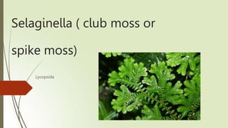
 šŸ ¶ It is commonly called as club moss and spike moss. šŸ ¶ It has world wide distribution šŸ ¶ Abundant in tropics and grows in ground and shady places šŸ ¶ Most common species is šŸ ¶ Selaginella kraussiana
  • 4. Vegetative morphology šŸ ¶ The plant body is sporophyte and it is differentiated in to 1. Root 2. Stem 3. Leaves 4. Ligules 5. rhizophores
  • 5. Root šŸ ¶ The root of young sporophyte is of primary root while others are adventitious šŸ ¶ The adventitious roots are at the tips of rhizophores šŸ ¶ Aerial roots have developed caps, and cutinized epidermal cells And enter soil.
  • 6. Stem šŸ ¶ Stem is green, dorsiventral and prostrate with short erect branches šŸ ¶ The branches are arranged dichotomously šŸ ¶ They are also pseudomonopodia ( false ,growth from one point) šŸ ¶ The shoot apex consists of a single apical cell in most cases
  • 7. Rhizophore šŸ ¶ In some species , leafless and colorless branches arise from the prostrate stem near point of branching. šŸ ¶ These grow downwards and have group of adventitious roots šŸ ¶ They are called as rhizophores šŸ ¶ Some scientist consider them branches and some consider them as roots and still other consider it as an organ for protection or other function. šŸ ¶ But recently they are known as adventitious roots that have dichotomous branches at tip.
  • 8. Leaves šŸ ¶ Microphylls are present. ( leaves are small and single veined. They are of 2 types 1. Isophyllous 2. Anisophyllous. The anisophyllous leaves are in pairs. They may be šŸ ¶ Small: these are inserted on the dorsal side of stem šŸ ¶ Larger: these leaves are inserted on the ventral side of stem
  • 9. Ligules šŸ ¶ Ligule: there is small outgrowth on adaxial side ( upper side) of the leaf near base. It is vestigial organ and provide water .
  • 10. Anatomy šŸ ¶ Stem anatomy šŸ ¶ Steler system šŸ ¶ Leaf anatomy šŸ ¶ Root anatomy
  • 11. Stem anatomy šŸ ¶ Epidermis : thick epidermis , thin walled, rectangular cells, covered with cuticle šŸ ¶ cortex : many layered , outer 2-4 are thick walled called as hypodermis Below is thin walled parenchyma having chloroplast, have small intercellular spaces. Central portion is separated from cortex by a cavity having air spaces šŸ ¶ endodermis :the cortex and central tissue is connected by radially elongated cells called trabeculae They contain casparian strips, trabuculae are modified endodermal cells.
  • 12.
  • 13. Steler system šŸ ¶ Pericycle : there is single layer of pericycle formed of thin walled cells, enclosing vascular tissue ( xylem and phloem) šŸ ¶ Phloem : there are phloem composed of sieve cells and phloem parenchyma, companion cells are absent , phloem surrounds the xylem completely šŸ ¶ Xylem : present in center , it consists of 1. Protoxylem : occupies two ends of meta xylem 2. Metaxylem: occupies the major portion of stele These are composed of tracheid and parenchyma cells Fibers are absent.
  • 14. Rhizophore šŸ ¶ Outermost layer is epidermis šŸ ¶ It is of thick walled and single layer cells šŸ ¶ Beneath the epidermis there is cortex 1. Hypodermis ( thick walled) 2. Thin walled parenchymatous region 3. Inner most layer is endodermis šŸ ¶ Thin walled pericycle is present around the vascular tissue šŸ ¶ Stele is protostele ( xylem is in center and phloem surrounds the xylem)
  • 15. Leaf anatomy šŸ ¶ The upper and lower epidermis are present šŸ ¶ They are one celled thick and contain chloroplast šŸ ¶ Stomata are present on upper and lower epidermis ( but majority have in lower side) šŸ ¶ Below the epidermis there is mesophyll tissue having thin walled parenchyma cells, these contain chloroplast and have small and large air spaces šŸ ¶ Vascular tissue is present in center šŸ ¶ Phloem has few sieve cells and many parenchyma šŸ ¶ Vascular bundle is surrounded by single layer forming bundle sheath.
  • 17. Root anatomy ļ‚“ by thin cuticle ļ‚“ šŸ ¶ Root hairs are present and arise from epidermis ļ‚“ šŸ ¶ Beneath the epidermis, wide zone of cortex is present 1. Outer hypodermis ( have sclerenchyma cells) 2. Endodermis ( inconspicuous ) ļ‚“ šŸ ¶ Single layered pericycle is present ļ‚“ šŸ ¶ Protostele is next ļ‚“ šŸ ¶ Xylem is surrounded by phloem šŸ ¶ Outermost layer is epidermis ( single layer ), covered
  • 18.
  • 19.
  • 20. Reproduction šŸ ¶ Life cycle in Selaginella is characterized by alternation of generation šŸ ¶ Both spore producing and gamete producing generations are independent . šŸ ¶ Some species reproduce by vegetative reproduction
  • 21. The sporophyte : Vegetative and Asexual reproduction There are following methods for vegetative reproduction šŸ ¶ Adventitious branches šŸ ¶ Tuber production during unfavorable conditions šŸ ¶ By production of resting buds at the ends of aerial branches. ( these are surrounded by leaves and can survive in adverse conditions, upon reaching suitable conditions they develop into new plants.
  • 22. Asexual reproduction šŸ ¶ These are hetrosporous 1. Microspore of smaller size produced in microsporangia 2. Macrospores of larger size produced in macrosporangia .
  • 23. Sporangioferous spike šŸ ¶ Sporangia are produced on the axils of ligulated leaves called sporophylls šŸ ¶ These sporophylls are organized to form strobili at the ends of shoots šŸ ¶ The sporophylls in strobilus is arranged just like bracts in angiosperm plants. Thatā€™s why it is also called as sporangioferous spike
  • 24. Structure of sporangia šŸ ¶ Microsporangia : they are small, stalked, oval and varying in shapes, šŸ ¶ Mega sporangia : they are stalked and 4 lobes, larger in size and present at base of strobilus , spores are of larger size. 1. Both consist of 2 layered sporangial wall surrounding the tapetum and sporogenous tissue. 2. Tapetum is developed from innermost layer of sporangial wall. 3. Both differ in their size, location, and number of spores 4. To release spore, both sporangia form vertical cleft in wall .
  • 25. Sexual reproduction ( gametophyte ) šŸ ¶ Microspores and macrospores develop into male and female gametophyte šŸ ¶ Germination is precocoious ( within the walls of sporangia) šŸ ¶ Male gametophyte is released at 13 celled stage while female gametophyte comes at various stages , depending upon the species.
  • 26. Microspore ( male gametophyte) The microspores are very minute in size and range in diameter from 0.015 to 0.05 mm. Soon after separation from the tetrad they will be triradiate but gradually assume a sub-spherical shape. The spore wall is two- layered. The outer exine (exospore) is very thick and is sculptured. The inner inline (endospore) is thin and delicate. The spore consists of reserve food material in the form of oil globules and nitrogenous material.
  • 27. Megaspores female gametophyte) The megaspores are much bigger in size than the microspores and range in diameter from 1.5-5 mm. When they are in tetrad the spores have a triadiate shape but become sub- spherical on separation. The wall of the megaspore is very thick and consists of a sculptured exine, a middle mesospore and a thin intine. The cytoplasm consists of reserve food in the form of oil globules and nitrogenous material. The amount of nitrogenous material present is considerably less in comparison with the microspore. Chemical analysis of the stored food in megaspores of Selaginella reveals that they have 48% fats, 0.43% nitrogenous matter and 1.26% mineral material.