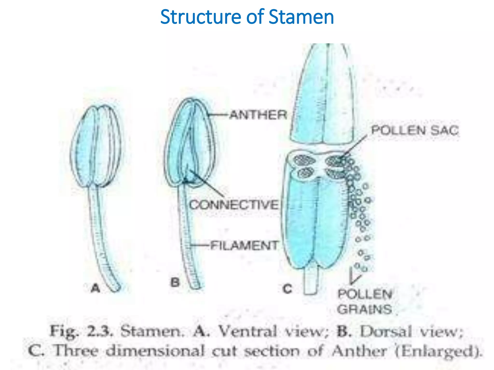
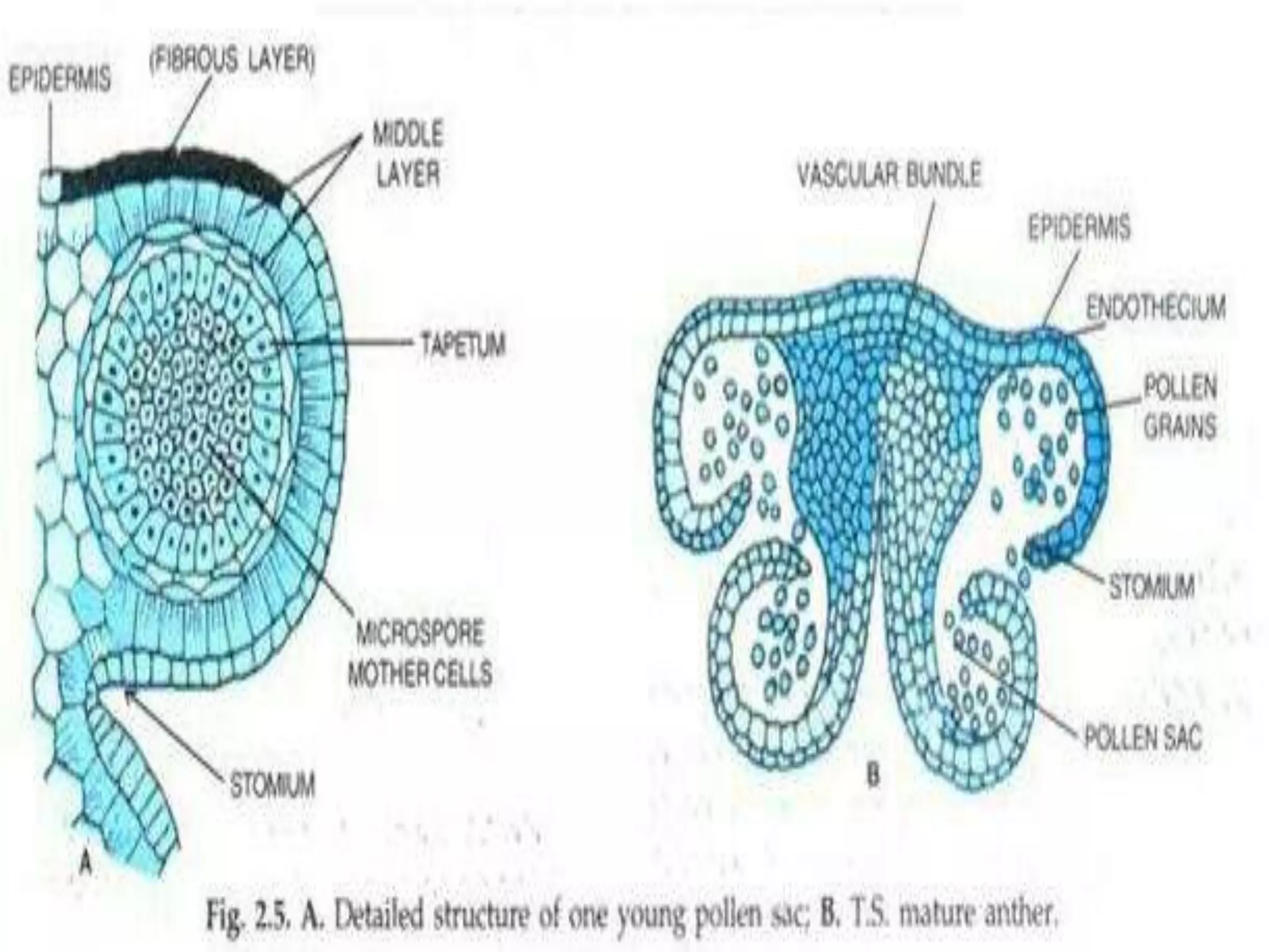
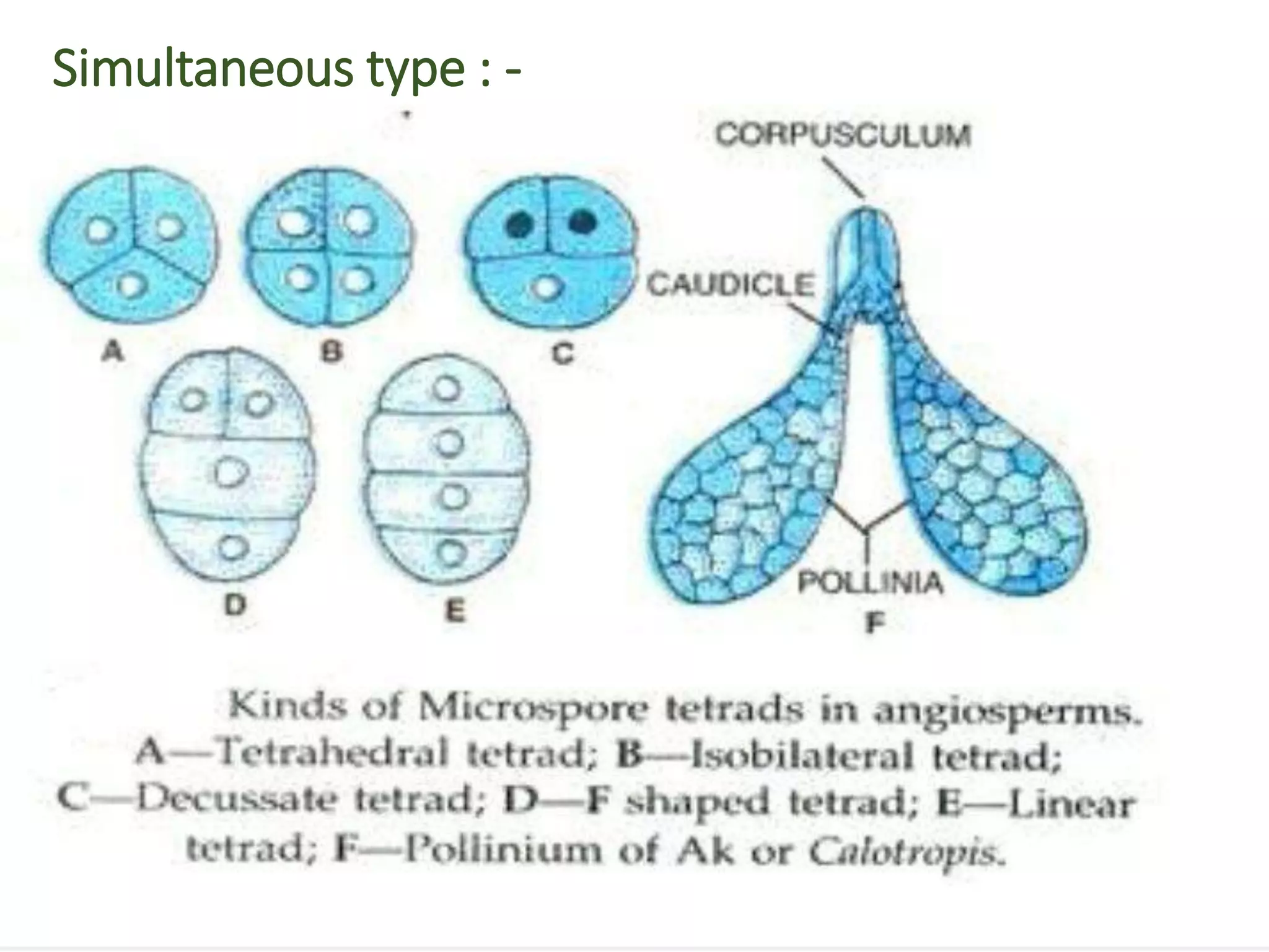
Microsporangium
Presented by : Salvi Pinky P.
M.sc sem - 2
Botany - 404
Department of Life Sciences ,
H. N. G. U., Patan
 Introduction
 Development of microsporangium
 Anther wall
 Sporogenous tissue
 Microsporogenesis
Content
Introduction
• Microsporophyll or the stamen is the male reproductive
part of the flower, which consists filament, anther and
connective.
• Anther is the fertile part of microsporophyll sometimes it
is sterile called staminode.
• The filament is the slender stalk of the stamen and the
anther is the expended head borne by the filament at its
tip.
Structure of Stamen
Development of microsporangium
• In each lobe a few cells in the hypodermal region become
differentiated by their large size, radial growth, dense cytoplasm
and conspicuous nuclei.
• There is much variation in the number of cells of archesporium.
• Generally, the archesporium consists of a two to three cell, wide
plate running along the entire length of the lobe.
• The extent of archesporium varies both length wise and breadth
wise.
The wall of microsporangium
• The epidermis along with the 3 – 5 layers derived from the
primary parietal layer form the wall of the sporangium.
• The cells of the epidermis devide anticlinally only.
• The cells thus formed become greatly stretched and flattened.
• Sometimes its cells may become greatly lignified or cutinized.
• A mature anther wall consists of 4 layers :
A) epidermis
B) endothecium
C) middle layer
D) tapetum
• Types of tapetum : -
1) Amoeboidal Tapetum : -
a) Sagittaria type
b) Butomus type
c) Sparaganium type
d) Triglochin type
2) Secretory or Glandular Tapetum
Sporogenous Tissue
• The primary sporogenous layer cells give rise to the microspore
mother cells or the pollen mother cells.
• The sporogenous cells in normal way divide several times
mitotically before functioning as pollen mother cells.
• In certain cases the primary sporogenous cells may either show
only few divisions or no divisions at cell, thus giving rise to a very
small group of sporogenous cells or the primary sporogenous cells
themselves function directly as the microspore mother cells.
Microsporogenesis
• The microspore mother cells which are at first polygonal and
closely packed gradually become rounded and loosely arranged in
the rapidly enlarging microsporangium.
• Each functional spore mother cell produces four microspore or
pollen grains.
• Types of microsporogenesis : -
A) Meiosis
B) Cytokinesis type : -
1) Successive type
2) Simultaneous type : -
a) Tetrahedral
b) Isobilateral tetrad
C) Decussate tetrad
d) T – shaped
e) Linear tetrad
3) The pollen grains
Cytokinesis type : -
• The division of the microspore mother cells may be for the
successive and simultaneous type.
• In the successive type, a cell plate is developed immediately after
the first meiotic division and another in each of the two daughter
cells after the second meiotic division.
Successive type : -
• In this type a cell wall is laid down between the two daughter
nuclei immediately after their formation during the first meiotic
division.
• The second meiotic division takes place forming two daughter
cells.
• And then again a wall is formed in if the two daughter cells.
Simultaneous type : -
• This type is commonly formed in dicotyledons plants.
• In this case the nucleus of the microspore mother cell divides
twice forming four haploid nuclei laying in the common mass of
cytoplasm of the mother cell.
• The 4 nuclei thus formed are being arranged in tetrahedral
manner and after cleavage of cytoplasm the 4 distinct segments
are formed.
The pollen grains : -
• The pollen grains or microspores are the male reproductive
bodies of a flower, and are contained in the pollen sac or
microsporangium.
• They are very minute in size, and are like particles of dust.
• Each pollen grains consists of a single microscopic cell, possessing
two coats : - 1) the exine
2) the intine
Structure of the pollen grains :
References
• A Taxt book Of botany : Angiosperms :
by : - B. P. Pandey
https://www.kullabs.com
https://en.m.wikipedia.org
https://www.pnas.org
THANK YOU

Microsporangium

  • 1.
    Microsporangium Presented by :Salvi Pinky P. M.sc sem - 2 Botany - 404 Department of Life Sciences , H. N. G. U., Patan
  • 2.
     Introduction  Developmentof microsporangium  Anther wall  Sporogenous tissue  Microsporogenesis Content
  • 3.
    Introduction • Microsporophyll orthe stamen is the male reproductive part of the flower, which consists filament, anther and connective. • Anther is the fertile part of microsporophyll sometimes it is sterile called staminode. • The filament is the slender stalk of the stamen and the anther is the expended head borne by the filament at its tip.
  • 4.
  • 5.
    Development of microsporangium •In each lobe a few cells in the hypodermal region become differentiated by their large size, radial growth, dense cytoplasm and conspicuous nuclei. • There is much variation in the number of cells of archesporium. • Generally, the archesporium consists of a two to three cell, wide plate running along the entire length of the lobe. • The extent of archesporium varies both length wise and breadth wise.
  • 7.
    The wall ofmicrosporangium • The epidermis along with the 3 – 5 layers derived from the primary parietal layer form the wall of the sporangium. • The cells of the epidermis devide anticlinally only. • The cells thus formed become greatly stretched and flattened. • Sometimes its cells may become greatly lignified or cutinized. • A mature anther wall consists of 4 layers : A) epidermis B) endothecium C) middle layer D) tapetum
  • 9.
    • Types oftapetum : - 1) Amoeboidal Tapetum : - a) Sagittaria type b) Butomus type c) Sparaganium type d) Triglochin type 2) Secretory or Glandular Tapetum
  • 10.
    Sporogenous Tissue • Theprimary sporogenous layer cells give rise to the microspore mother cells or the pollen mother cells. • The sporogenous cells in normal way divide several times mitotically before functioning as pollen mother cells. • In certain cases the primary sporogenous cells may either show only few divisions or no divisions at cell, thus giving rise to a very small group of sporogenous cells or the primary sporogenous cells themselves function directly as the microspore mother cells.
  • 11.
    Microsporogenesis • The microsporemother cells which are at first polygonal and closely packed gradually become rounded and loosely arranged in the rapidly enlarging microsporangium. • Each functional spore mother cell produces four microspore or pollen grains. • Types of microsporogenesis : - A) Meiosis B) Cytokinesis type : - 1) Successive type 2) Simultaneous type : - a) Tetrahedral b) Isobilateral tetrad C) Decussate tetrad d) T – shaped e) Linear tetrad 3) The pollen grains
  • 13.
    Cytokinesis type :- • The division of the microspore mother cells may be for the successive and simultaneous type. • In the successive type, a cell plate is developed immediately after the first meiotic division and another in each of the two daughter cells after the second meiotic division. Successive type : - • In this type a cell wall is laid down between the two daughter nuclei immediately after their formation during the first meiotic division. • The second meiotic division takes place forming two daughter cells. • And then again a wall is formed in if the two daughter cells.
  • 14.
  • 15.
    • This typeis commonly formed in dicotyledons plants. • In this case the nucleus of the microspore mother cell divides twice forming four haploid nuclei laying in the common mass of cytoplasm of the mother cell. • The 4 nuclei thus formed are being arranged in tetrahedral manner and after cleavage of cytoplasm the 4 distinct segments are formed. The pollen grains : - • The pollen grains or microspores are the male reproductive bodies of a flower, and are contained in the pollen sac or microsporangium. • They are very minute in size, and are like particles of dust. • Each pollen grains consists of a single microscopic cell, possessing two coats : - 1) the exine 2) the intine
  • 16.
    Structure of thepollen grains :
  • 17.
    References • A Taxtbook Of botany : Angiosperms : by : - B. P. Pandey https://www.kullabs.com https://en.m.wikipedia.org https://www.pnas.org
  • 18.