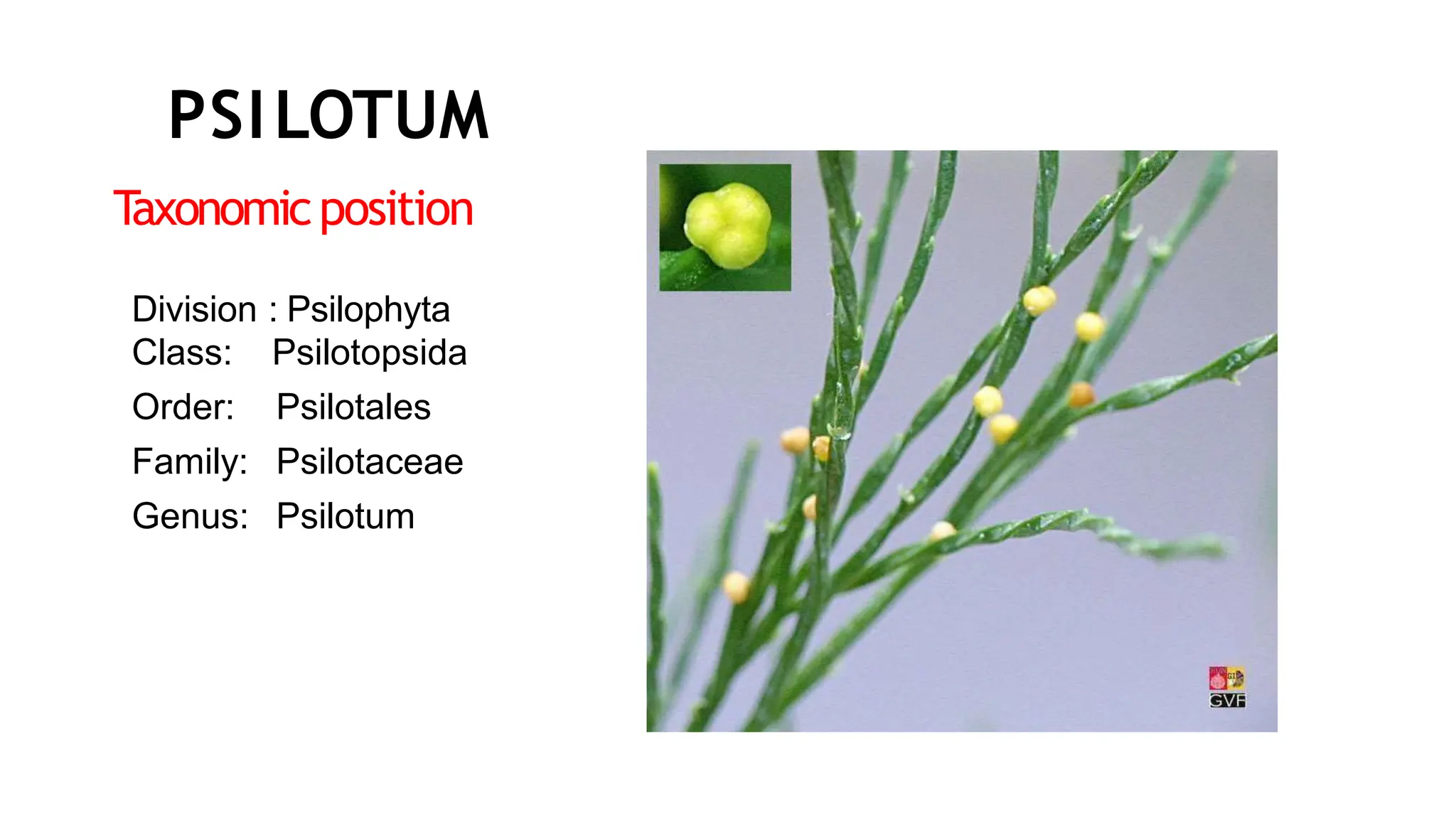
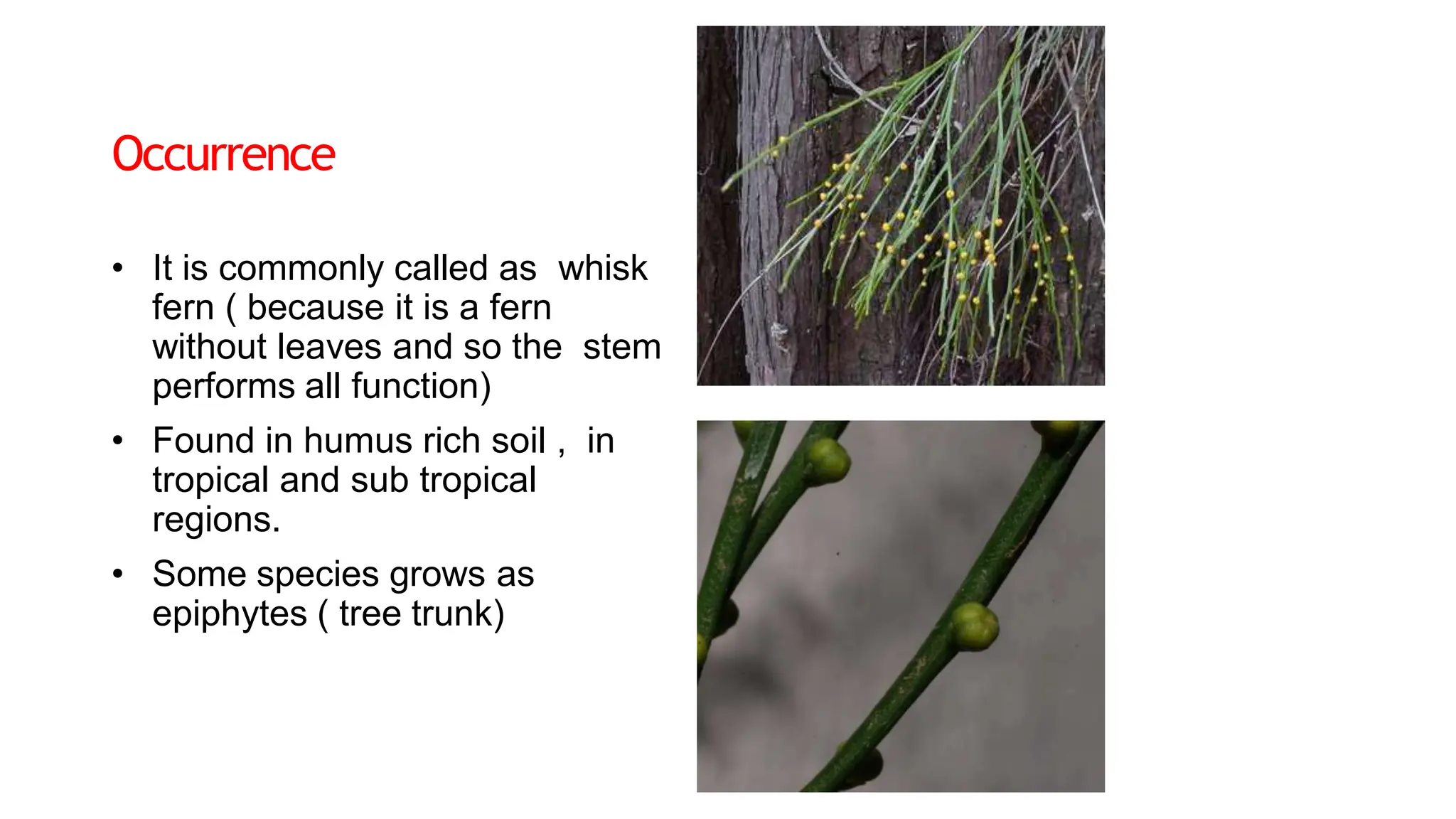
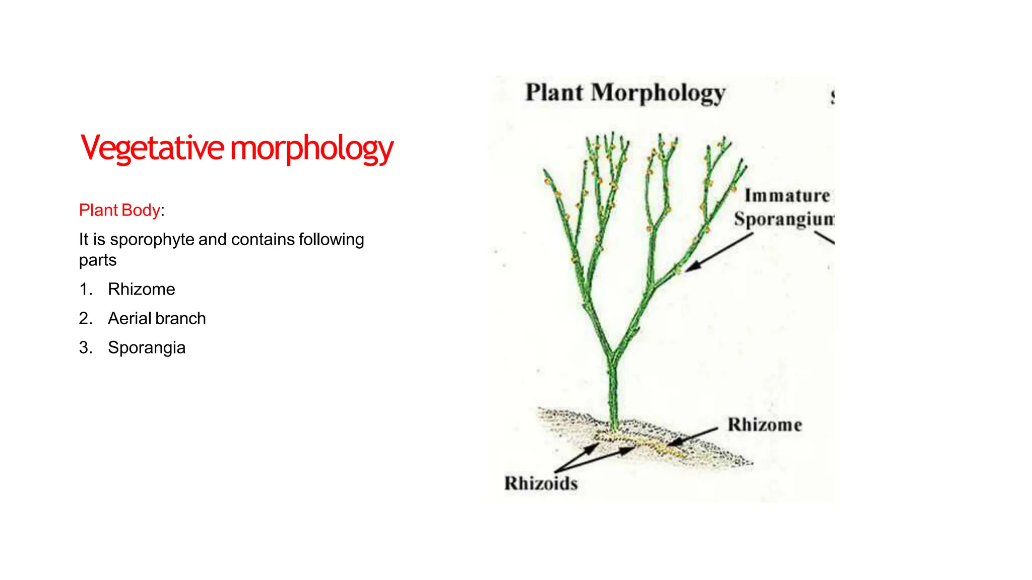
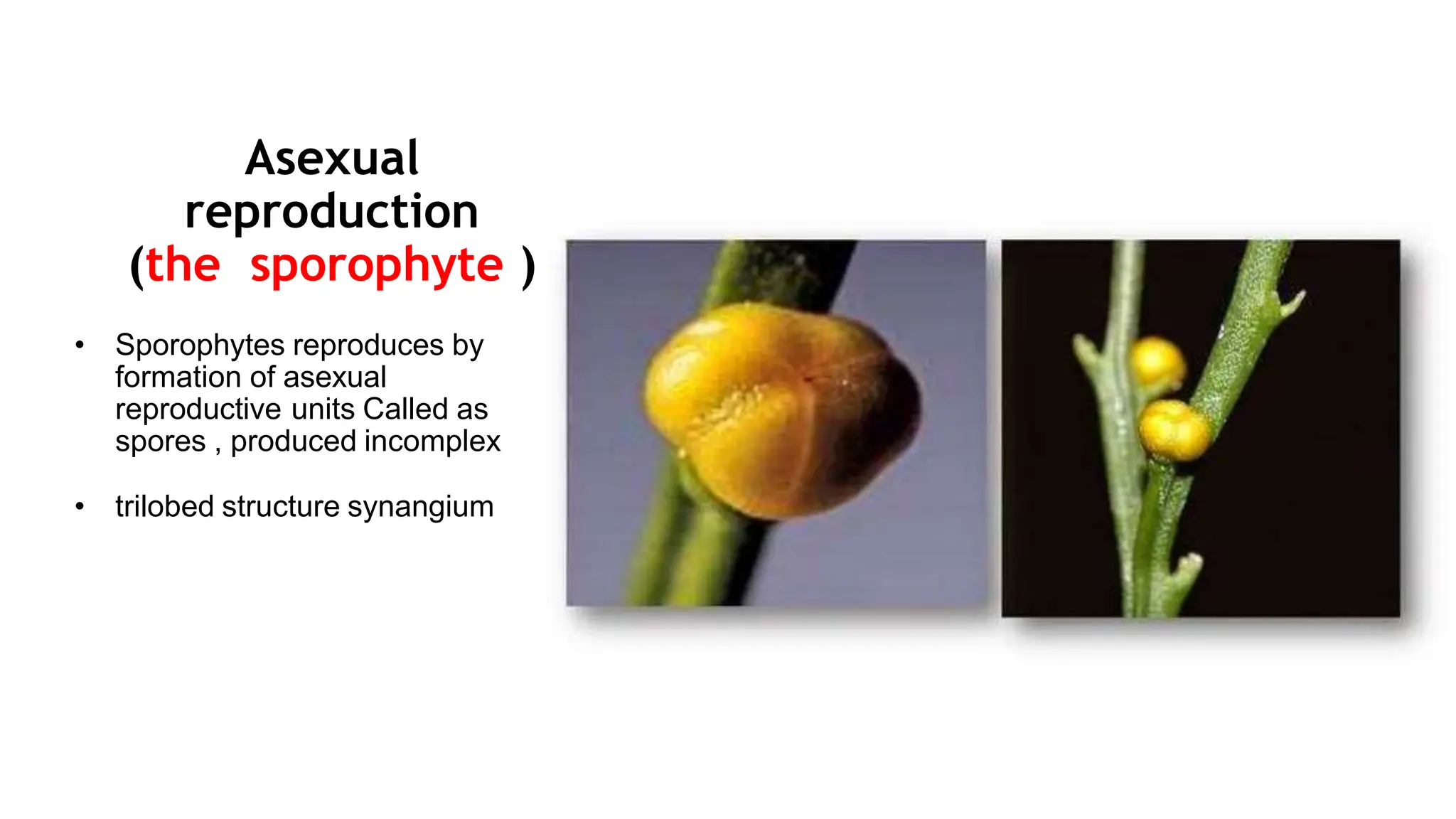
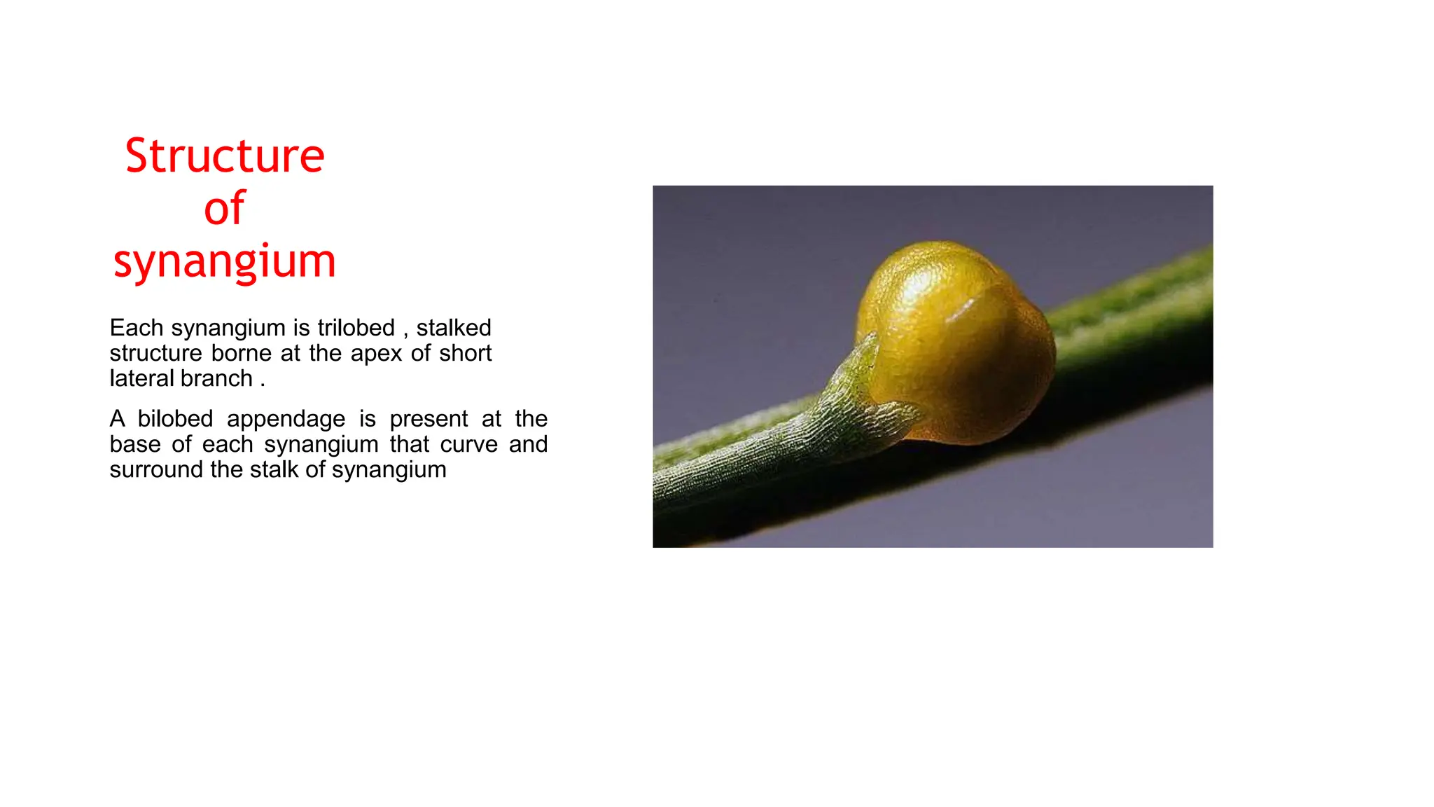
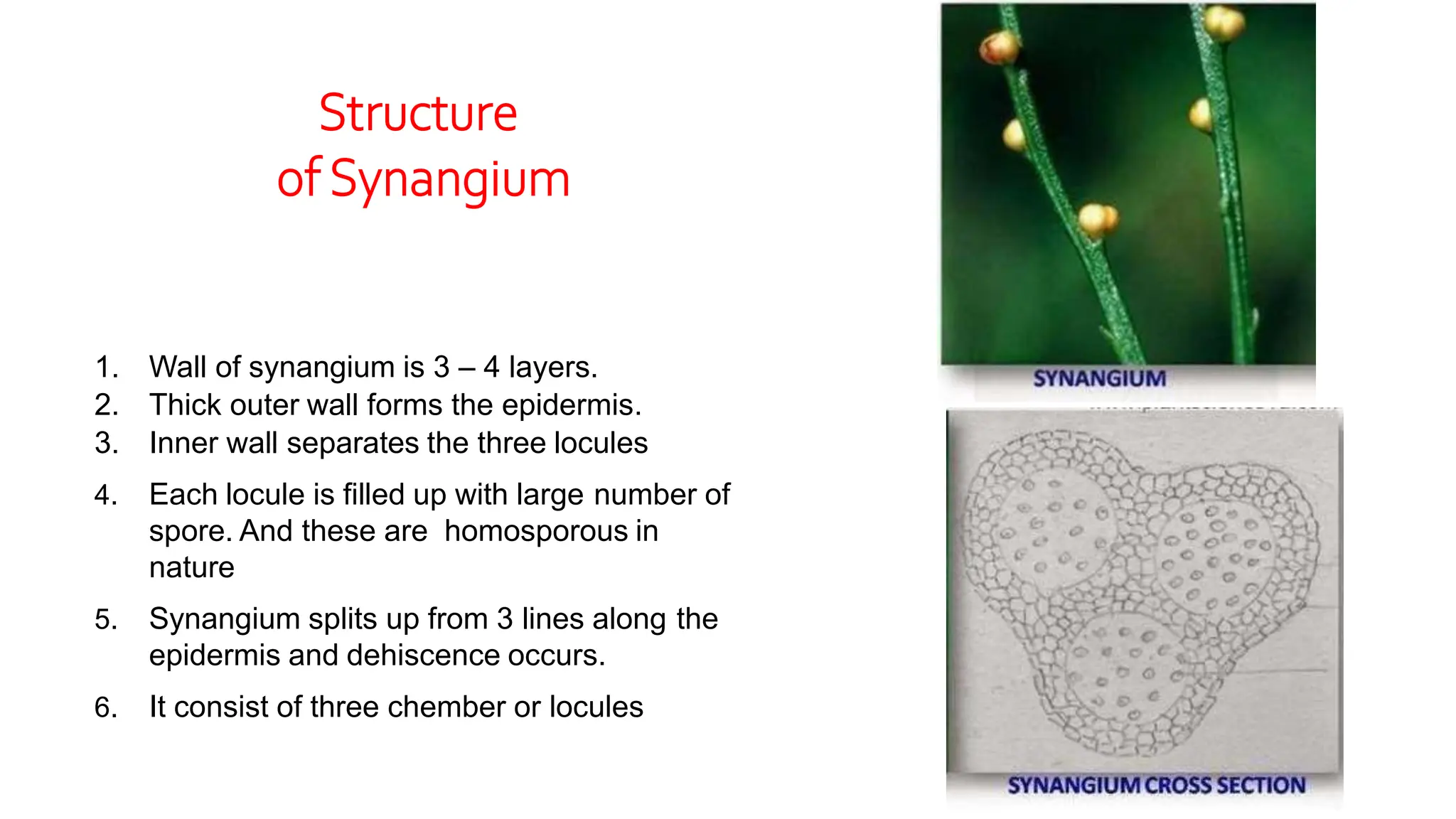
T
axonomicposition
Division : Psilophyta
Class:
Order:
Family:
Genus:
Psilotopsida
Psilotales
Psilotaceae
Psilotum
PSILOTUM
Occurrence
• It is commonly called as whisk
fern ( because it is a fern
without leaves and so the stem
performs all function)
• Found in humus rich soil , in
tropical and sub tropical
regions.
• Some species grows as
epiphytes ( tree trunk)
Vegetativemorphology
Plant Body:
It is sporophyte and contains following
parts
1. Rhizome
2. Aerial branch
3. Sporangia
Stelersystem
The stele of Psilotum consists of
following parts a, while pericycle and
pith are usually absent.
1. Endodermis
There is well developed endodermis
between the stele and the cortex. These
cells has casparian bands on their radial
walls
2: Xylem :
The xylem is actinostelic and radial in
outside in 6 rays, the protoxylem is
located at the tip of the rays. In the
center the metaxylem xore is present
The cells of xylem are thick walled and
their main function is transport of
nutrients
Stelersystem
4: Phloem
Between the endodermis and the xylem
there is phloem.
It is of thin walled cells
It consist of sieve cells and sieve areas in
their oblique end walls. Nuclei
disintegrate at maturity
Reproduction
• It is characterized by alternation
of
generation
• Both spore producing and
gamete producing regeneration
are independent
• Sporophyte reproduces by
asexual
reproduction
• Gametophyte reproduces by
sexual reproduction
Asexual
reproduction
(the sporophyte )
• Sporophytes reproduces by
formation of asexual
reproductive units Called as
spores , produced incomplex
• trilobed structure synangium
Structure
of
synangium
Each synangium is trilobed , stalked
structure borne at the apex of short
lateral branch .
A bilobed appendage is present at the
base of each synangium that curve and
surround the stalk of synangium
1. Wall of synangium is 3 – 4 layers.
2. Thick outer wall forms the epidermis.
3. Inner wall separates the three locules
4. Each locule is filled up with large number of
spore. And these are homosporous in
nature
5. Synangium splits up from 3 lines along the
epidermis and dehiscence occurs.
6. It consist of three chember or locules
Structure
of Synangium

psilotum structure and reproduction.pptx

  • 1.
  • 2.
    Occurrence • It iscommonly called as whisk fern ( because it is a fern without leaves and so the stem performs all function) • Found in humus rich soil , in tropical and sub tropical regions. • Some species grows as epiphytes ( tree trunk)
  • 3.
    Vegetativemorphology Plant Body: It issporophyte and contains following parts 1. Rhizome 2. Aerial branch 3. Sporangia
  • 5.
    Stelersystem The stele ofPsilotum consists of following parts a, while pericycle and pith are usually absent. 1. Endodermis There is well developed endodermis between the stele and the cortex. These cells has casparian bands on their radial walls 2: Xylem : The xylem is actinostelic and radial in outside in 6 rays, the protoxylem is located at the tip of the rays. In the center the metaxylem xore is present The cells of xylem are thick walled and their main function is transport of nutrients
  • 6.
    Stelersystem 4: Phloem Between theendodermis and the xylem there is phloem. It is of thin walled cells It consist of sieve cells and sieve areas in their oblique end walls. Nuclei disintegrate at maturity
  • 7.
    Reproduction • It ischaracterized by alternation of generation • Both spore producing and gamete producing regeneration are independent • Sporophyte reproduces by asexual reproduction • Gametophyte reproduces by sexual reproduction
  • 8.
    Asexual reproduction (the sporophyte ) •Sporophytes reproduces by formation of asexual reproductive units Called as spores , produced incomplex • trilobed structure synangium
  • 9.
    Structure of synangium Each synangium istrilobed , stalked structure borne at the apex of short lateral branch . A bilobed appendage is present at the base of each synangium that curve and surround the stalk of synangium
  • 10.
    1. Wall ofsynangium is 3 – 4 layers. 2. Thick outer wall forms the epidermis. 3. Inner wall separates the three locules 4. Each locule is filled up with large number of spore. And these are homosporous in nature 5. Synangium splits up from 3 lines along the epidermis and dehiscence occurs. 6. It consist of three chember or locules Structure of Synangium