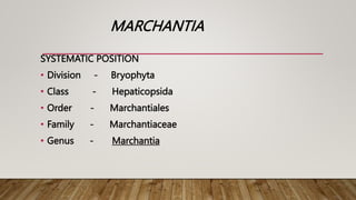
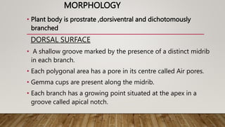
- Marchantia is a genus of liverworts that includes about 65 species. It grows in cool, moist, shady places and its thallus is dorsiventral and dichotomously branched.
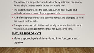
- The life cycle of Marchantia involves alternation of generations between a dominant haploid gametophyte phase and a short diploid sporophyte phase. The gametophyte produces male and female sex organs that generate gametes via antherozoids and archegonia. Fertilization leads to a diploid zygote that develops into the sporophyte.
- The sporophyte is differentiated into a foot, seta, and capsule. Inside the


![bryophytes.pptxforbotany [Autosaved].pptx](https://cdn.slidesharecdn.com/ss_thumbnails/bryophytes-241024055212-3ccb7683-thumbnail.jpg?width=640&height=640&fit=bounds)


