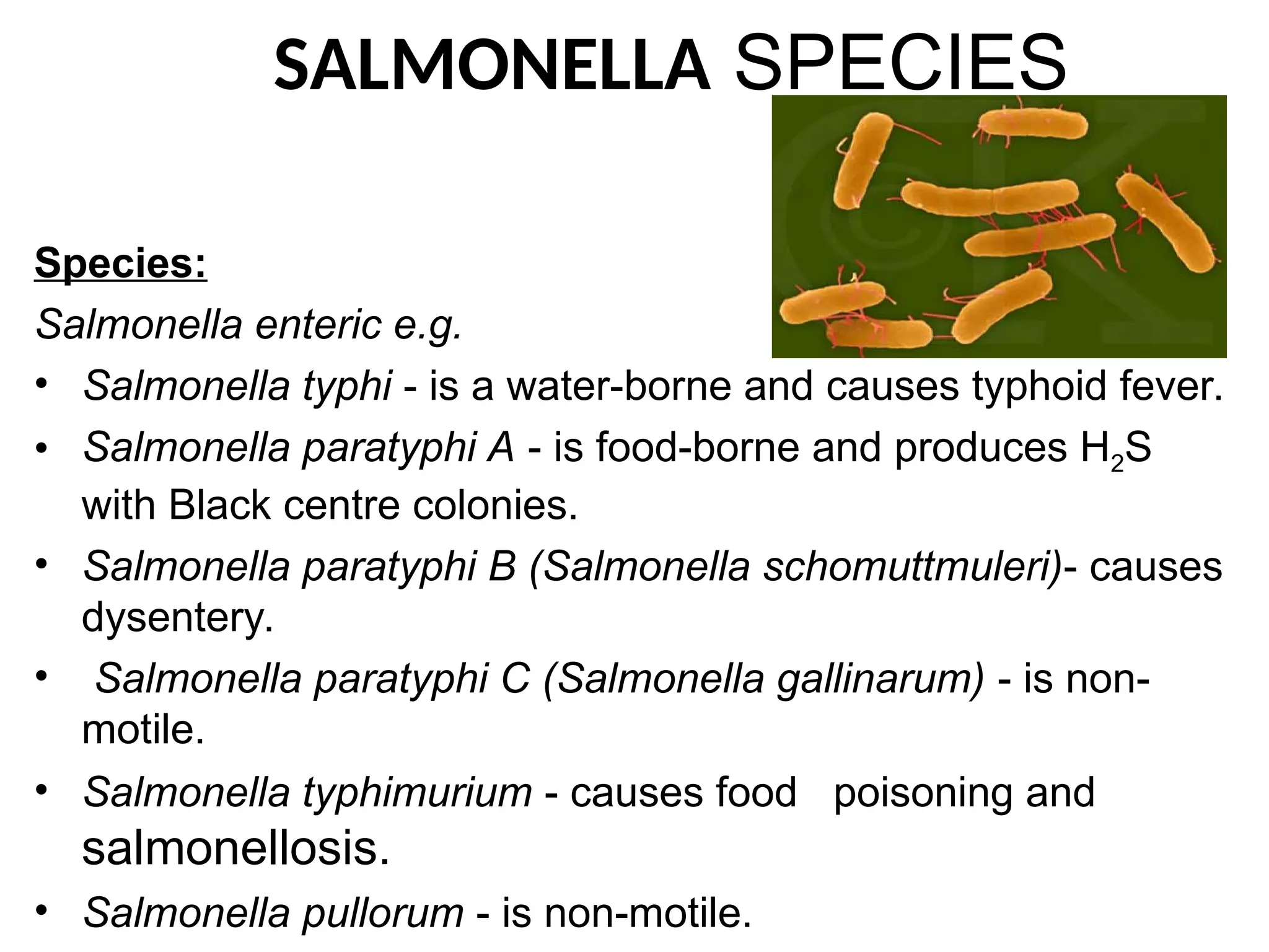
SALMONELLA SPECIES
Species:
Salmonella enterice.g.
• Salmonella typhi - is a water-borne and causes typhoid fever.
• Salmonella paratyphi A - is food-borne and produces H2S
with Black centre colonies.
• Salmonella paratyphi B (Salmonella schomuttmuleri)- causes
dysentery.
• Salmonella paratyphi C (Salmonella gallinarum) - is non-
motile.
• Salmonella typhimurium - causes food poisoning and
salmonellosis.
• Salmonella pullorum - is non-motile.
2.
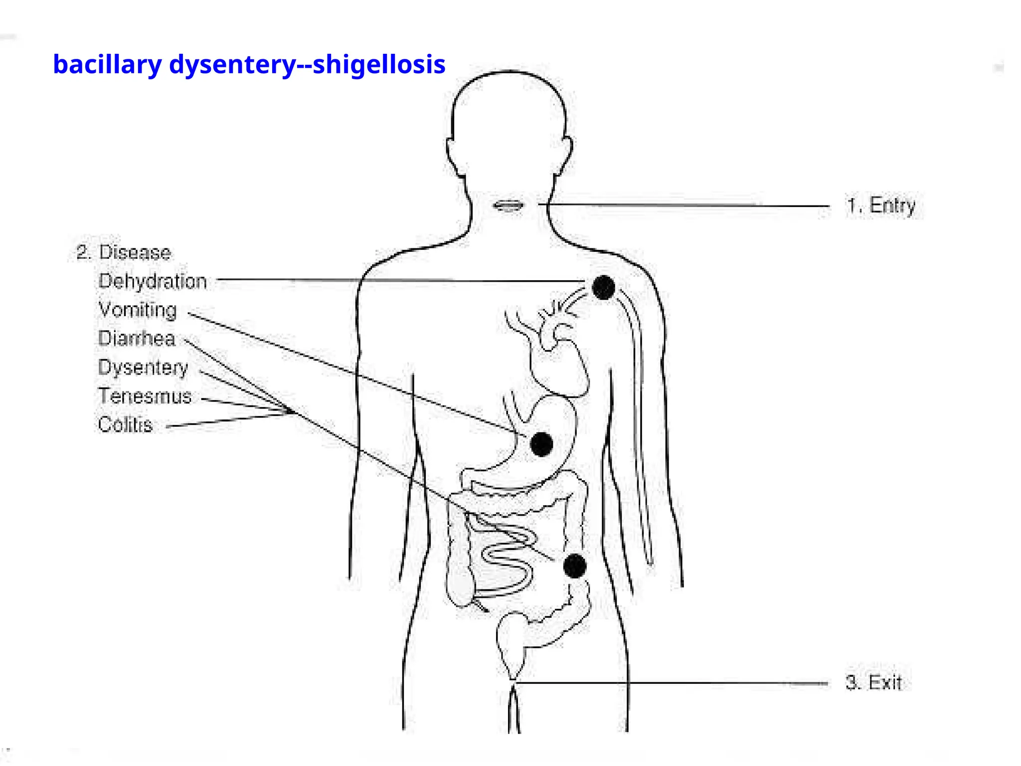
Habitat.
• Exclusively parasitesof human and animal
intestines causing bacillary dysentery in man.
• Dysentery is characterized with passage of
loose stool mixed with blood and mucus.
3.
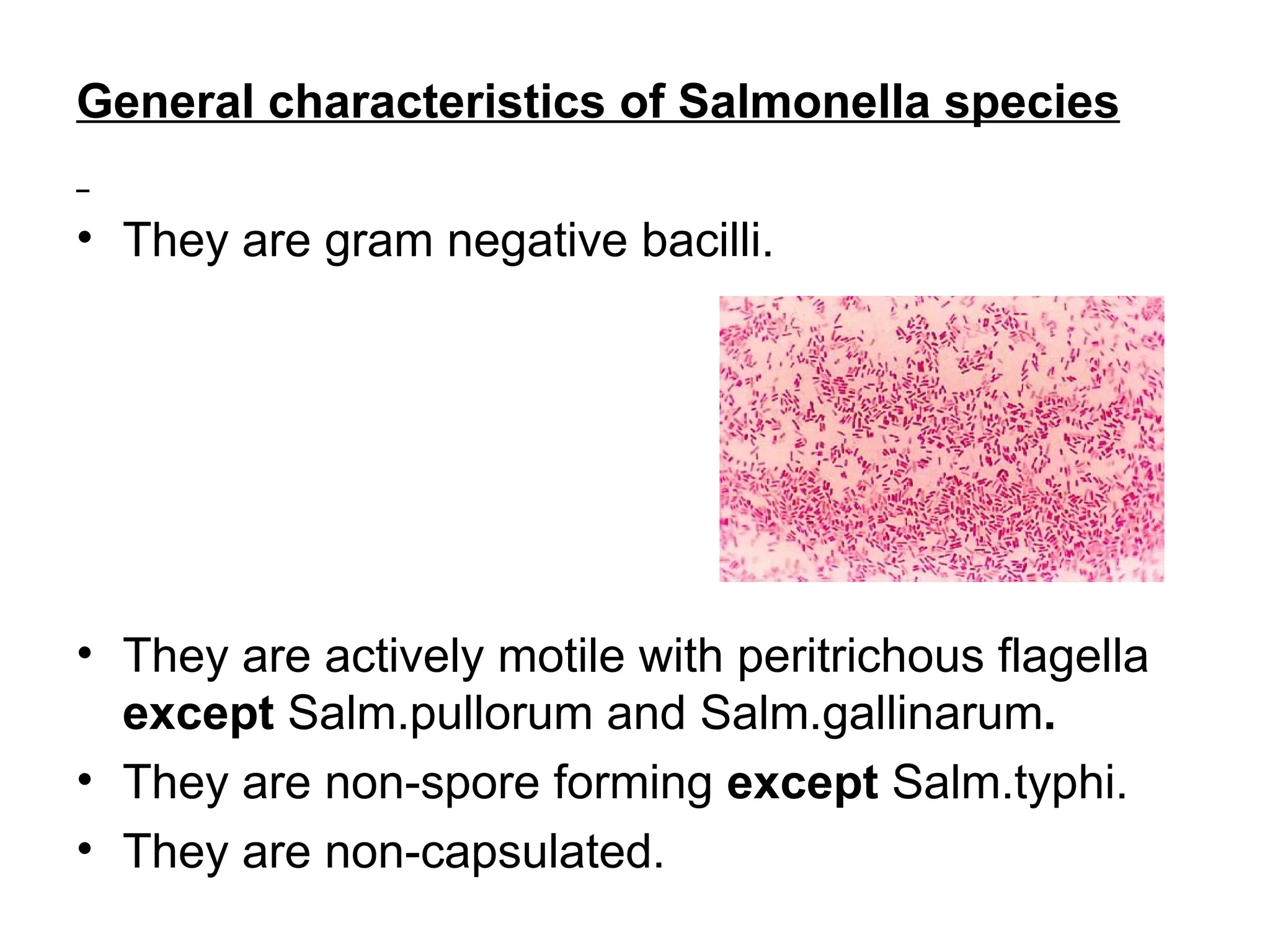
General characteristics ofSalmonella species
• They are gram negative bacilli.
• They are actively motile with peritrichous flagella
except Salm.pullorum and Salm.gallinarum.
• They are non-spore forming except Salm.typhi.
• They are non-capsulated.
4.
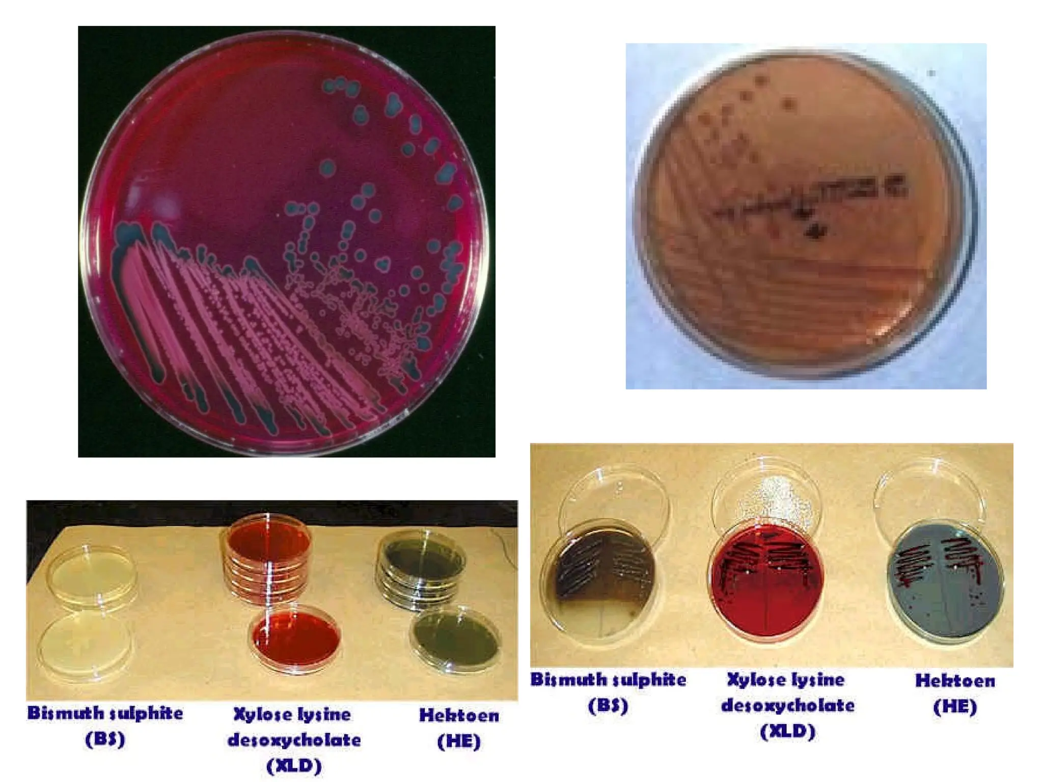
Cultural characteristics.
• Aerobesand facultative anaerobes with an optimum of 36-
370
C.
• Culture media: XLD agar medium, MacConkey agar and DCA
agar and SS medium
• Colonies after 18-48hrs incubation:
Colony morphology
• In XLD agar medium: Red-pink colonies, circular, convex,
compact, smooth edges and large measuring 2-4mm in
diameter and with black centre’s due H2S production.
• In MacConkey agar and DCA agar medium: pale colored
colonies, 1-2 mm in diameter with black centres in DCA
(H2S producing Salmonellae).
• In Nutrient agar and Blood agar medium: smooth, greyish or
colorless, translucent colonies that measures 2-3mm in
diameter
6.
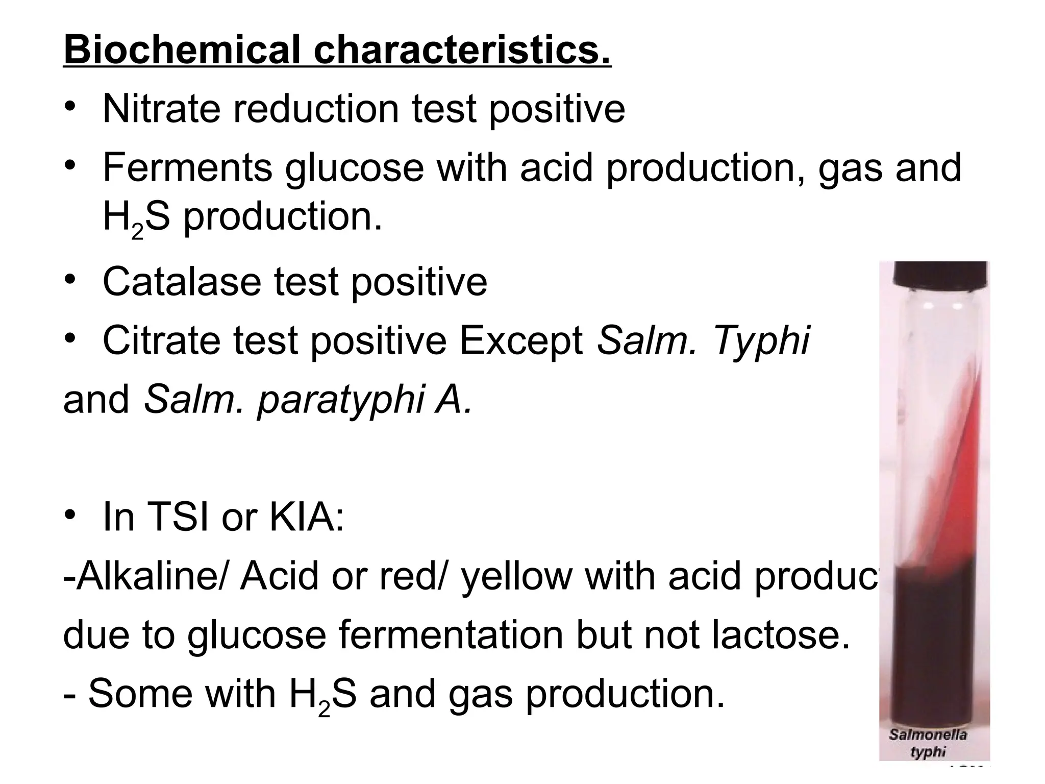
Biochemical characteristics.
• Nitratereduction test positive
• Ferments glucose with acid production, gas and
H2S production.
• Catalase test positive
• Citrate test positive Except Salm. Typhi
and Salm. paratyphi A.
• In TSI or KIA:
-Alkaline/ Acid or red/ yellow with acid production
due to glucose fermentation but not lactose.
- Some with H2S and gas production.
7.
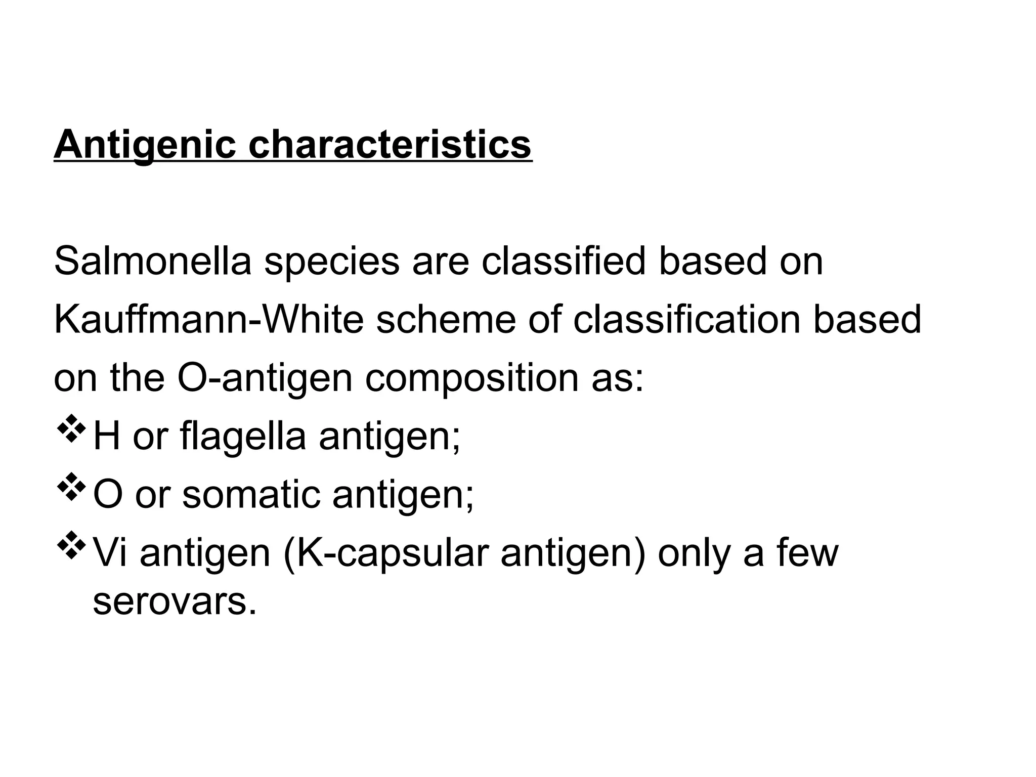
Antigenic characteristics
Salmonella speciesare classified based on
Kauffmann-White scheme of classification based
on the O-antigen composition as:
H or flagella antigen;
O or somatic antigen;
Vi antigen (K-capsular antigen) only a few
serovars.
8.
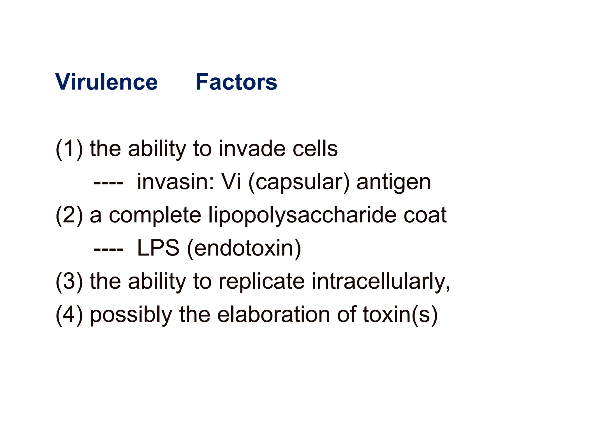
Virulence Factors
(1) theability to invade cells
---- invasin: Vi (capsular) antigen
(2) a complete lipopolysaccharide coat
---- LPS (endotoxin)
(3) the ability to replicate intracellularly,
(4) possibly the elaboration of toxin(s)
9.
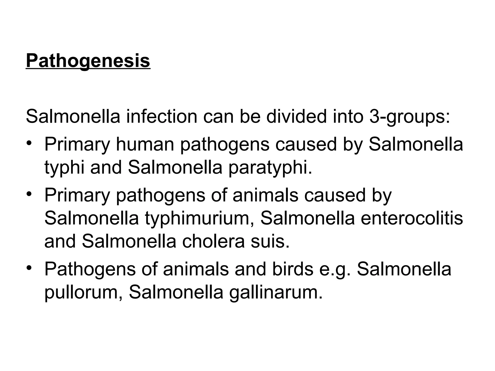
Pathogenesis
Salmonella infection canbe divided into 3-groups:
• Primary human pathogens caused by Salmonella
typhi and Salmonella paratyphi.
• Primary pathogens of animals caused by
Salmonella typhimurium, Salmonella enterocolitis
and Salmonella cholera suis.
• Pathogens of animals and birds e.g. Salmonella
pullorum, Salmonella gallinarum.
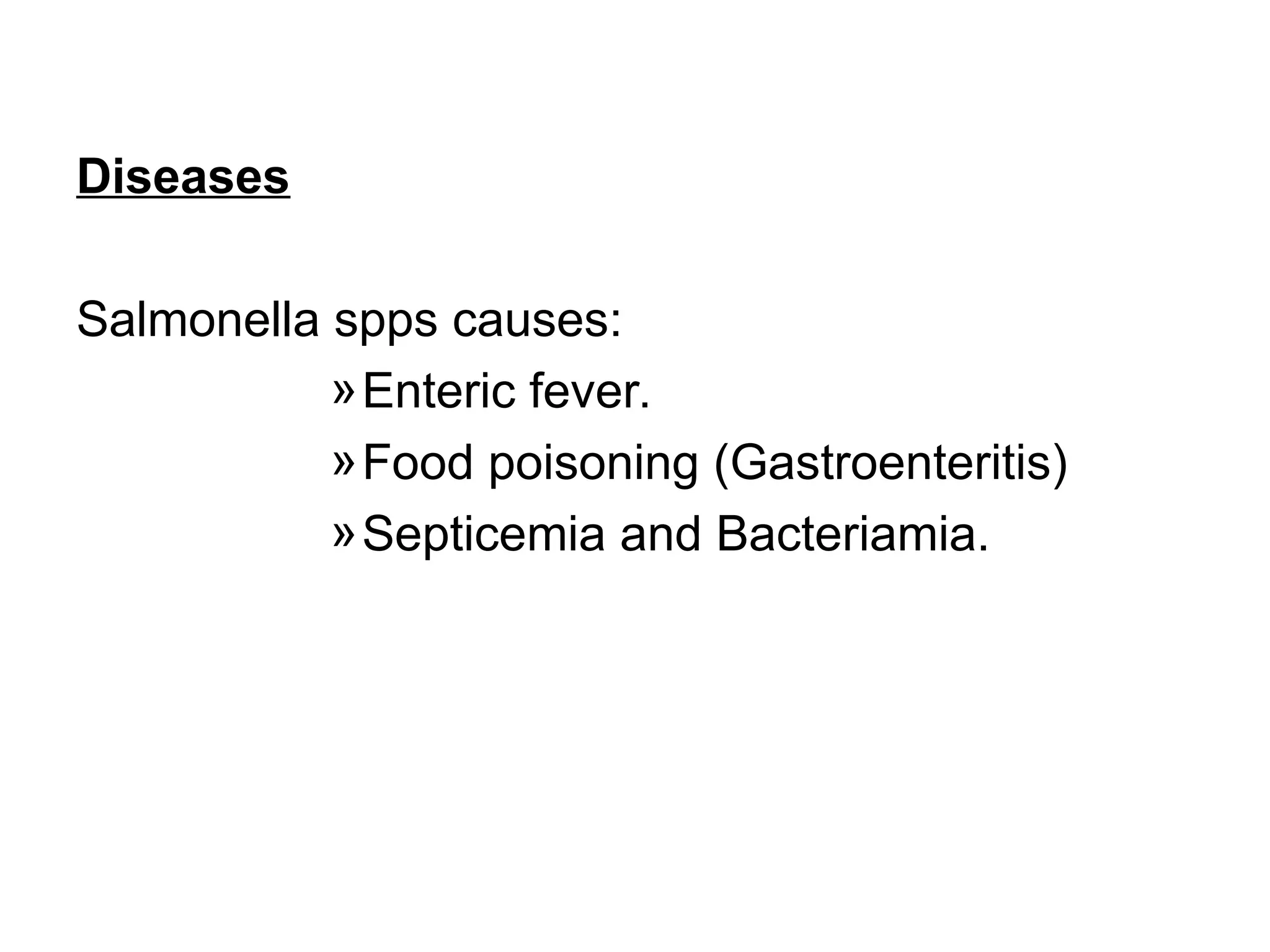
Salmonella -- ClinicalManifestations
1) Enteric fevers ---- typhoid
• Is a severe systemic form of fever which may be fatal.
• The best studied enteric fever is typhoid fever, mainly
caused by S. typhi
• Has an incubation period of 10 to 14 days with non-
specific symptoms such as :fever, anorexia, headache
etc.
• It has two bacteraemic phases:
primary bacteraemic phase:
secondary and heavier bacteraemic phase (2-wks)
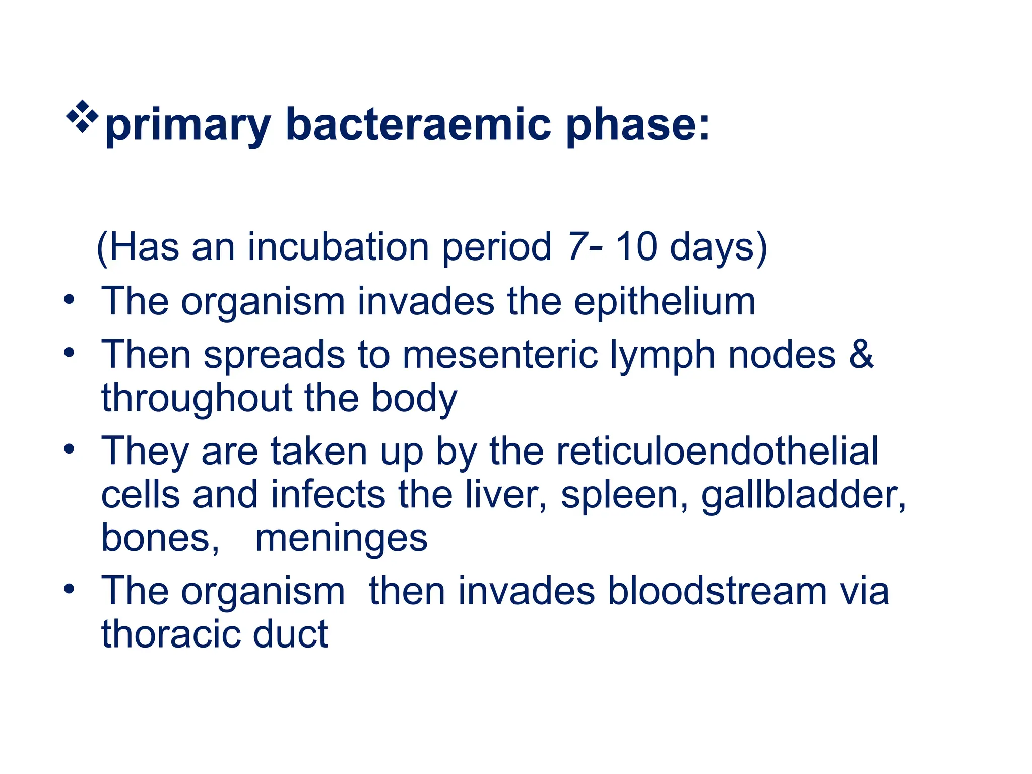
15.
primary bacteraemic phase:
(Hasan incubation period 7- 10 days)
• The organism invades the epithelium
• Then spreads to mesenteric lymph nodes &
throughout the body
• They are taken up by the reticuloendothelial
cells and infects the liver, spleen, gallbladder,
bones, meninges
• The organism then invades bloodstream via
thoracic duct
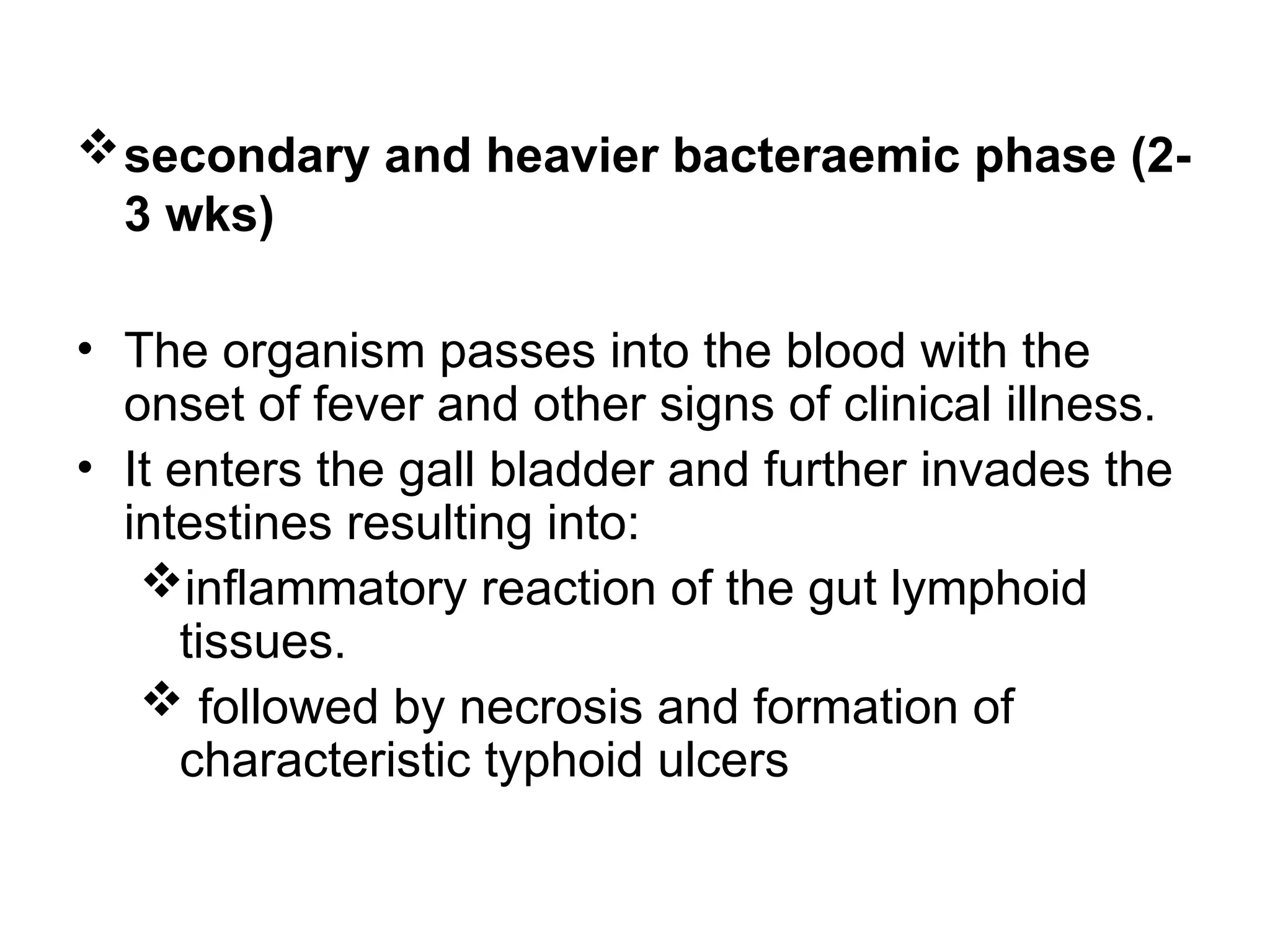
16.
secondary and heavierbacteraemic phase (2-
3 wks)
• The organism passes into the blood with the
onset of fever and other signs of clinical illness.
• It enters the gall bladder and further invades the
intestines resulting into:
inflammatory reaction of the gut lymphoid
tissues.
followed by necrosis and formation of
characteristic typhoid ulcers
17.
• Onset: 2weeks with early symptoms
• Progression :
It progresses with temperature rise over the 1st
week of the illness and remains high for 7-10
days and then falls by lysis during the 3rd or 4th
week.
• physical signs include: fever, hepatomegaly,
splenomegaly and often a rash of rose spots.
• Relapse: shorter and of milder.
• Complications: severe intestinal haemorrhage
18.
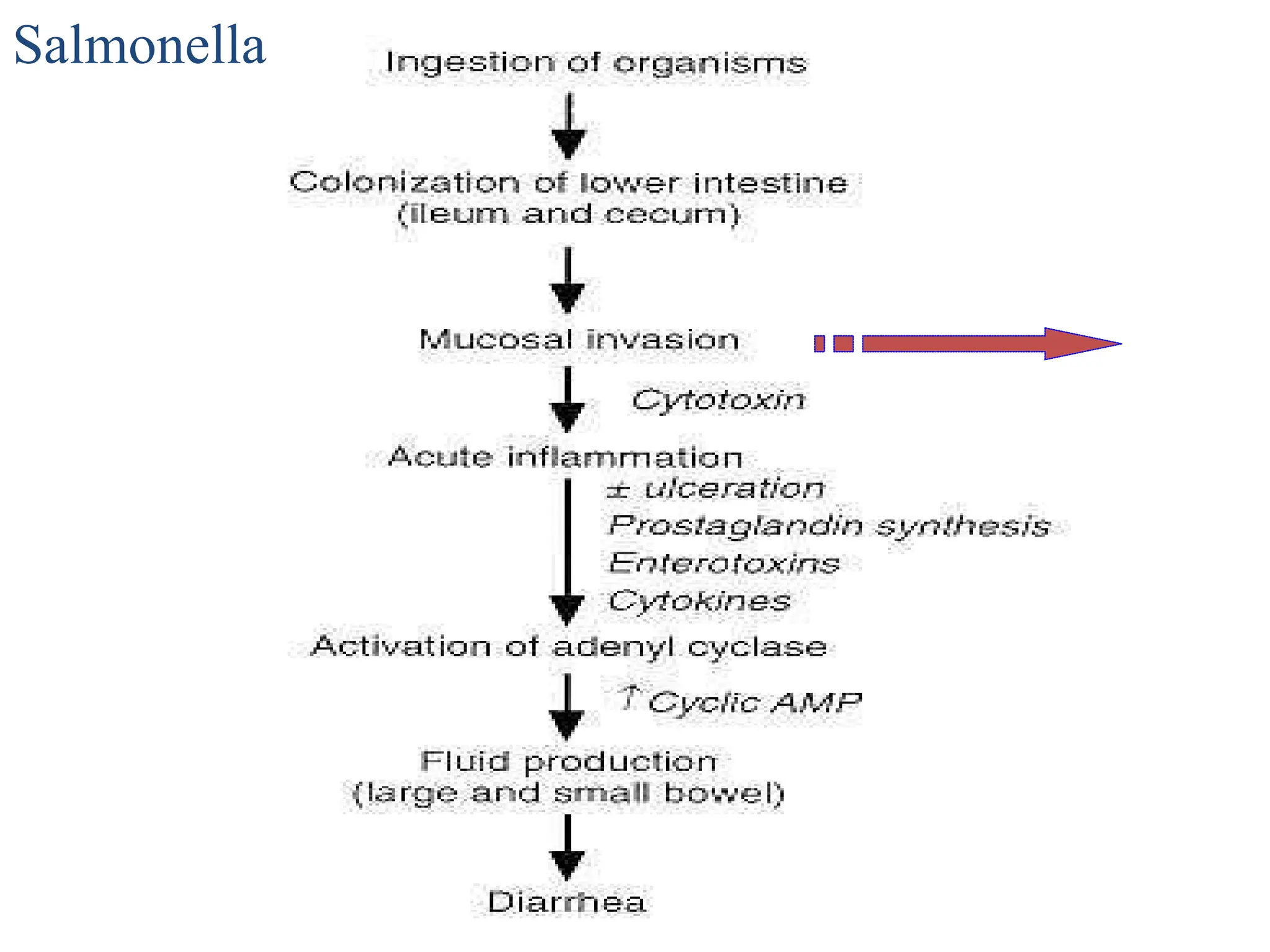
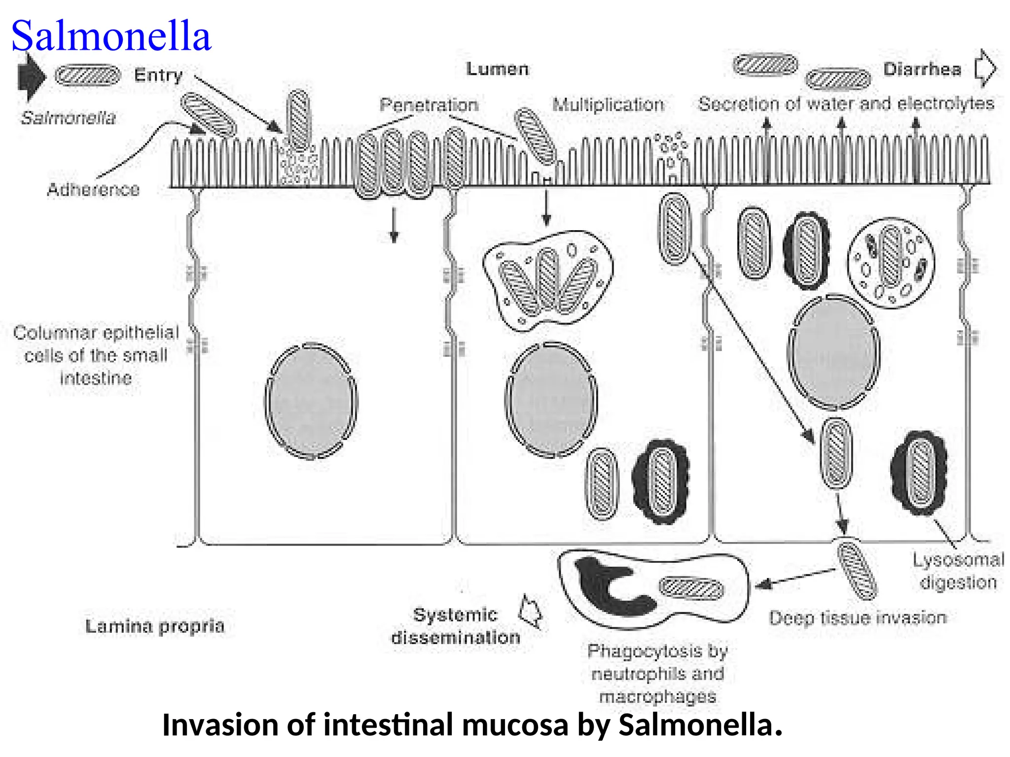
2) Gastroenteritis--food poisoning
Symptoms usually begin 6 to 48 hours after
ingestion of contaminated food or water
the cardinal manifestation is diarrhea.
nausea, vomiting, abdominal cramps,
headache, fever (38o
C to 39o
C) and chills are
common
The duration of fever and diarrhea is usually 2
to 7 days
19.
3) Septicemia
• anintermediate stage of infection –
no intestinal symptoms and the bacteria cannot
be isolated from fecal specimens.
• it may remain localized in the intestine or
disseminate to the blood streams
20.
4) The prolongedcarrier state
• continue to excrete the salmonellae for a year or
more
• The bacilli are most commonly present in the
gallbladder
21.
Laboratory diagnosis
Specimens:
Depends onthe site of infection e.g.
Blood culture for diagnosis of enteric fever
commonly found during the first 7-10 days and
during relapses
Blood serological tests.
Stool and urine culture for diagnosis of typhoid
fever:
• stool cultures are usually positive from the 2nd
week.
• urine cultures are usually positive from the 3rd
week of the infection.
22.
Day 1
Direct cultureof the specimen onto:
• XLD agar, MacConkey agar, DCA agar and
SS medium, BA, Nutrient agar medium.
• Incubate at 370
C for 18-24hrs aerobically.
1st
. gram stain for gram negative rods or
bacilli. (Brief procedure of gram staining
technique).
23.
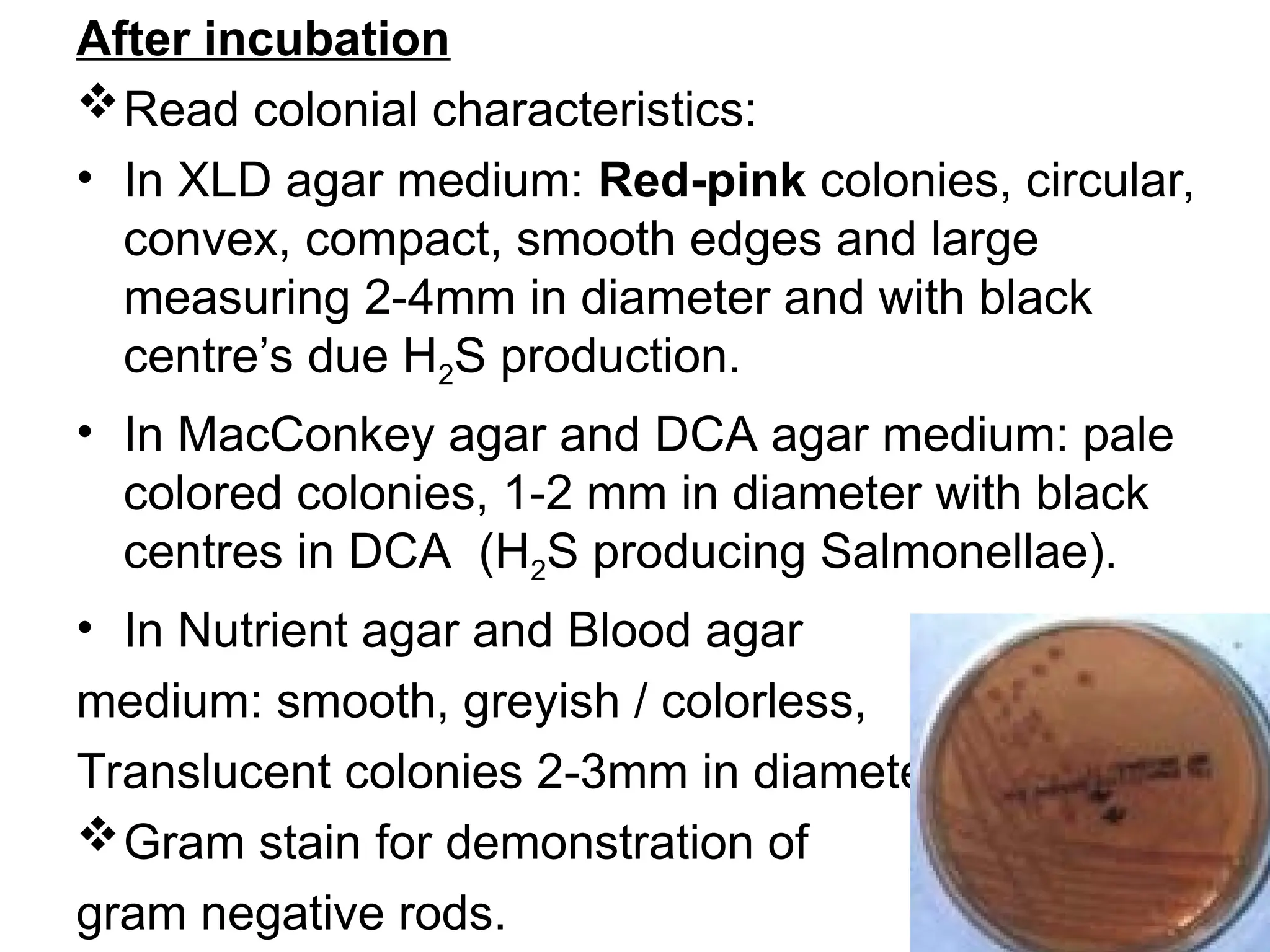
After incubation
Read colonialcharacteristics:
• In XLD agar medium: Red-pink colonies, circular,
convex, compact, smooth edges and large
measuring 2-4mm in diameter and with black
centre’s due H2S production.
• In MacConkey agar and DCA agar medium: pale
colored colonies, 1-2 mm in diameter with black
centres in DCA (H2S producing Salmonellae).
• In Nutrient agar and Blood agar
medium: smooth, greyish / colorless,
Translucent colonies 2-3mm in diameter
Gram stain for demonstration of
gram negative rods.
24.
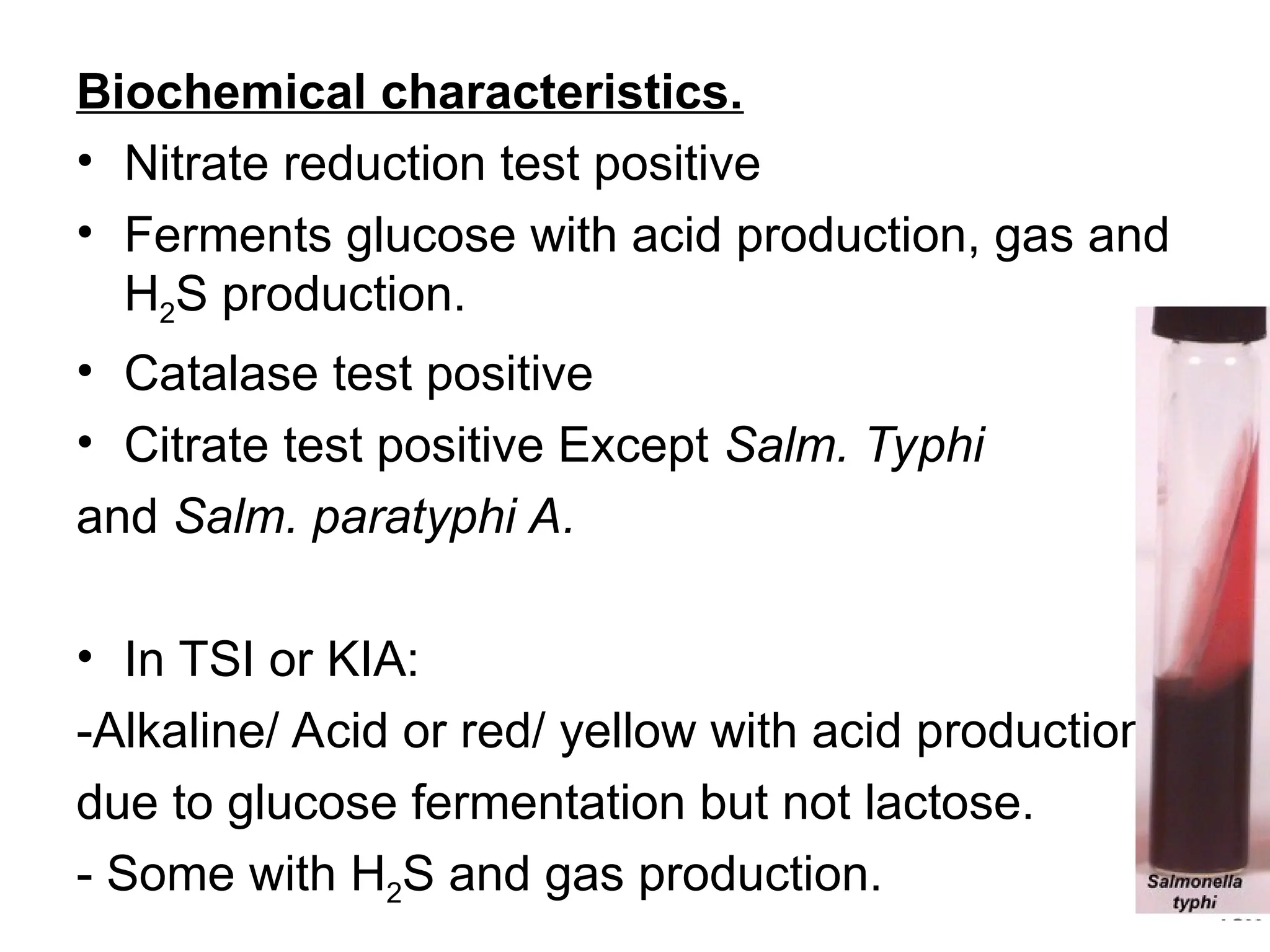
Biochemical characteristics.
• Nitratereduction test positive
• Ferments glucose with acid production, gas and
H2S production.
• Catalase test positive
• Citrate test positive Except Salm. Typhi
and Salm. paratyphi A.
• In TSI or KIA:
-Alkaline/ Acid or red/ yellow with acid production
due to glucose fermentation but not lactose.
- Some with H2S and gas production.
25.
Set drug sensitivity(susceptibility) tests using gram
negative drug discs; incubate aerobically at 370
C for
18-24hrs.
After setting drug sensitivity test.
• Read both sensitive and resistant drugs from the drug
susceptibility test, record the results and dispatch
them to the clinician for further management of the
patient.
Treatment
• Vaccines are available for typhoid fever and are
partially effective.
• Typhoid fever and enteric fevers should be treated
with antibiotics e.g. Chloromphinical, Sulphanomides,
Tetracycline, Streptomycin, Neomycin
26.
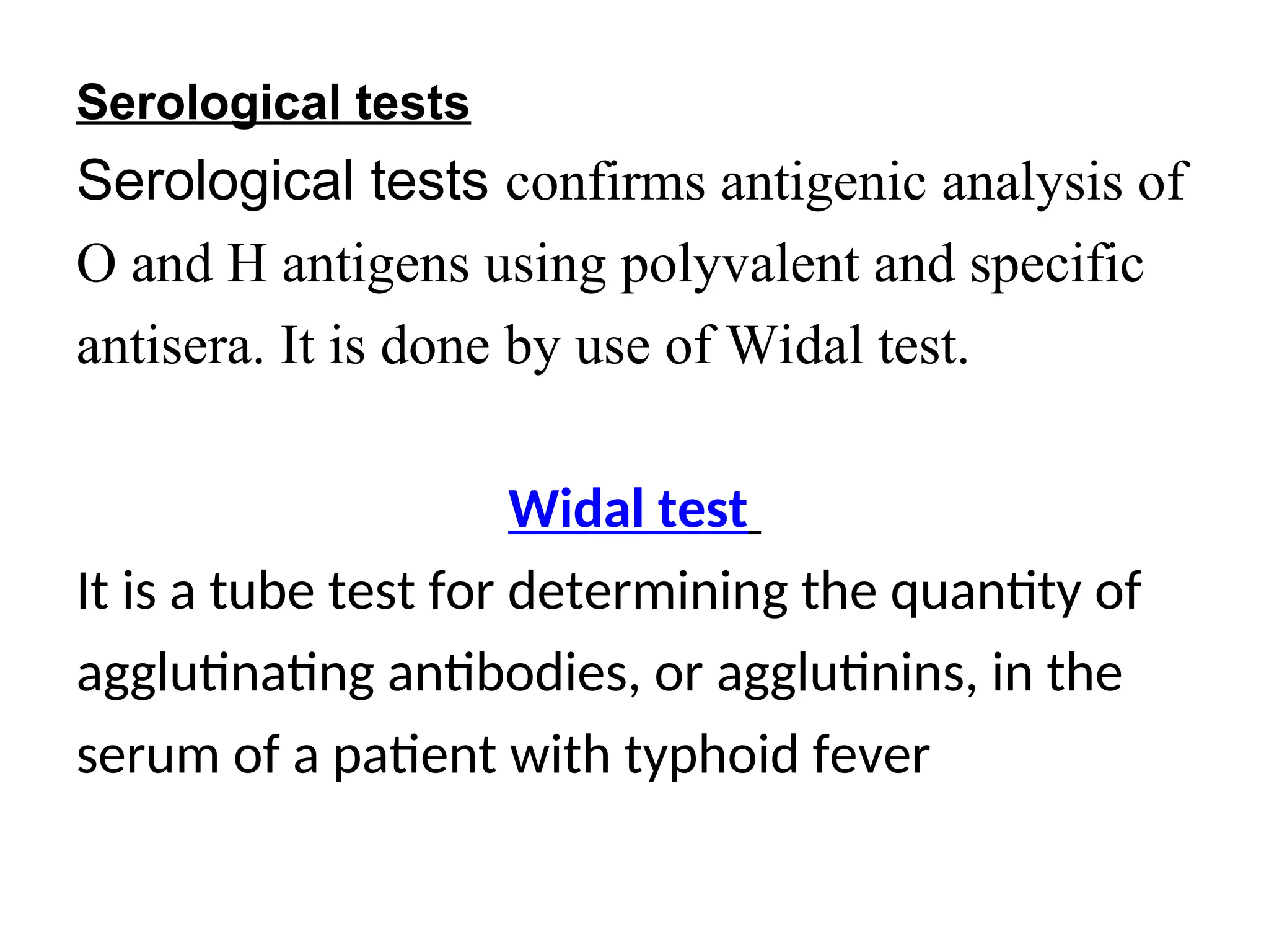
Serological tests
Serological testsconfirms antigenic analysis of
O and H antigens using polyvalent and specific
antisera. It is done by use of Widal test.
Widal test
It is a tube test for determining the quantity of
agglutinating antibodies, or agglutinins, in the
serum of a patient with typhoid fever
27.
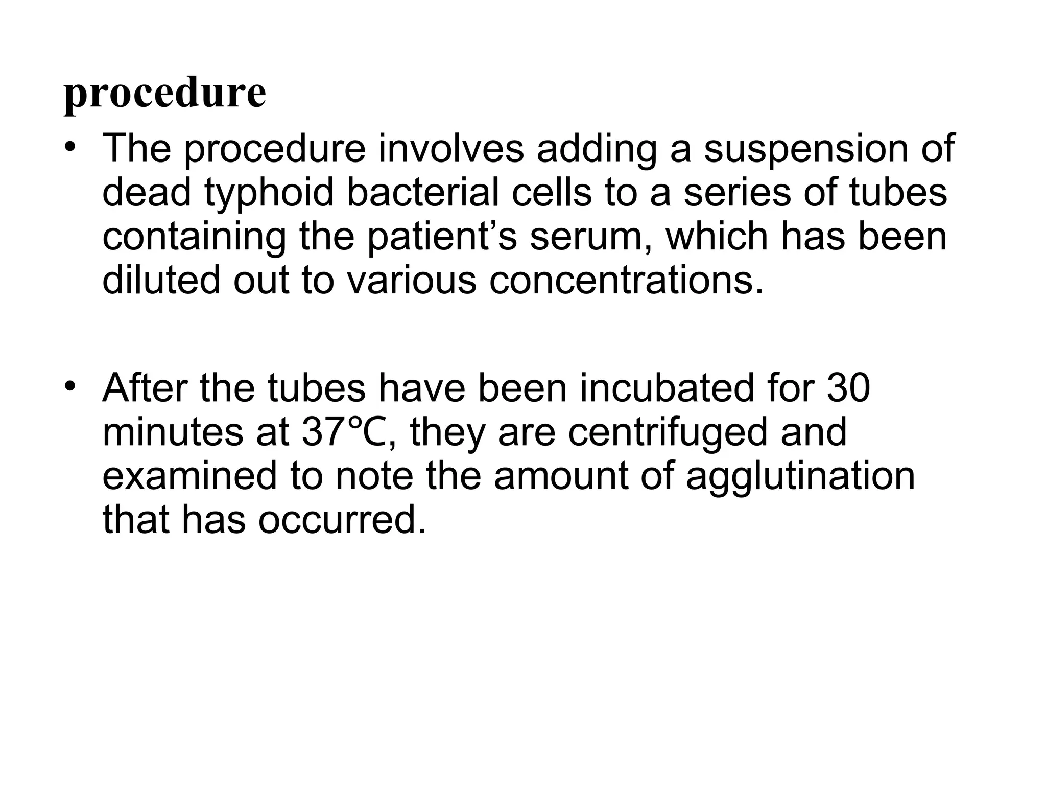
procedure
• The procedureinvolves adding a suspension of
dead typhoid bacterial cells to a series of tubes
containing the patient’s serum, which has been
diluted out to various concentrations.
• After the tubes have been incubated for 30
minutes at 37 , they are centrifuged and
℃
examined to note the amount of agglutination
that has occurred.
28.
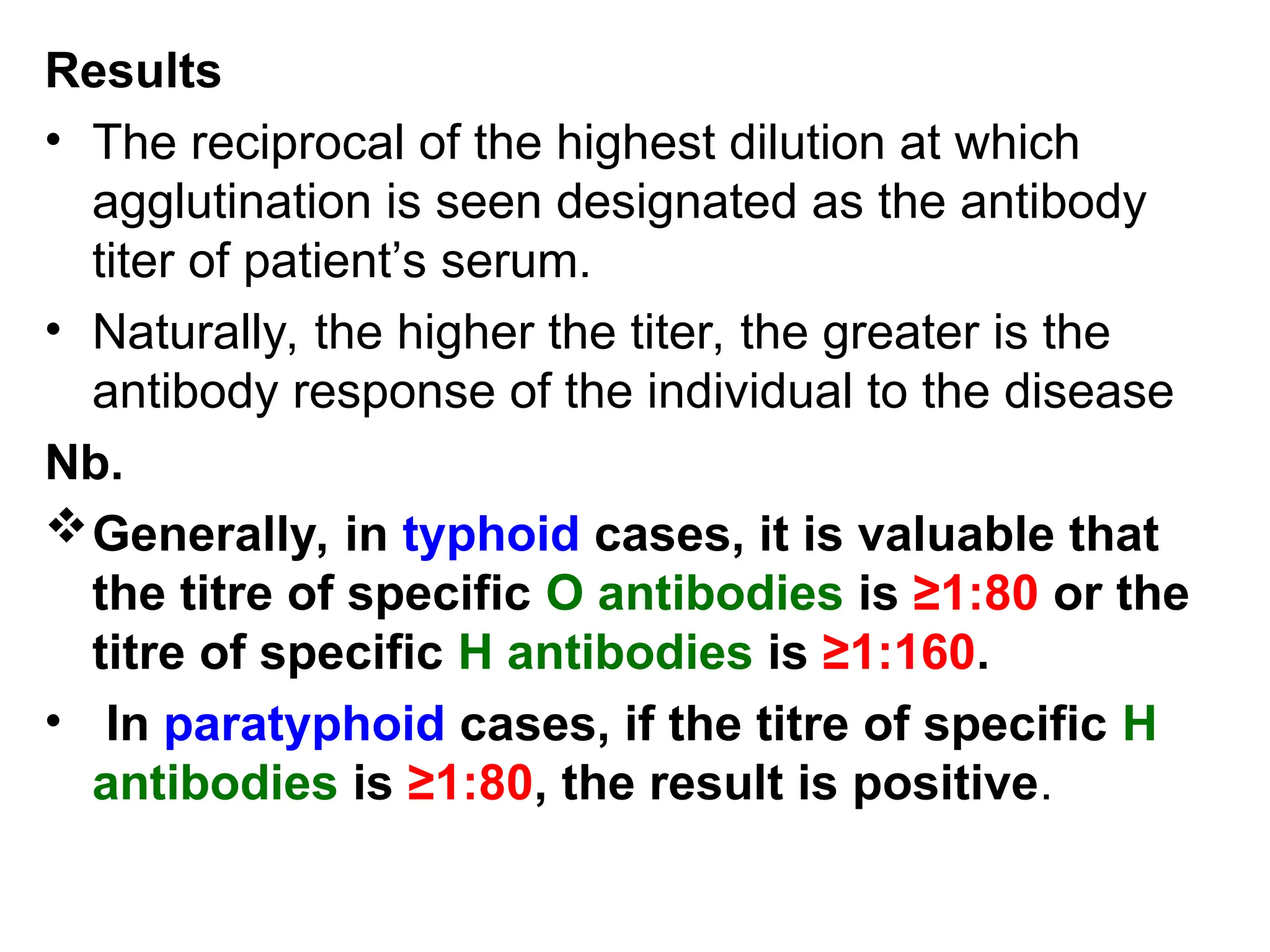
Results
• The reciprocalof the highest dilution at which
agglutination is seen designated as the antibody
titer of patient’s serum.
• Naturally, the higher the titer, the greater is the
antibody response of the individual to the disease
Nb.
Generally, in typhoid cases, it is valuable that
the titre of specific O antibodies is ≥1:80 or the
titre of specific H antibodies is ≥1:160.
• In paratyphoid cases, if the titre of specific H
antibodies is ≥1:80, the result is positive.
29.
Interpretation
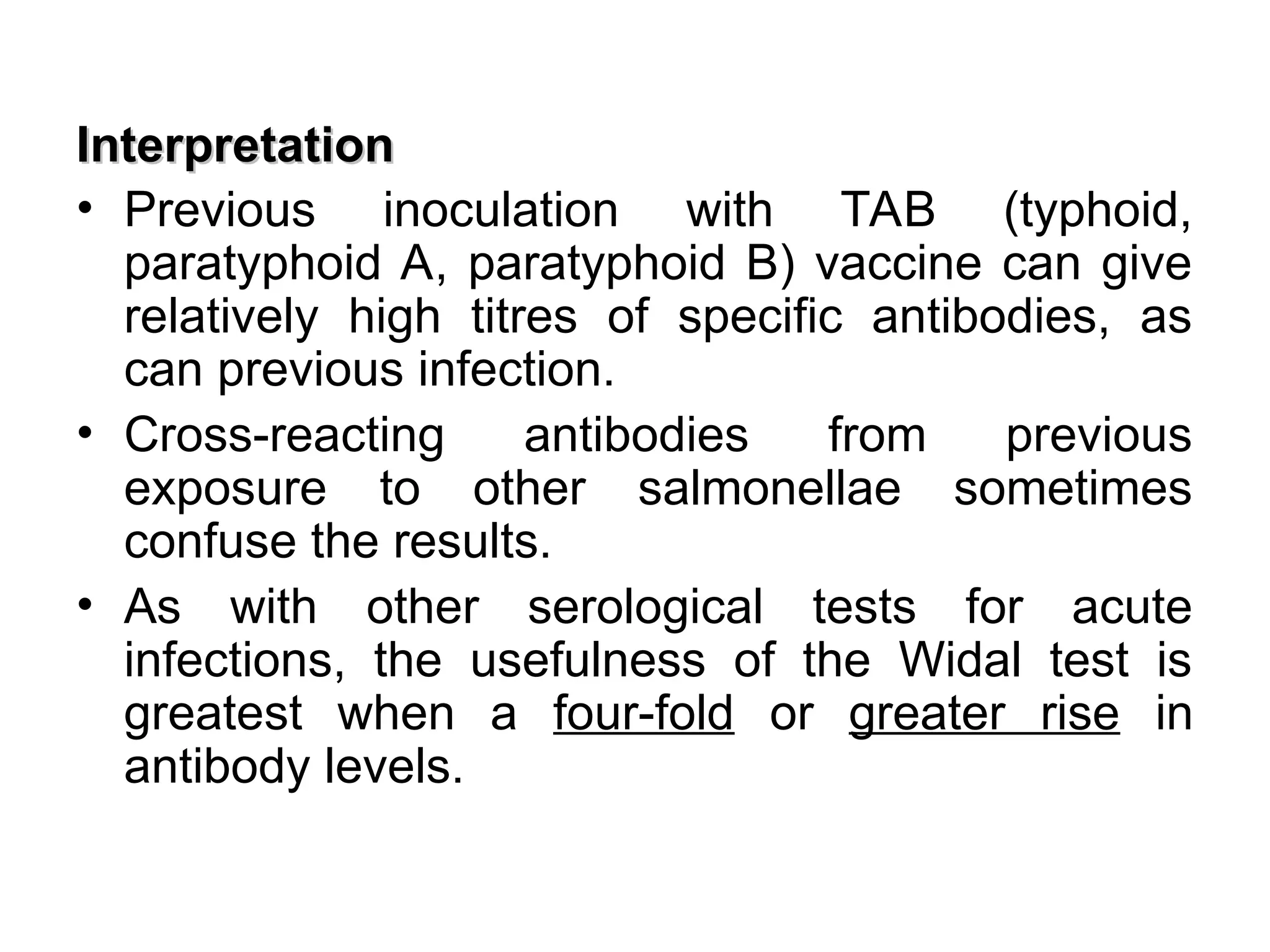
Interpretation
• Previous inoculationwith TAB (typhoid,
paratyphoid A, paratyphoid B) vaccine can give
relatively high titres of specific antibodies, as
can previous infection.
• Cross-reacting antibodies from previous
exposure to other salmonellae sometimes
confuse the results.
• As with other serological tests for acute
infections, the usefulness of the Widal test is
greatest when a four-fold or greater rise in
antibody levels.
30.
Other causes ofhigh titres of ‘O’ and ‘H’
antibodies.
• Chronic Salmonellosis associated with
schistosomal infections.
• Vaccines with Salm. paratyphi A and B vaccines.
• Chronic liver diseases.
• Immunological disorders e.g. multiple myeloma,
nephritic syndromes, arthritis and Rheumatoid
arthritis.
• Infection with other Salmonella species.
31.
ENTEROBACTER
• Enterobacter ispart of the commensal enteric flora, but it
is also found in water, sewage, soil, and plants.
• Organism posses a capsule and they are motile.
• Eleven species of Enterobacter have been described, but E.
aerogenes and E. cloacae cause most human infections.
• Enterobacter species cause opportunistic infections; most
often they cause urinary tract infections in debilitated or
catheterized patients.
• Occasionally they cause pneumonia, wound infections, and
sepsis in hospitalized patients.
32.
SERRATIA
• Nine speciesof Serratia have been described, but
most human disease is due to a single species, S.
marcescens.
• Between 75-90 % of all Serratia infections are
nosocomial.
• Serratia causes pneumonia and sepsis particularly in
patients with cancer and are receiving chemotherapy.
• It also causes occasionally urinary tract infections and
wound infections in hospitalized patients.
33.
PROTEUS
• Proteus speciesare part of the normal human gastrointestinal flora
and exist in water and soil as saprophytic organisms.
• Members of the genus Proteus are gram negative, motile bacilli.
• They exhibit strong urease activity.
• Although four species of Proteus have been identified, only two
cause human disease: P. mirabilis and P. vulgaris.
• P.mirabilis is the more common of the two pathogens.
• Many isolates of Proteus are extremely motile and exhibit “swarming
motility” on blood agar plates.
• Swarming Proteus isolates spread in waves across the agar surface
and sometimes makes isolation of other organisms on the plate
extremely difficult or impossible.
34.
Pathogenicity
• The principalvirulence determinants of Proteus
species are lipopolysaccharide, pili, urease activity,
and capsule.
• The lipopolysaccharide of Proteus, like that of
other enteric organisms, exhibits endotoxic
activity.
• The Proteus pili promote colonization of the
kidney, and the Proteus urease converts urea to
NH4 and CO2.
35.
• These productsof urea hydrolysis alkalinize the
urine, and this precipitates Mg2+ and Ca2+ and leads
to the formation of renal calculi (kidney stones).
• NH4 also protects Proteus in the kidney from classic
complement pathway by splitting C4.
• Studies have shown that the Proteus capsule not
only protects the organisms from phagocytosis, but
also precipitates MgNH4PO4 6H2O (stuvite).
• Struvite calculi are a frequent complication of P.
mirabilis urinary tract infection
36.
Diseases
• Proteus isa common cause of nosocomial and
community-acquired urinary tract infections,
including pyelonephritis and cystitis.
• Pyelonephritis may lead to sepsis.
• Other infections include wound infections and
pneumonia