Download as PPSX, PPTX


The document provides detailed information about the characteristics, growth conditions, and biochemical tests of gram-negative bacteria, particularly within the Enterobacteriaceae family. It includes specifics on fermentation processes, staining techniques, and identification methods based on various culture media. Additionally, it discusses the clinical significance, common infections, and identification challenges associated with specific species like Escherichia coli and Salmonella.
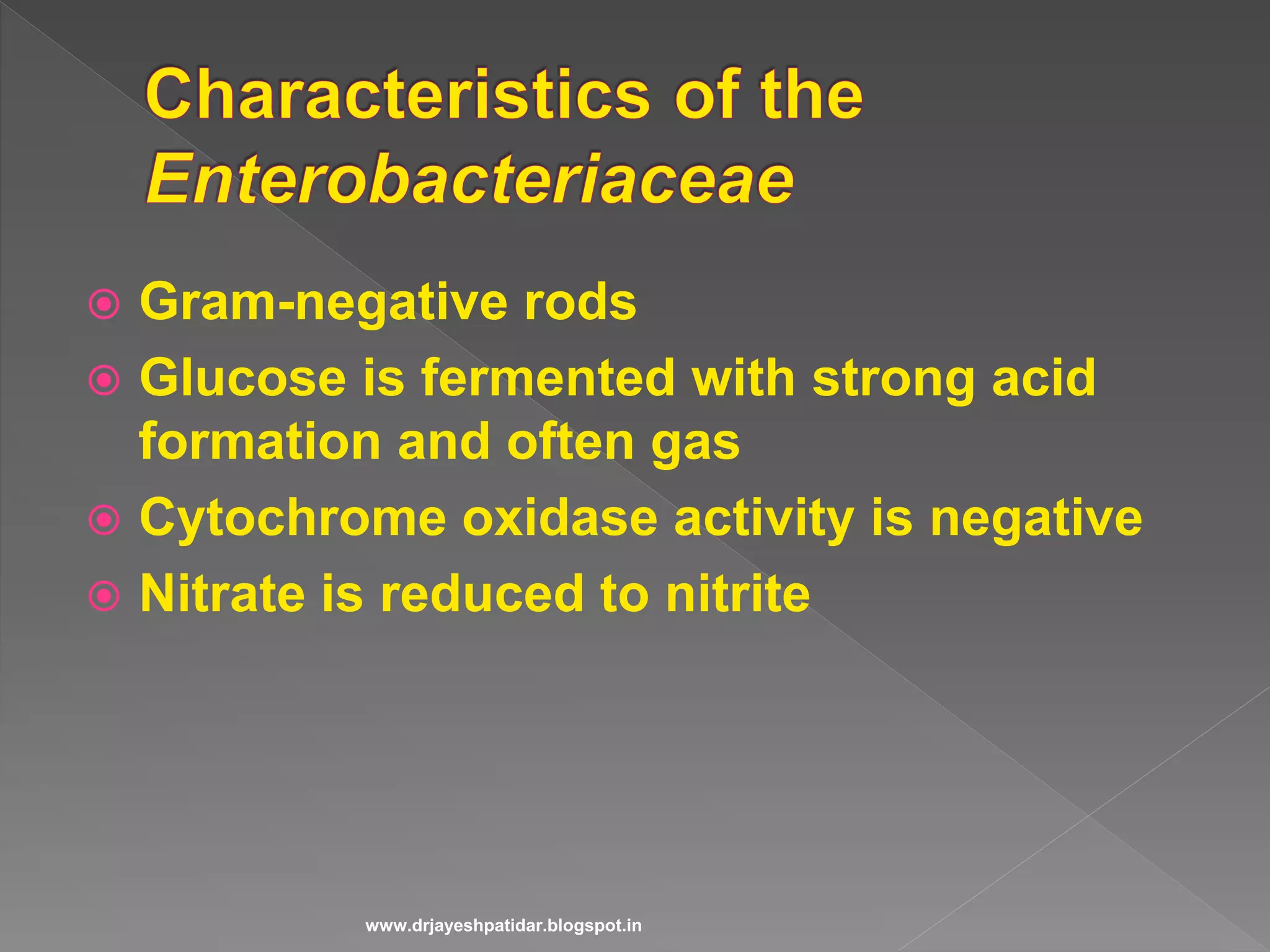
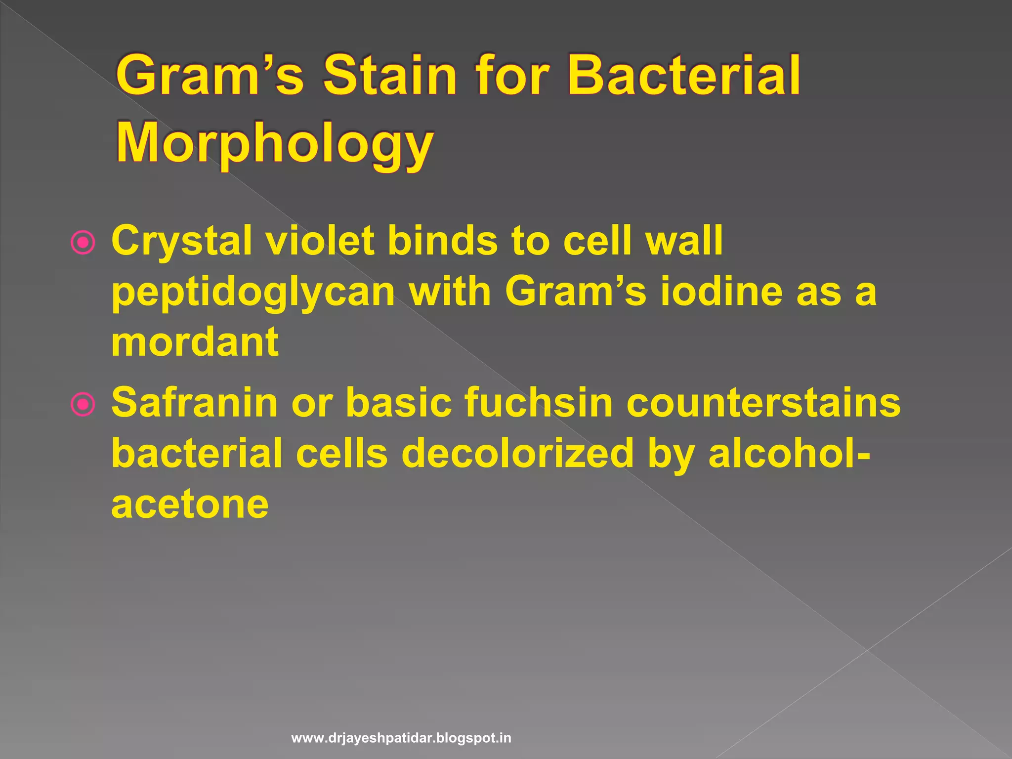
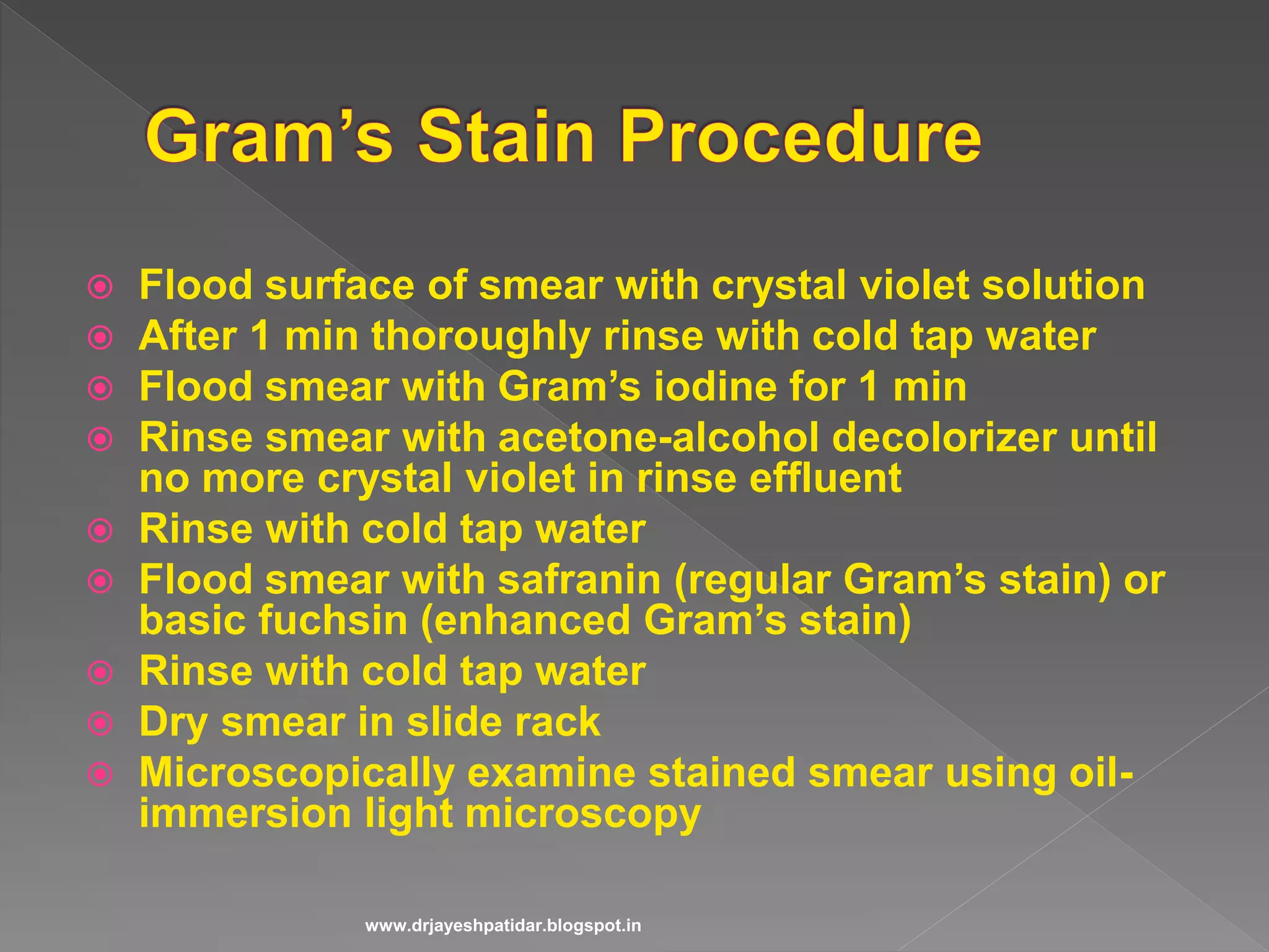
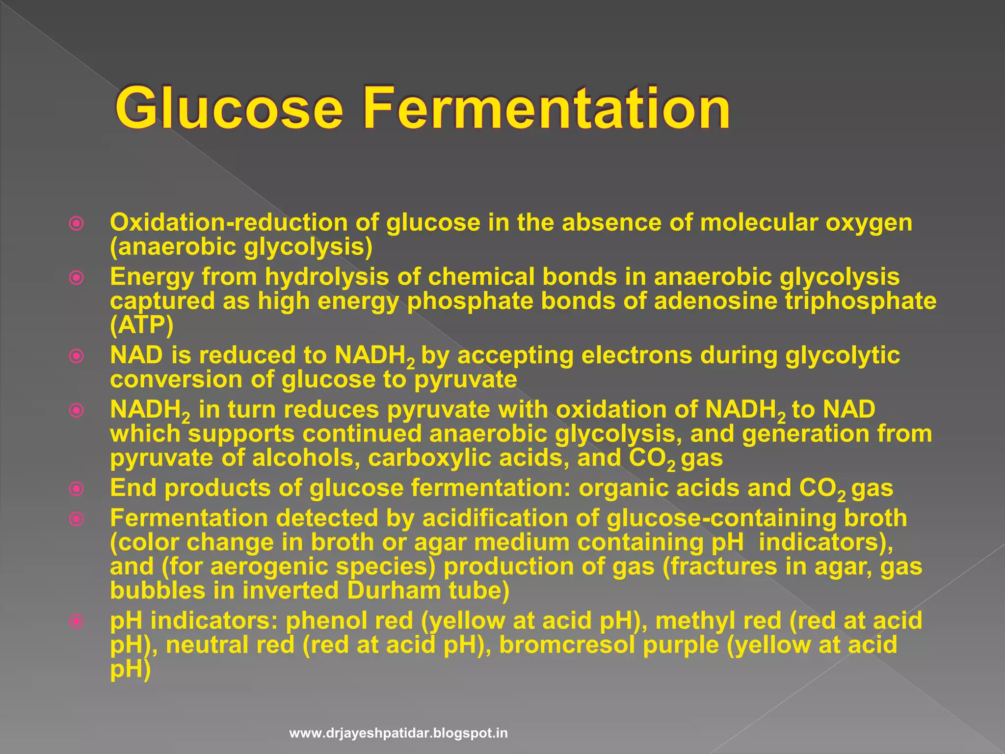
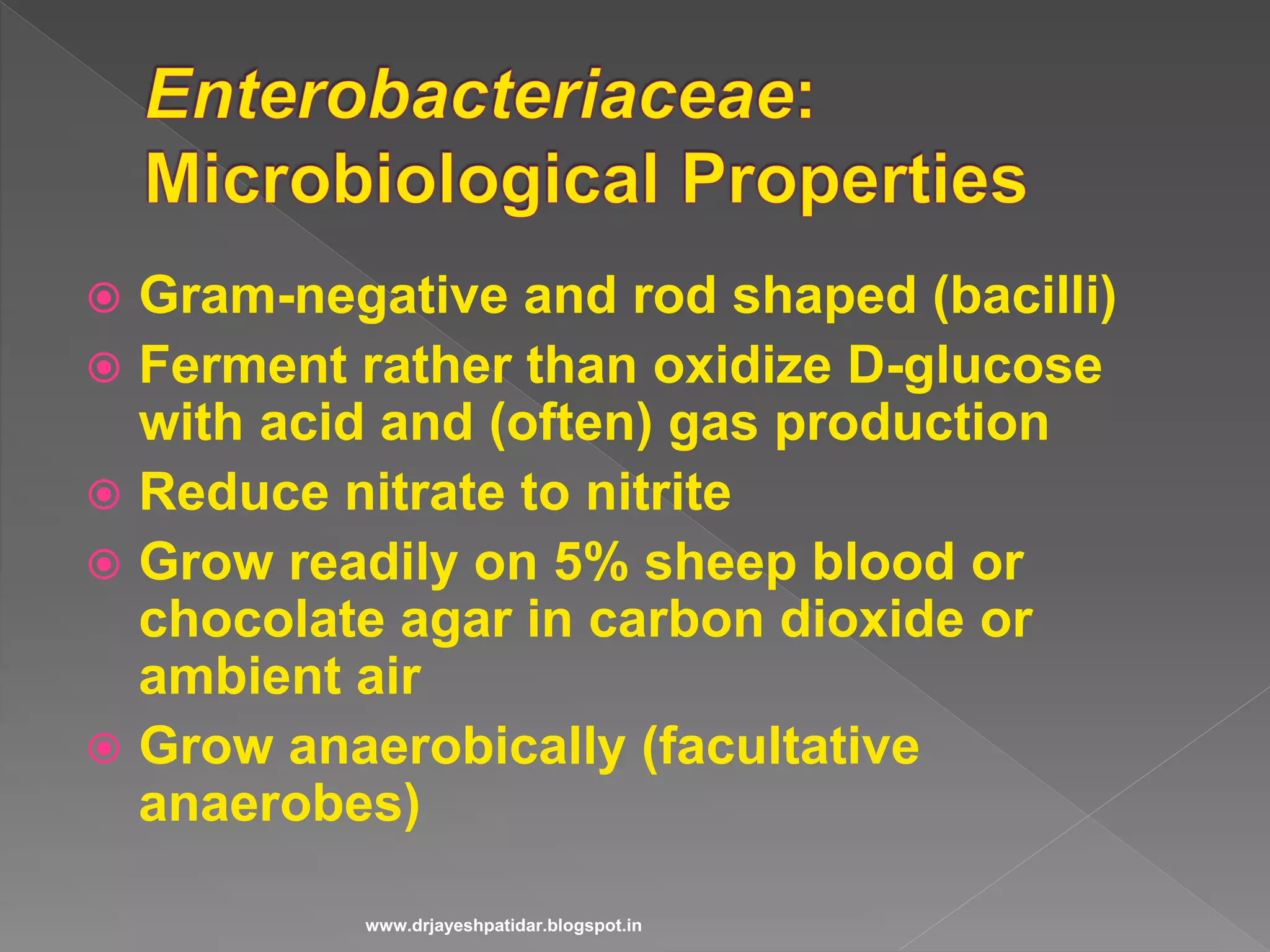
Discusses Gram-negative rods, glucose fermentation, and the Gram staining process.
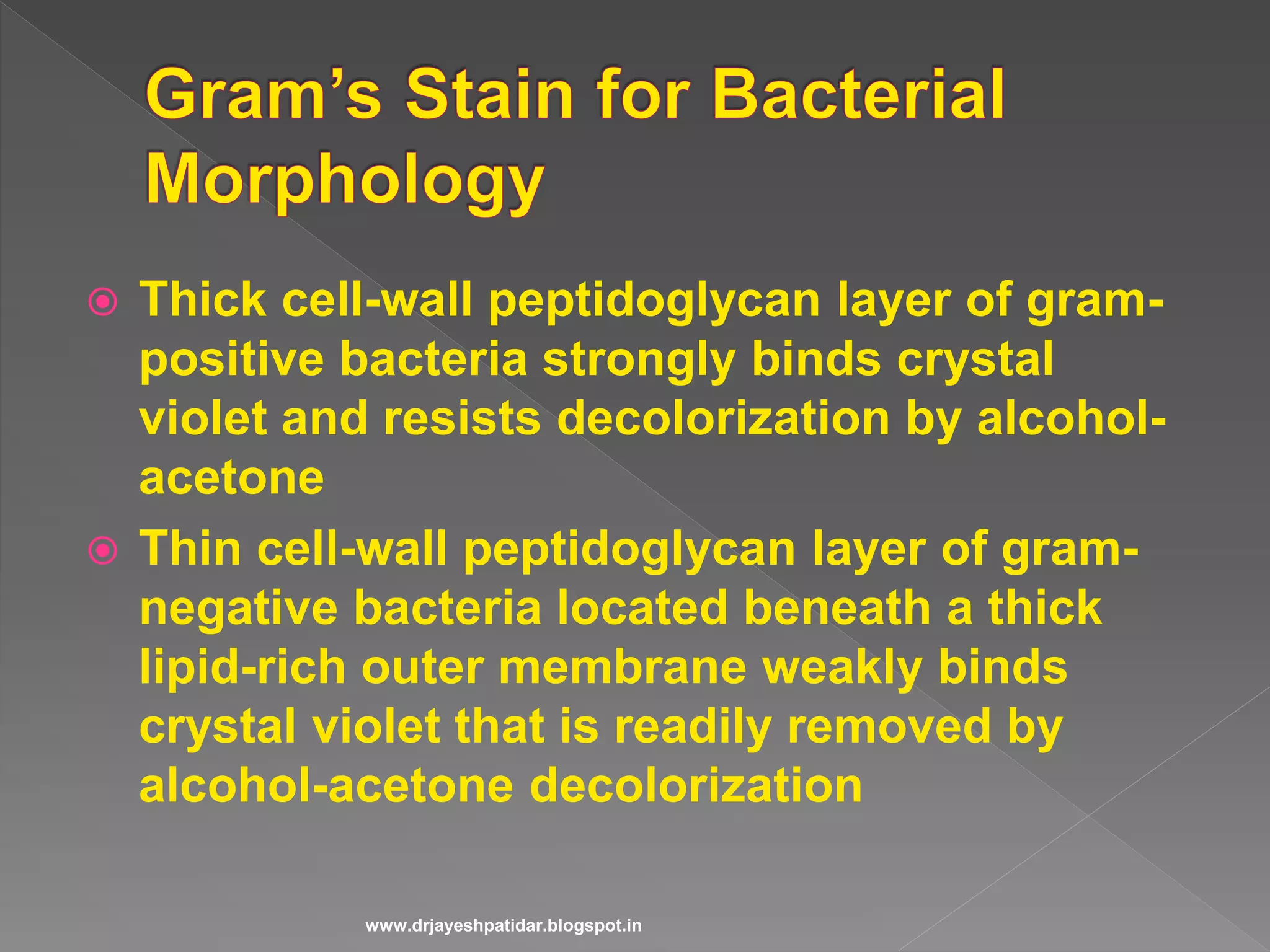
Details the Gram staining technique, highlighting differences in cell wall structure.
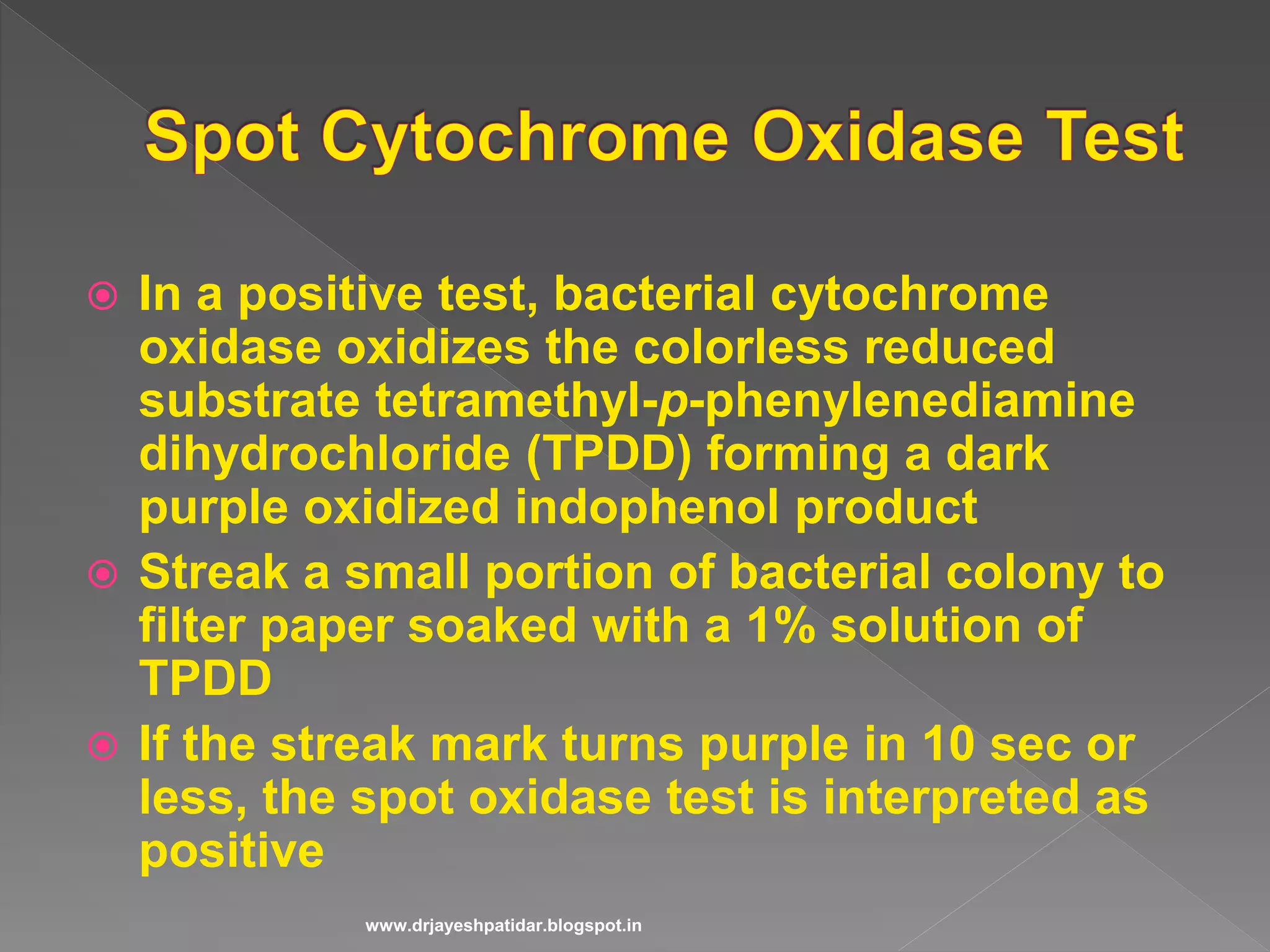
Covers anaerobic glycolysis, ATP production, and tests for nitrate reduction.
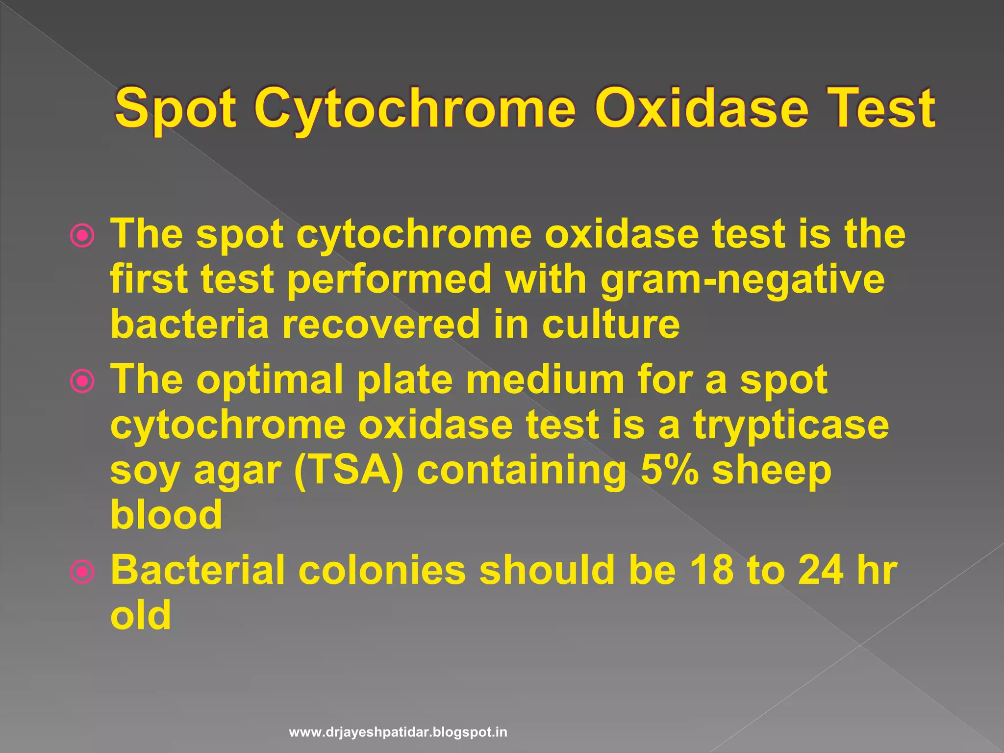
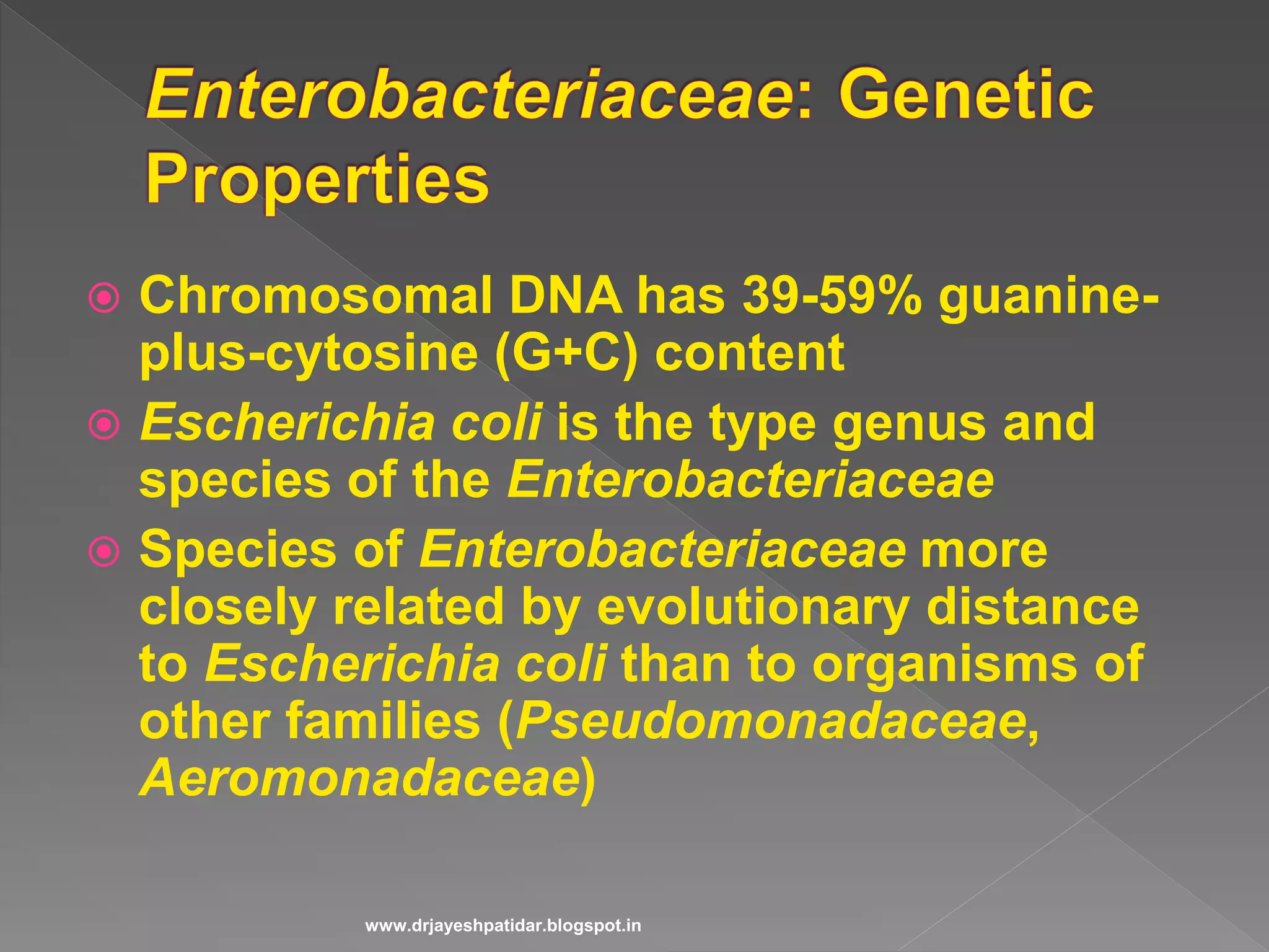
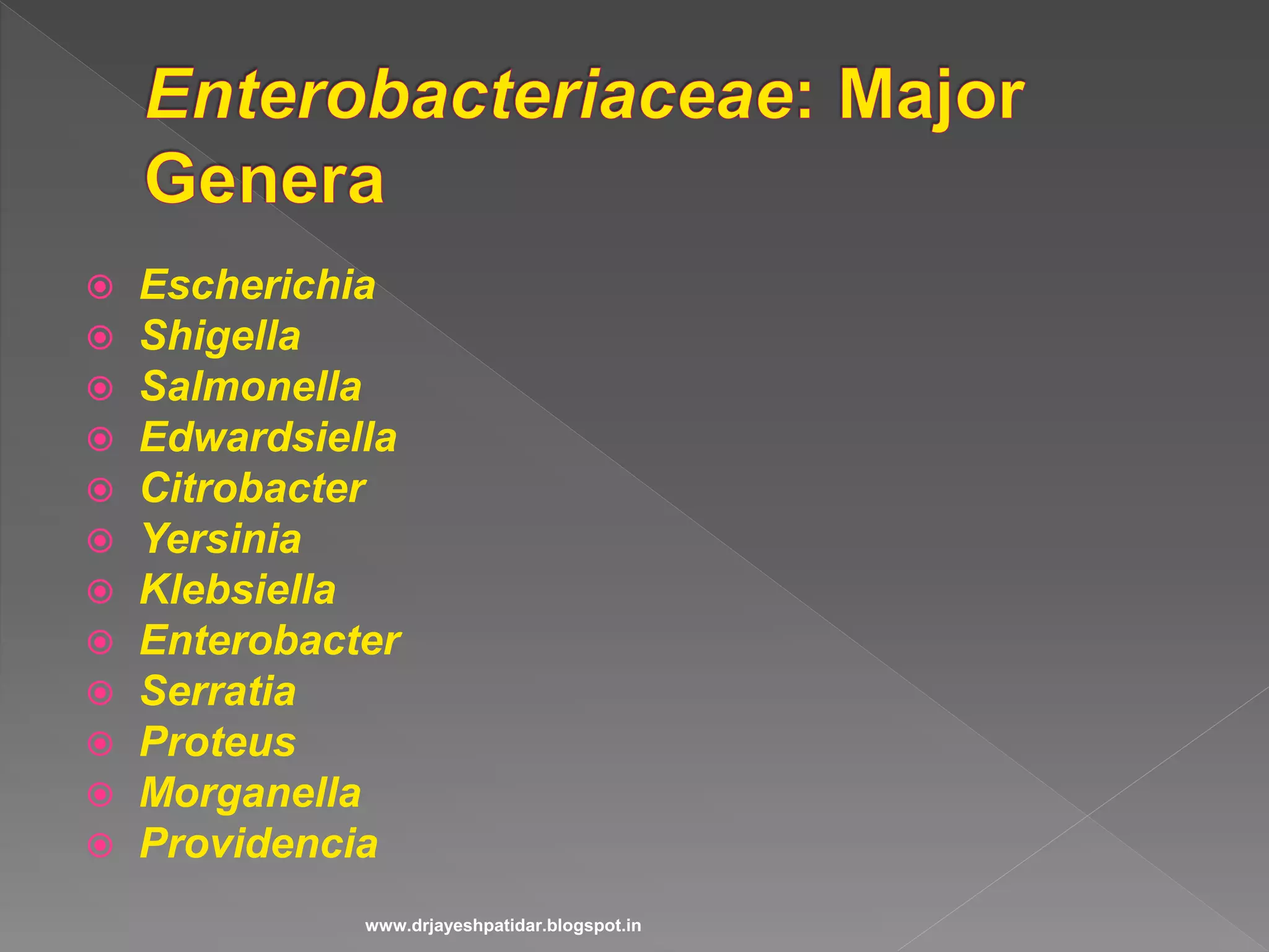
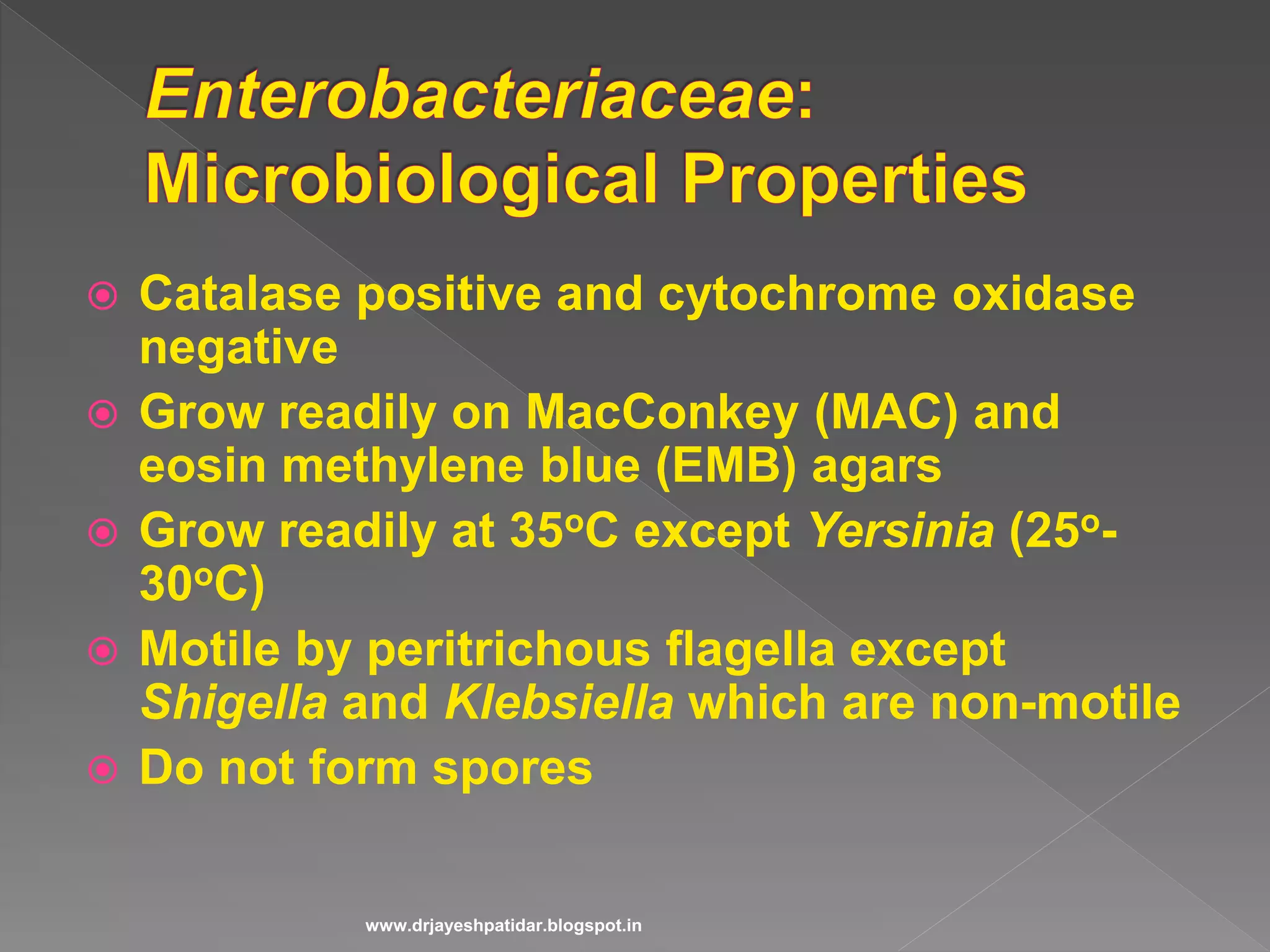
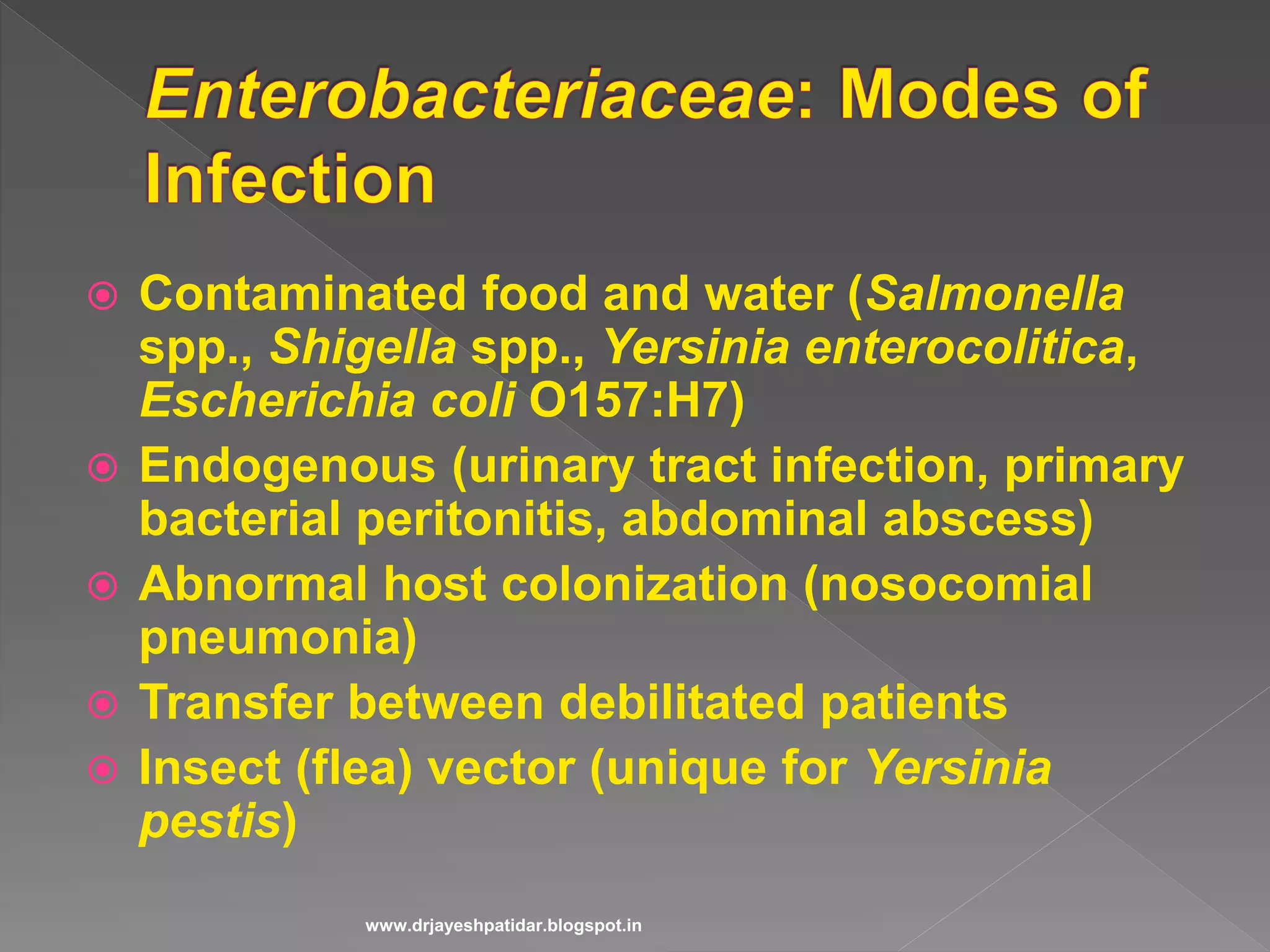
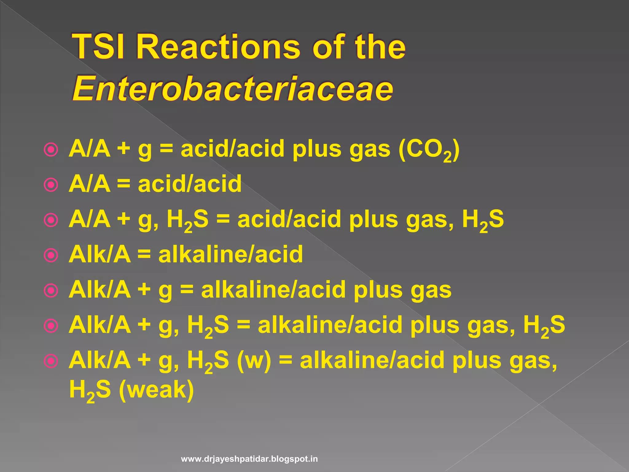
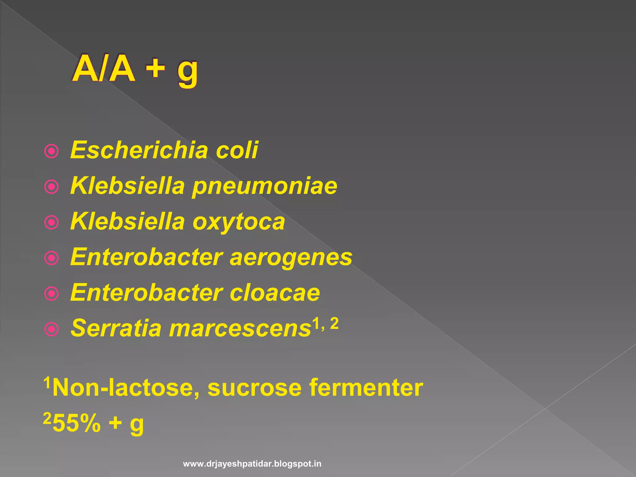
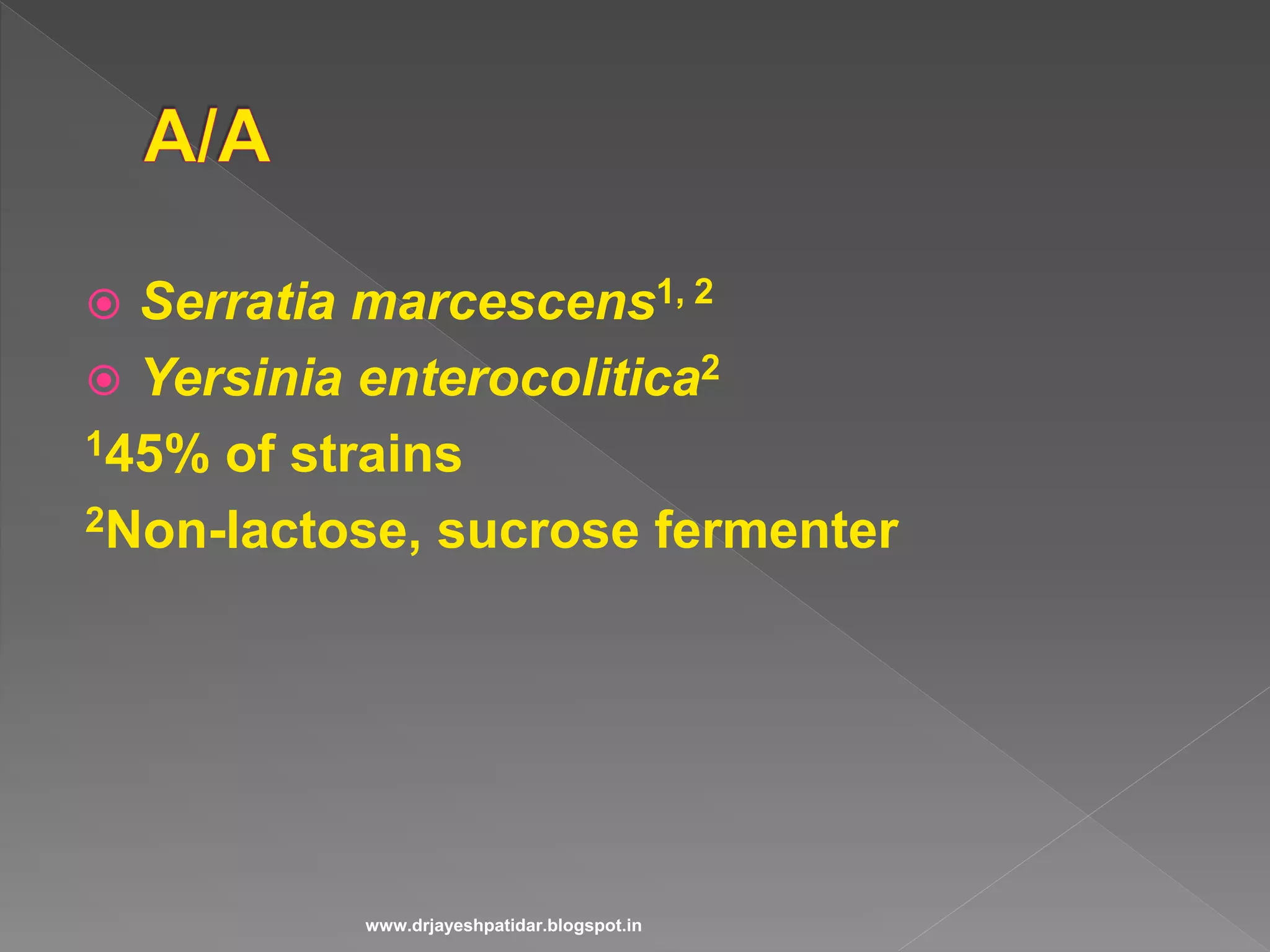
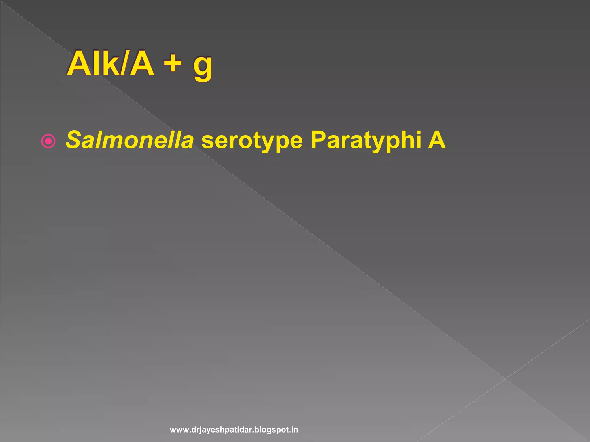
Identifies Enterobacteriaceae members, their characteristics, and growth requirements.
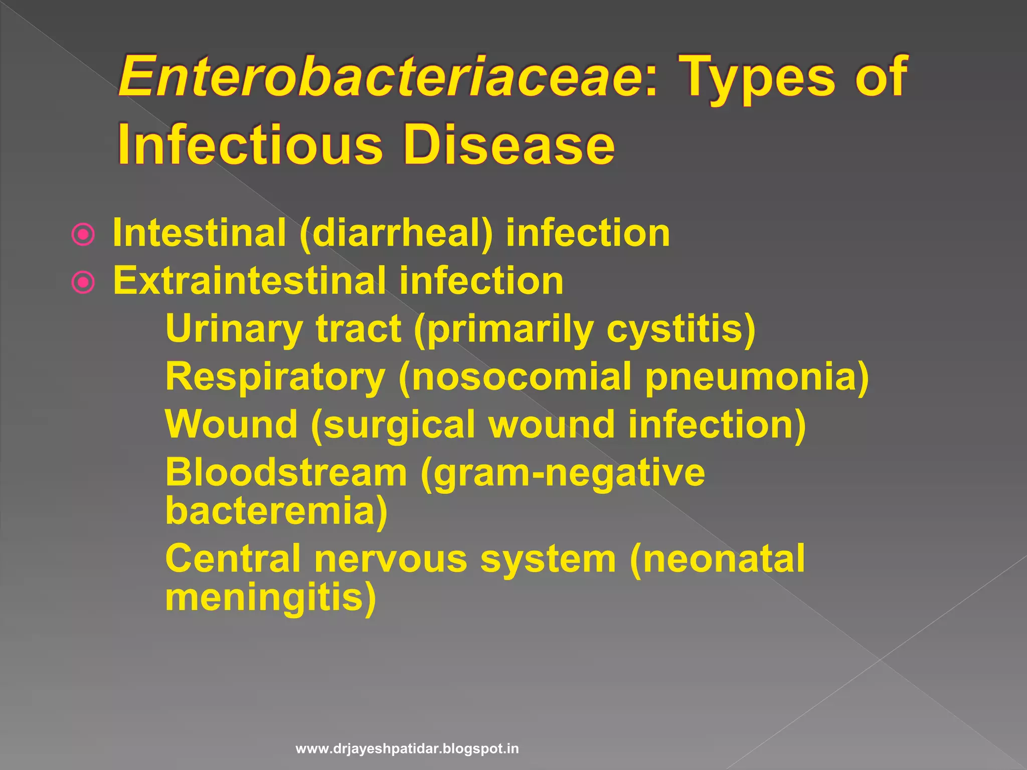
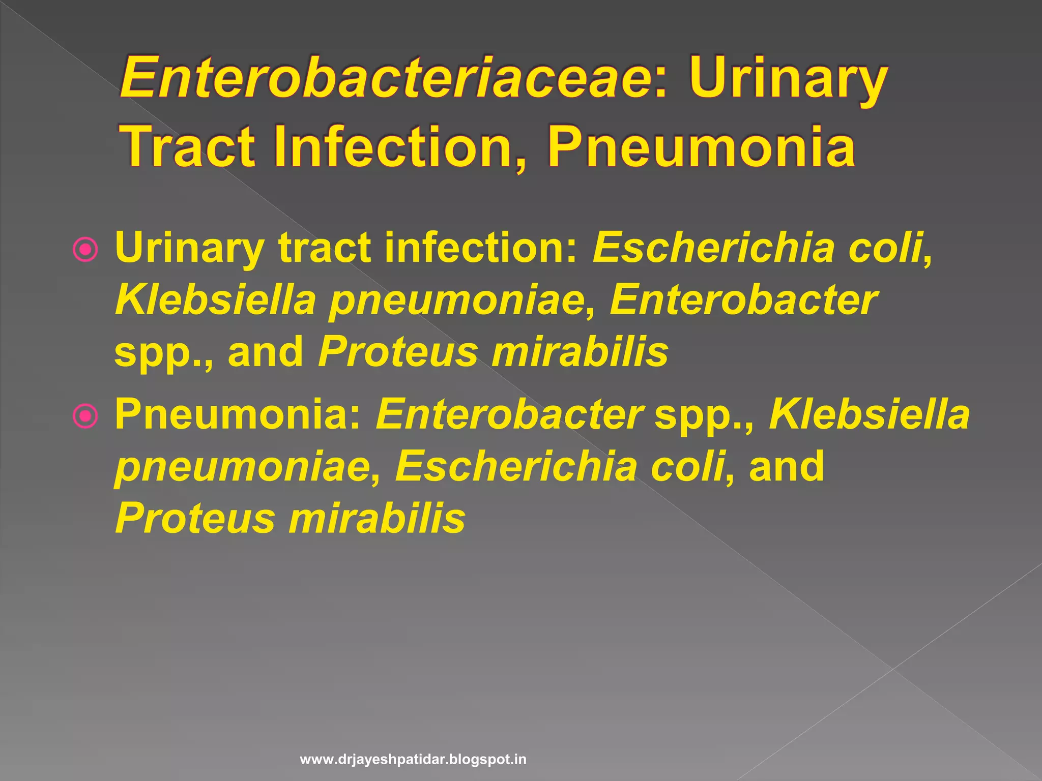
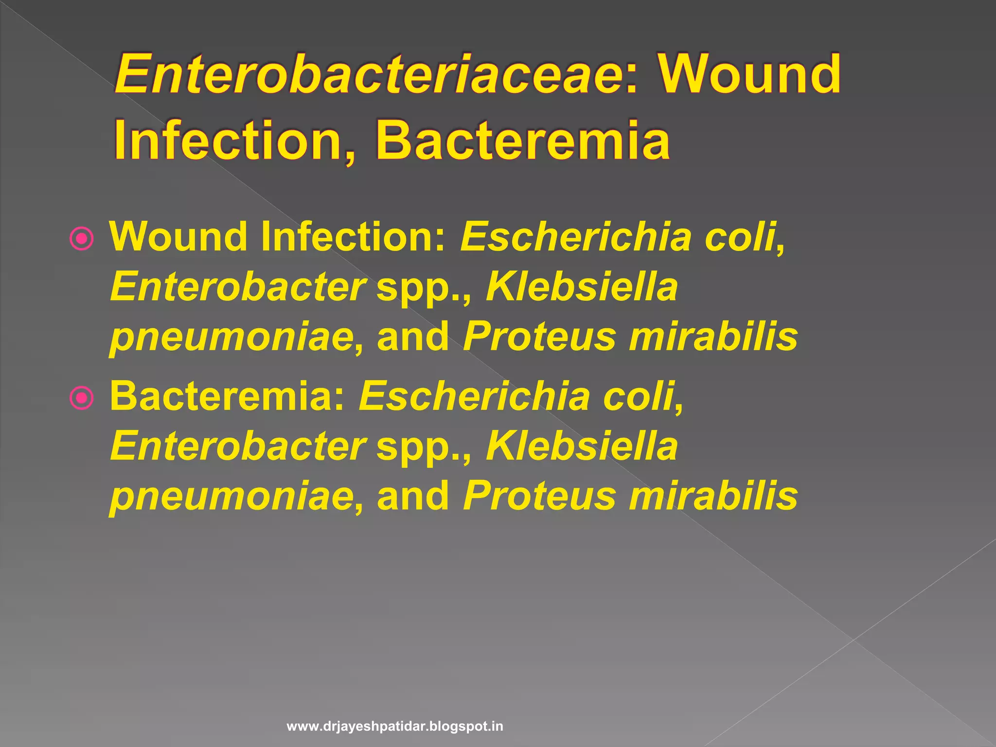
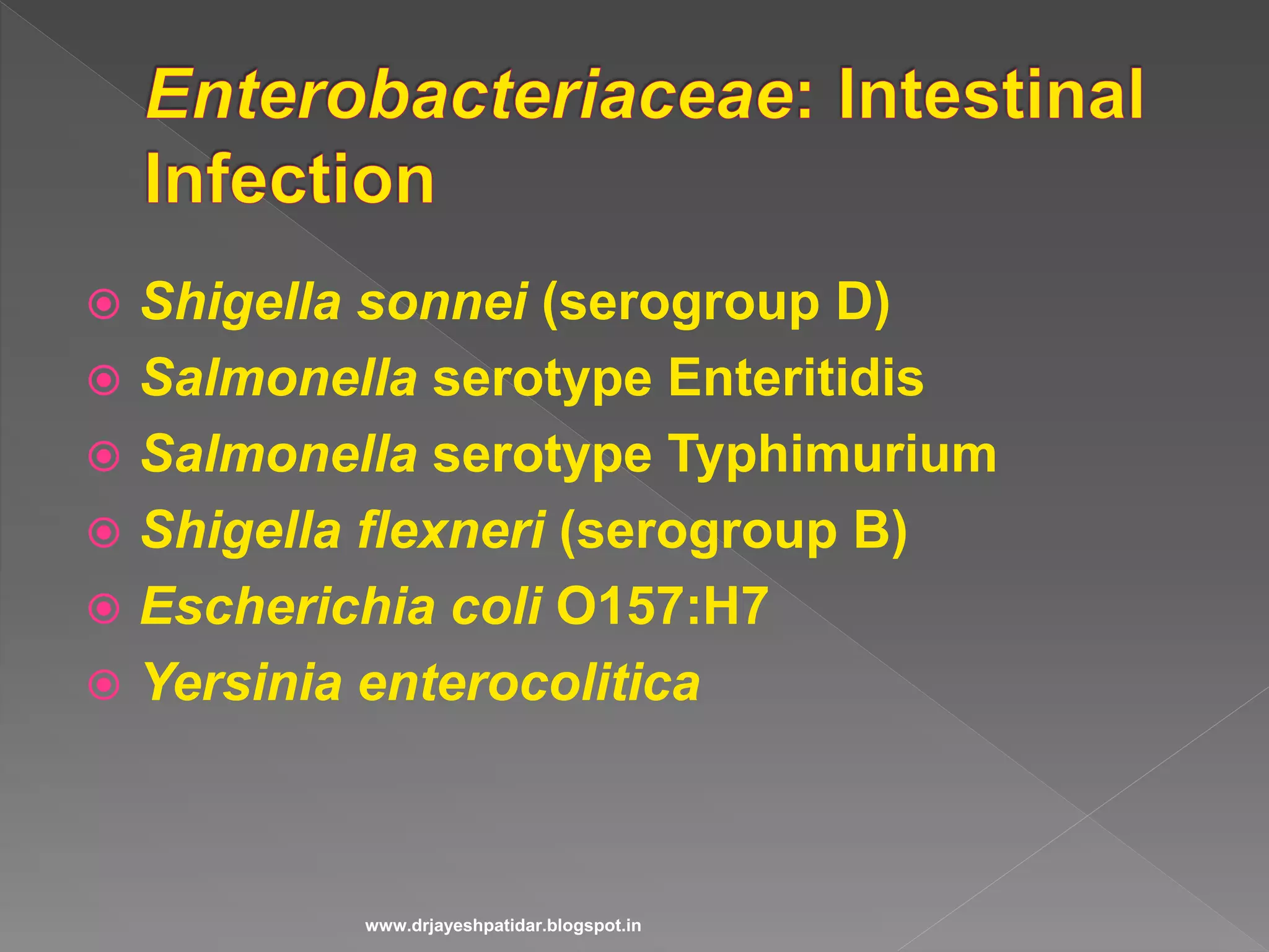
Discusses various infections such as UTIs, pneumonia, and bacteremia, with specific bacteria.
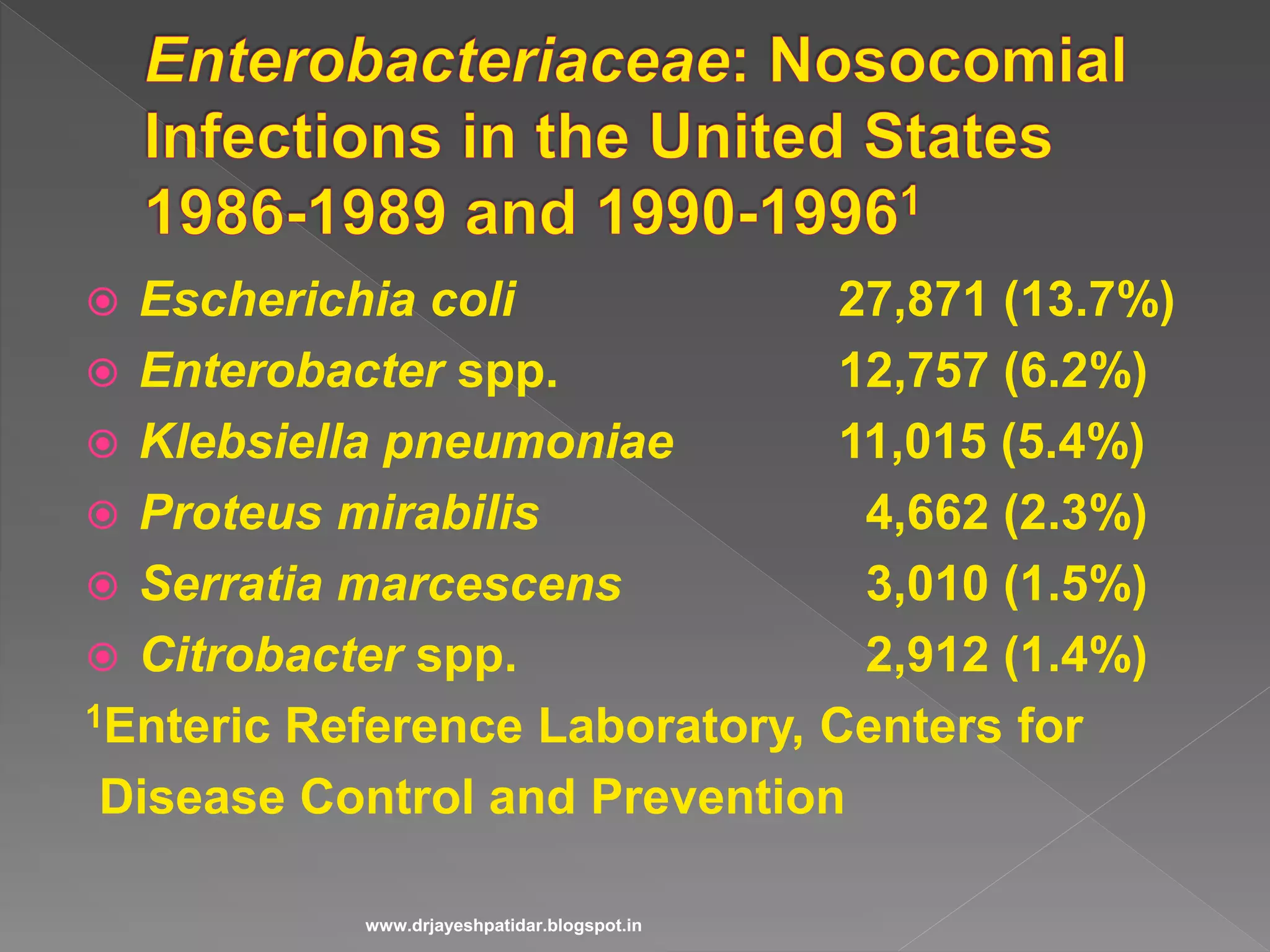
Presents infection rates and specific pathogens responsible, including E. coli and Salmonella.
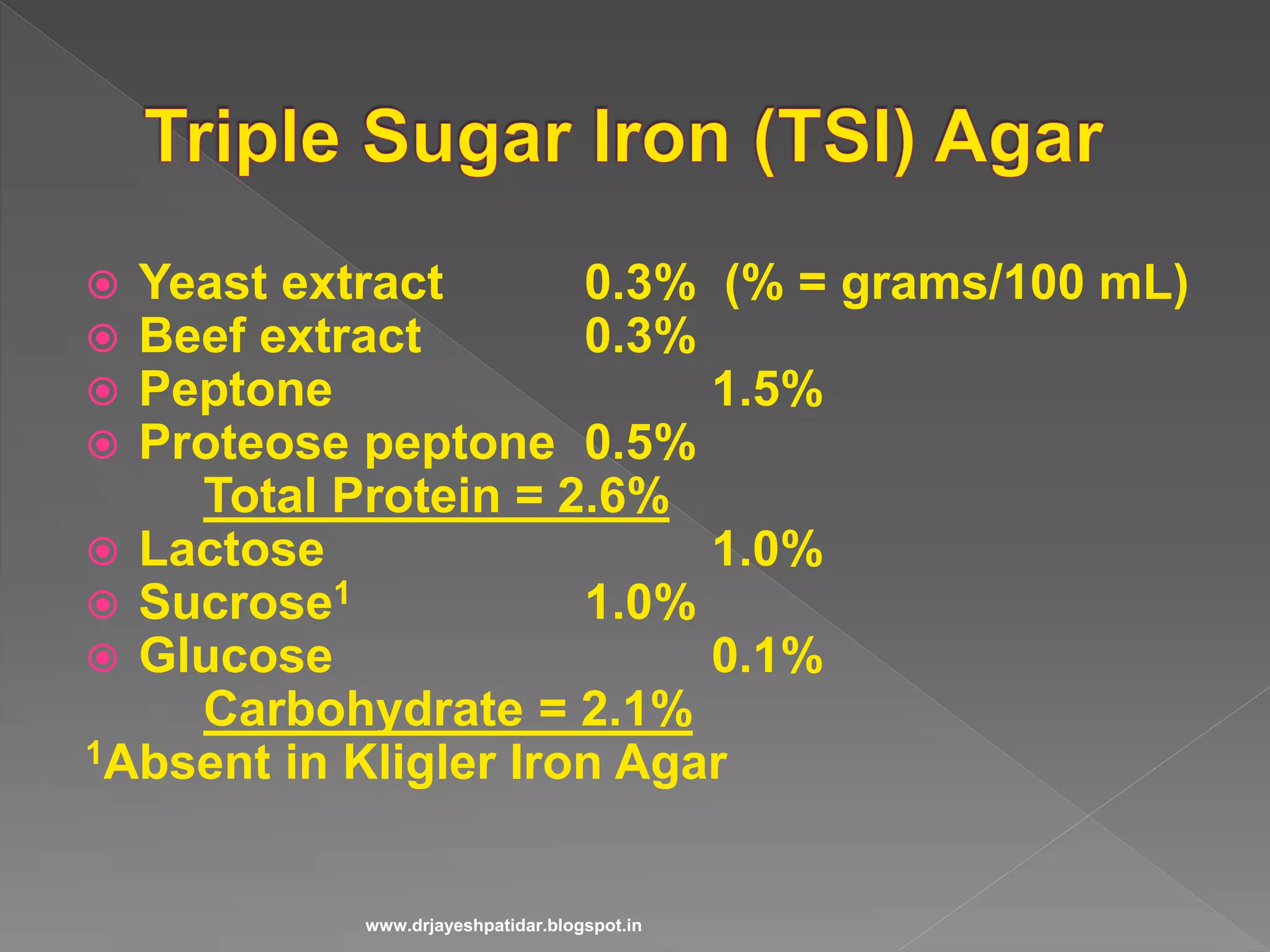
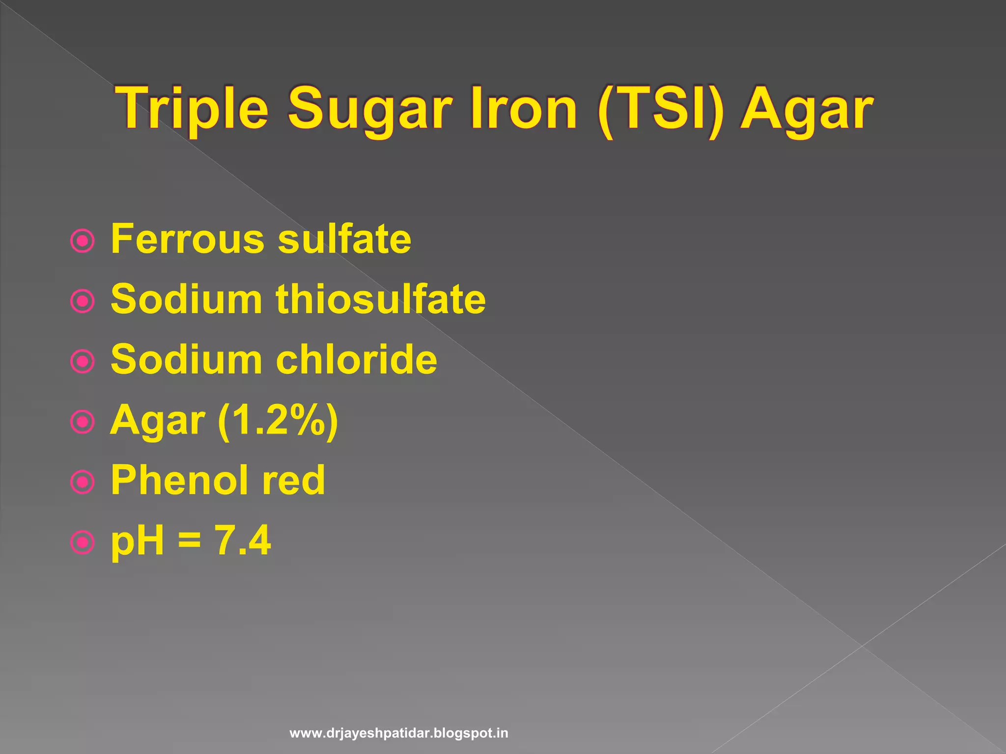
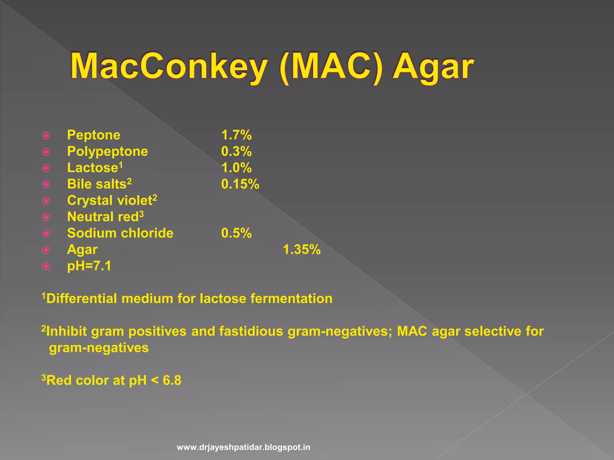
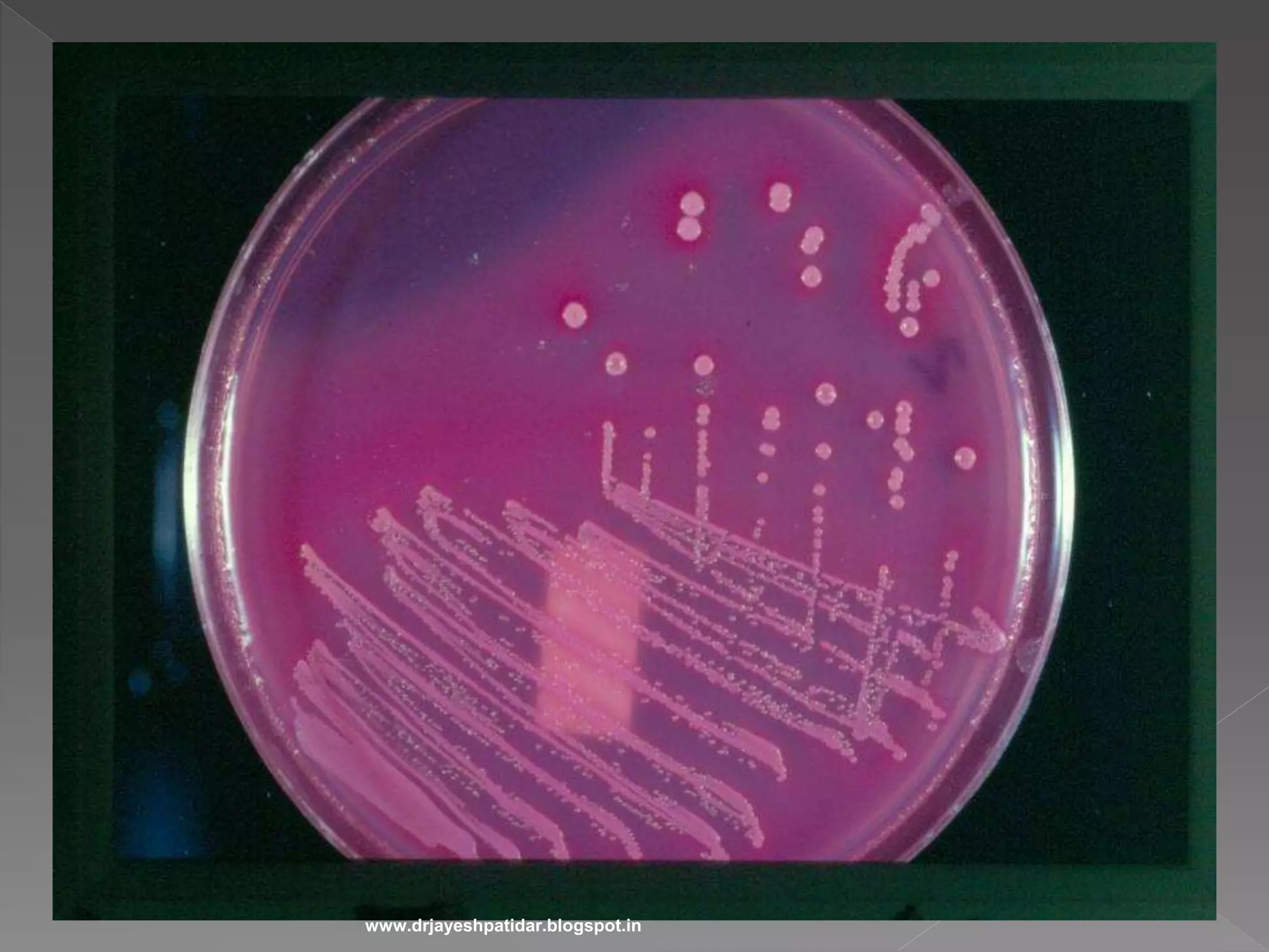
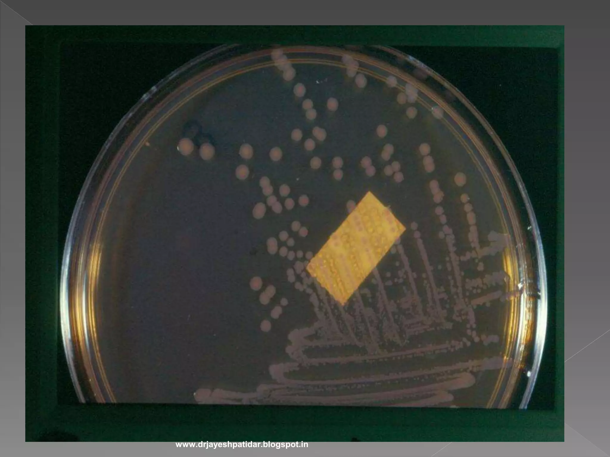
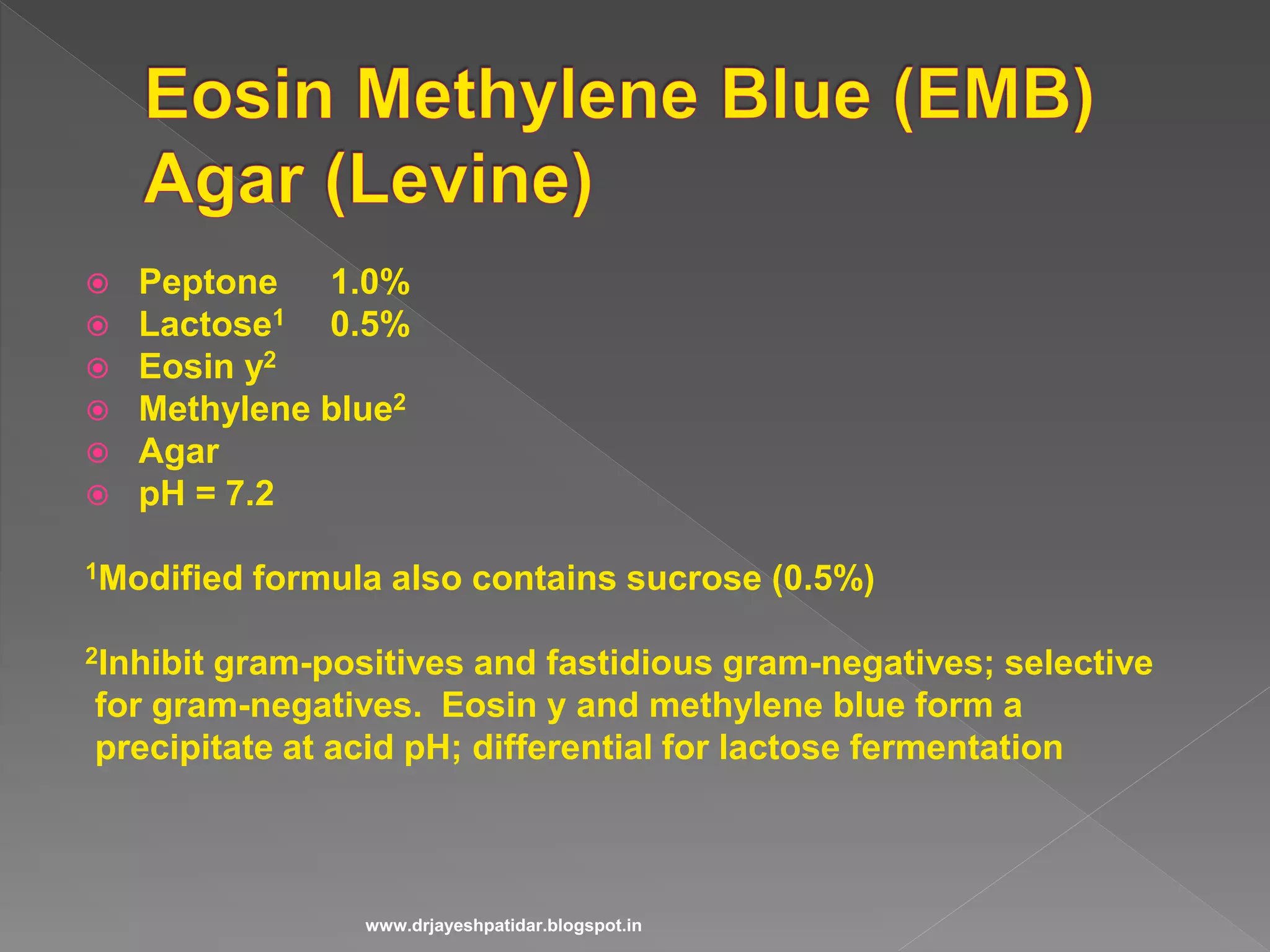
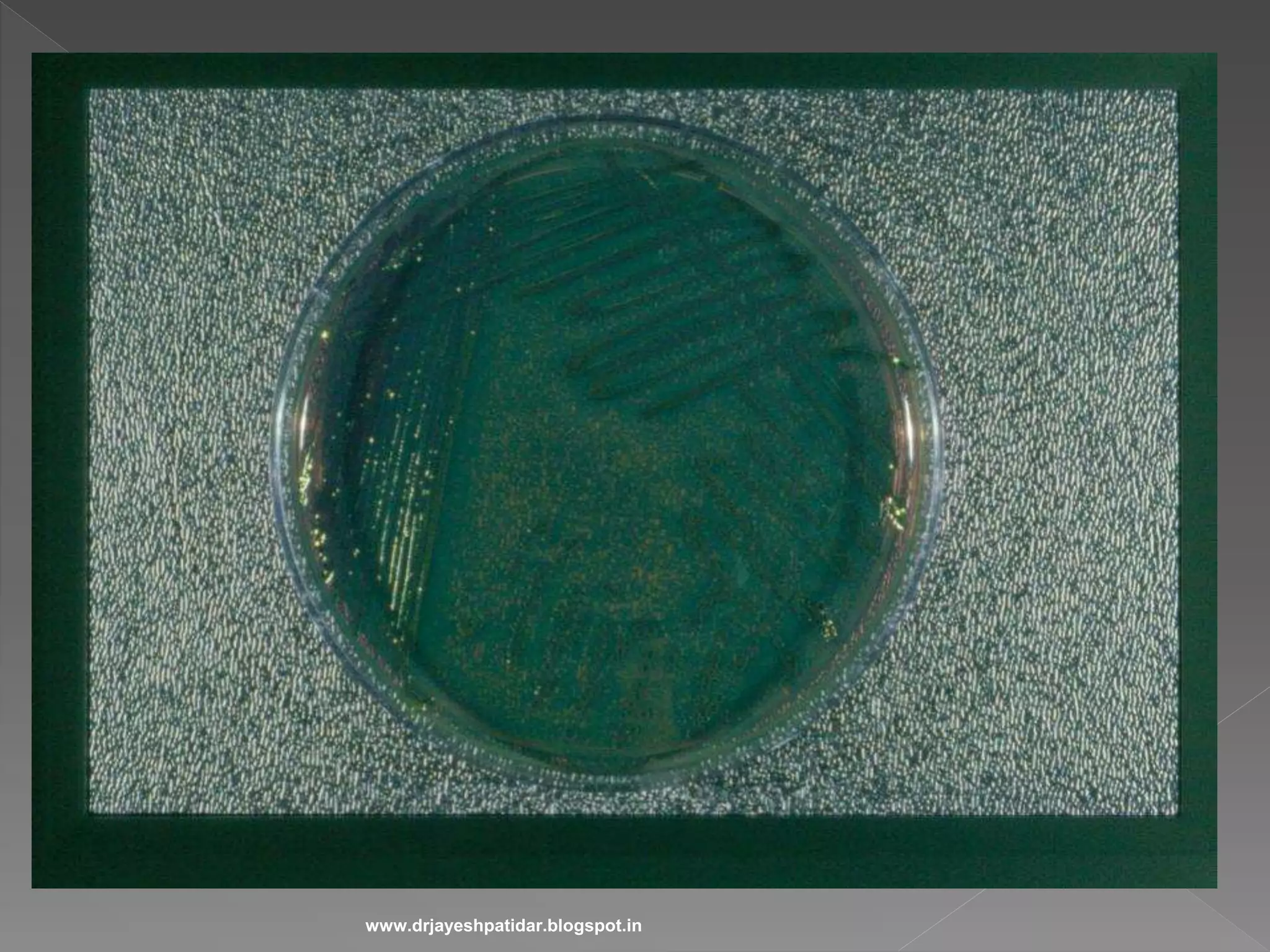
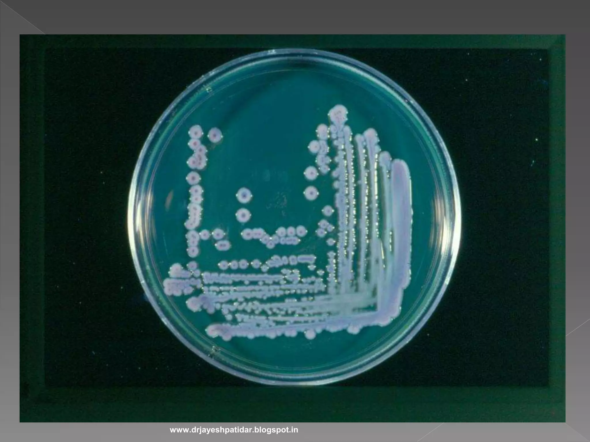
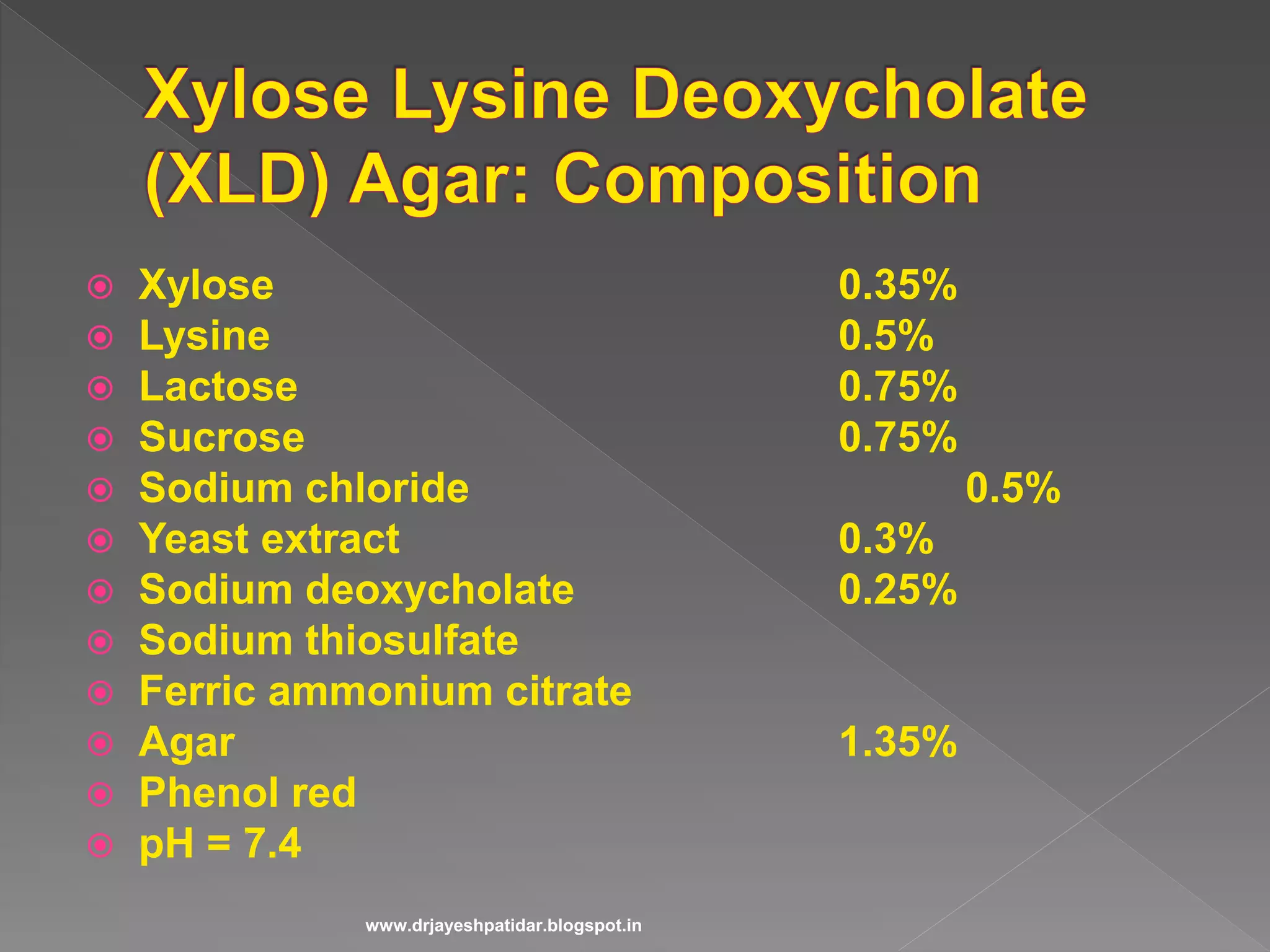
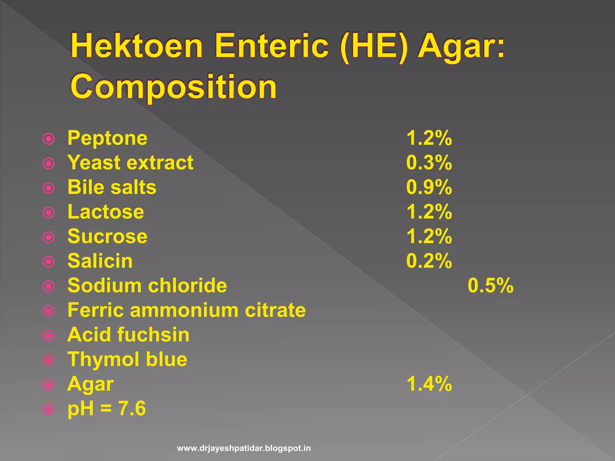
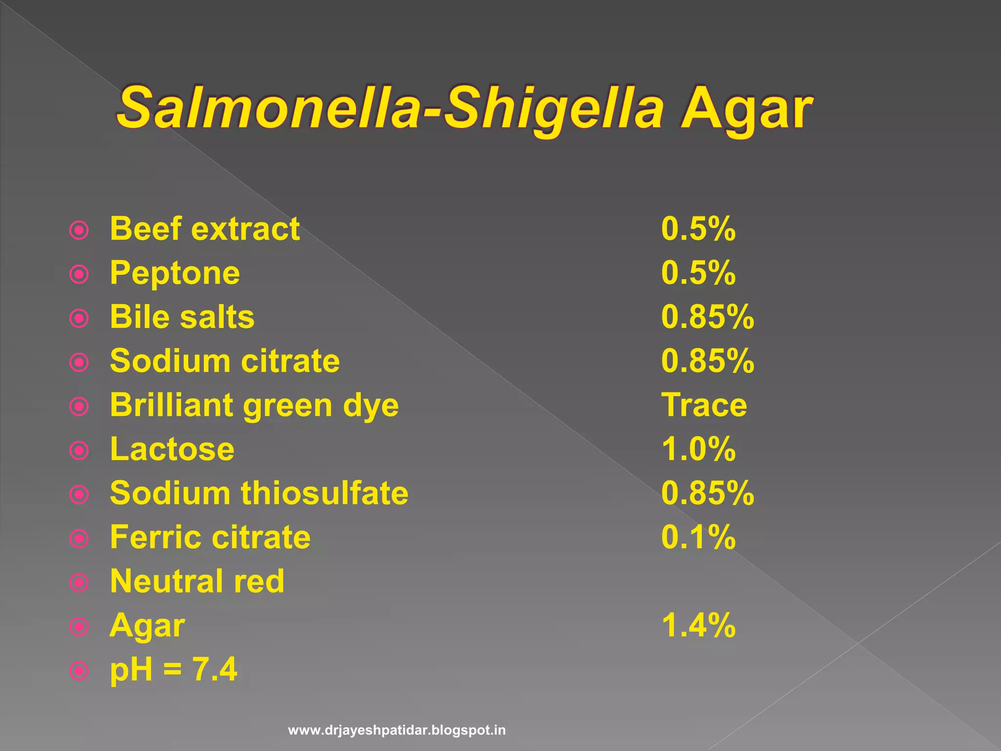
Details the components of media used for bacterial culture in laboratory settings.
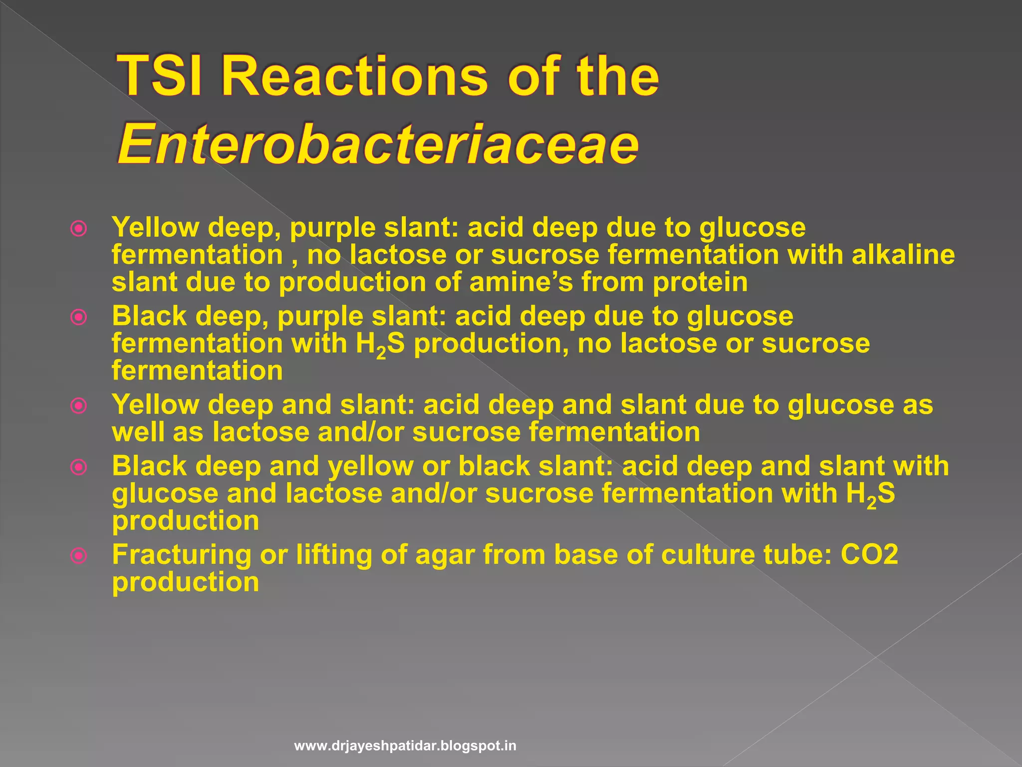
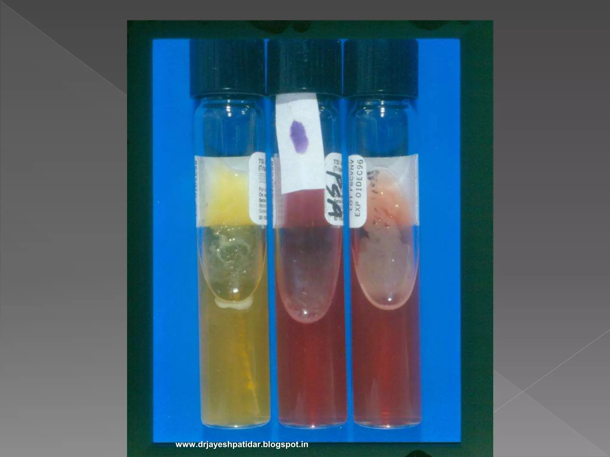
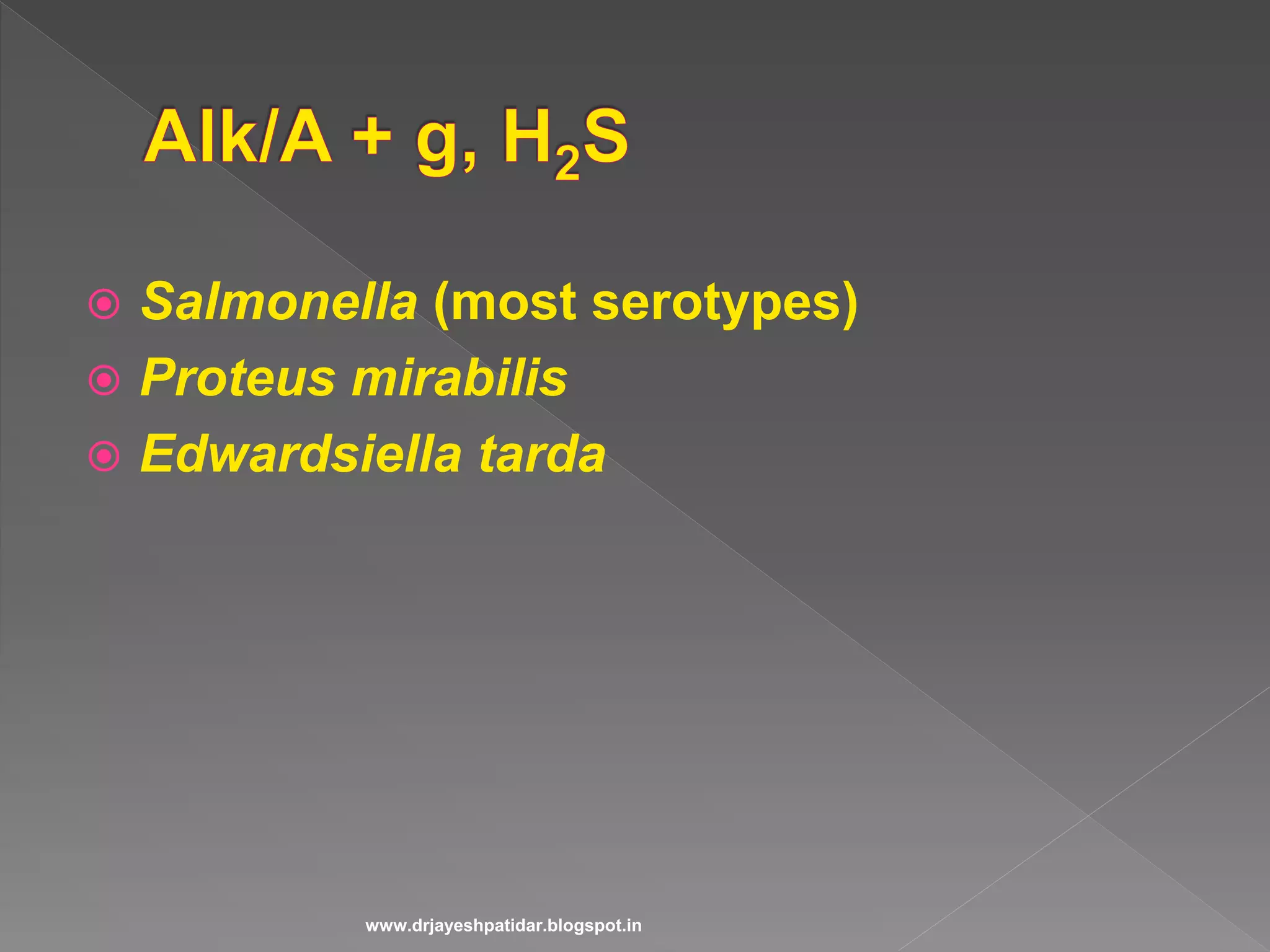
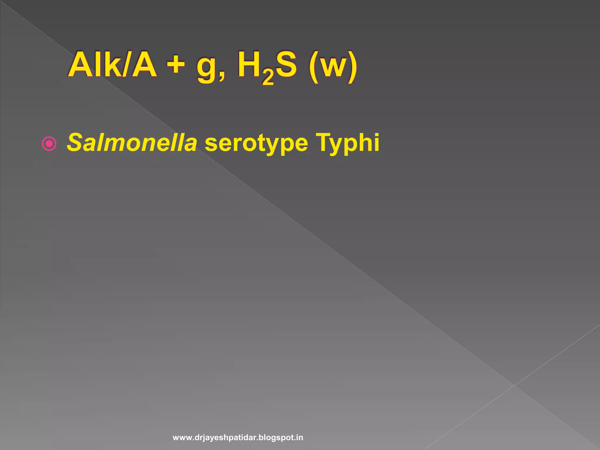
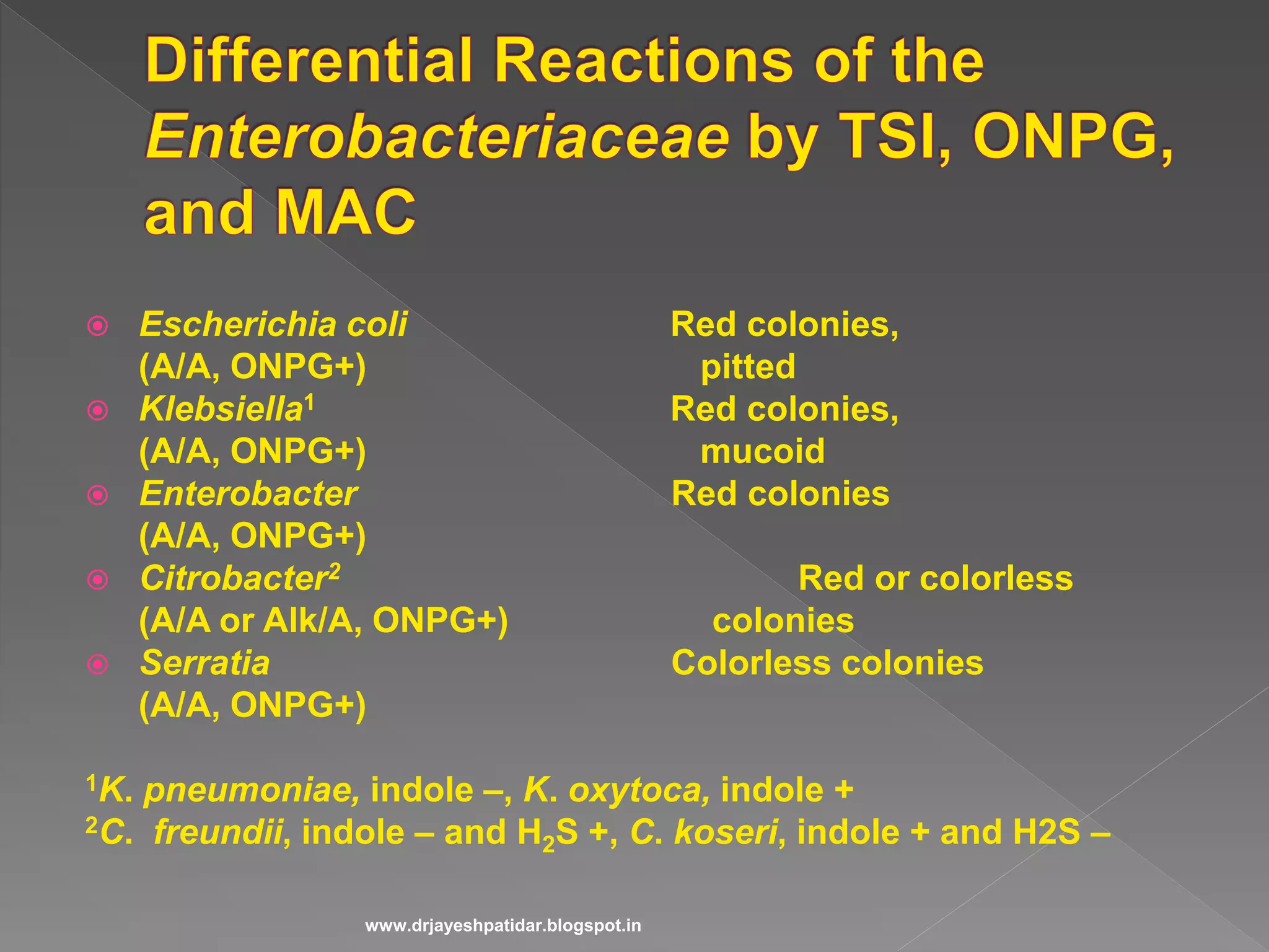
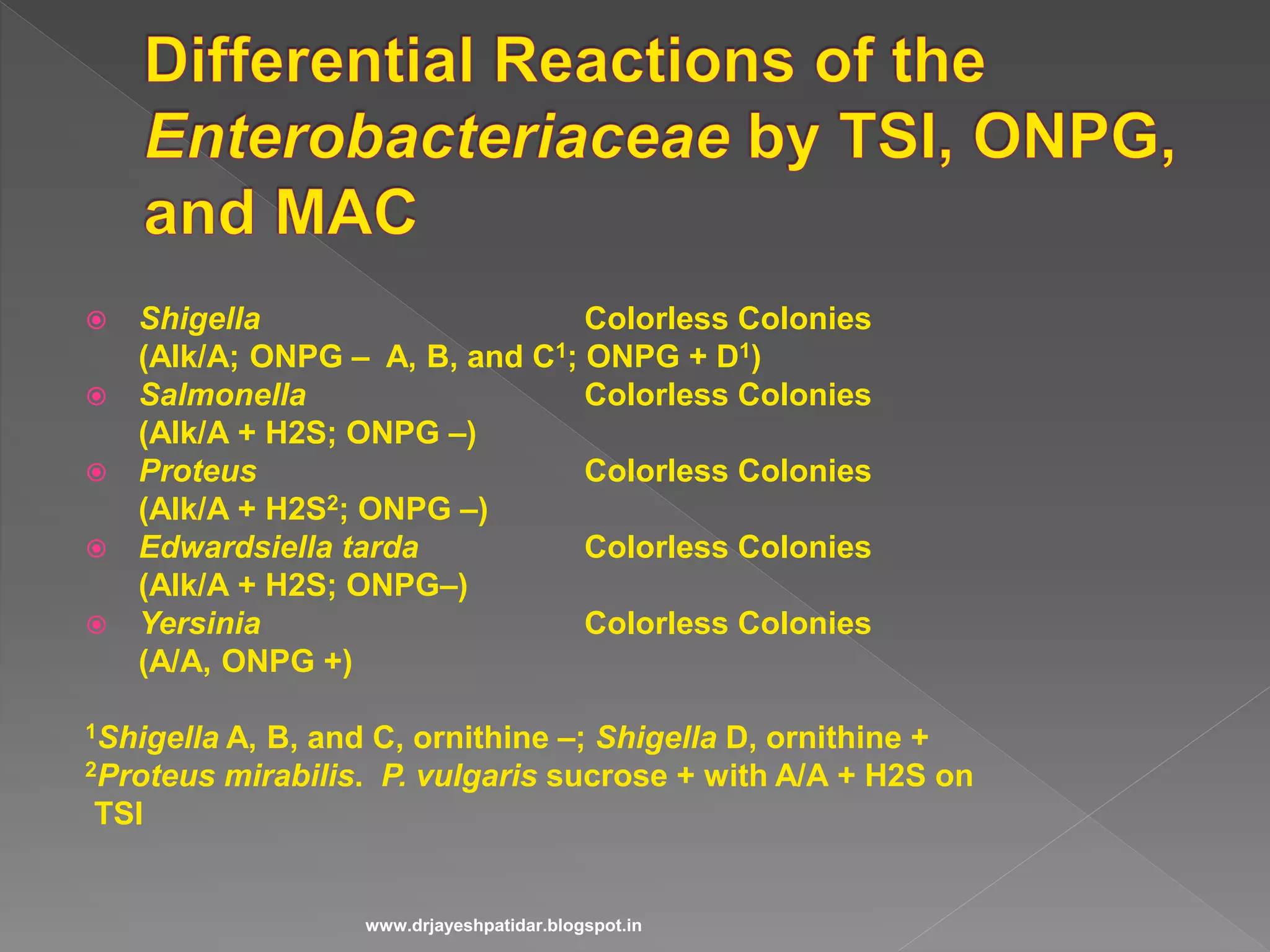
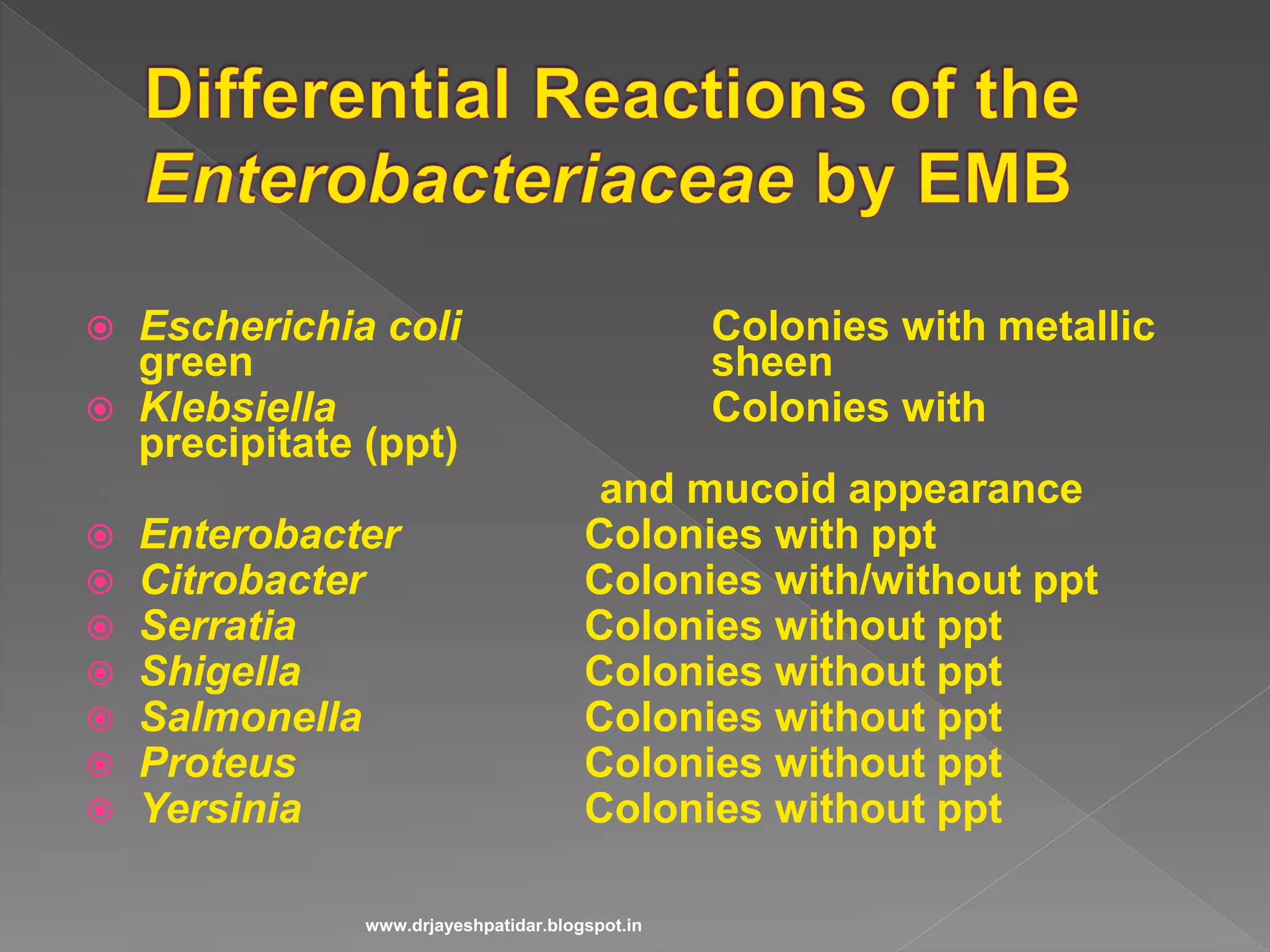
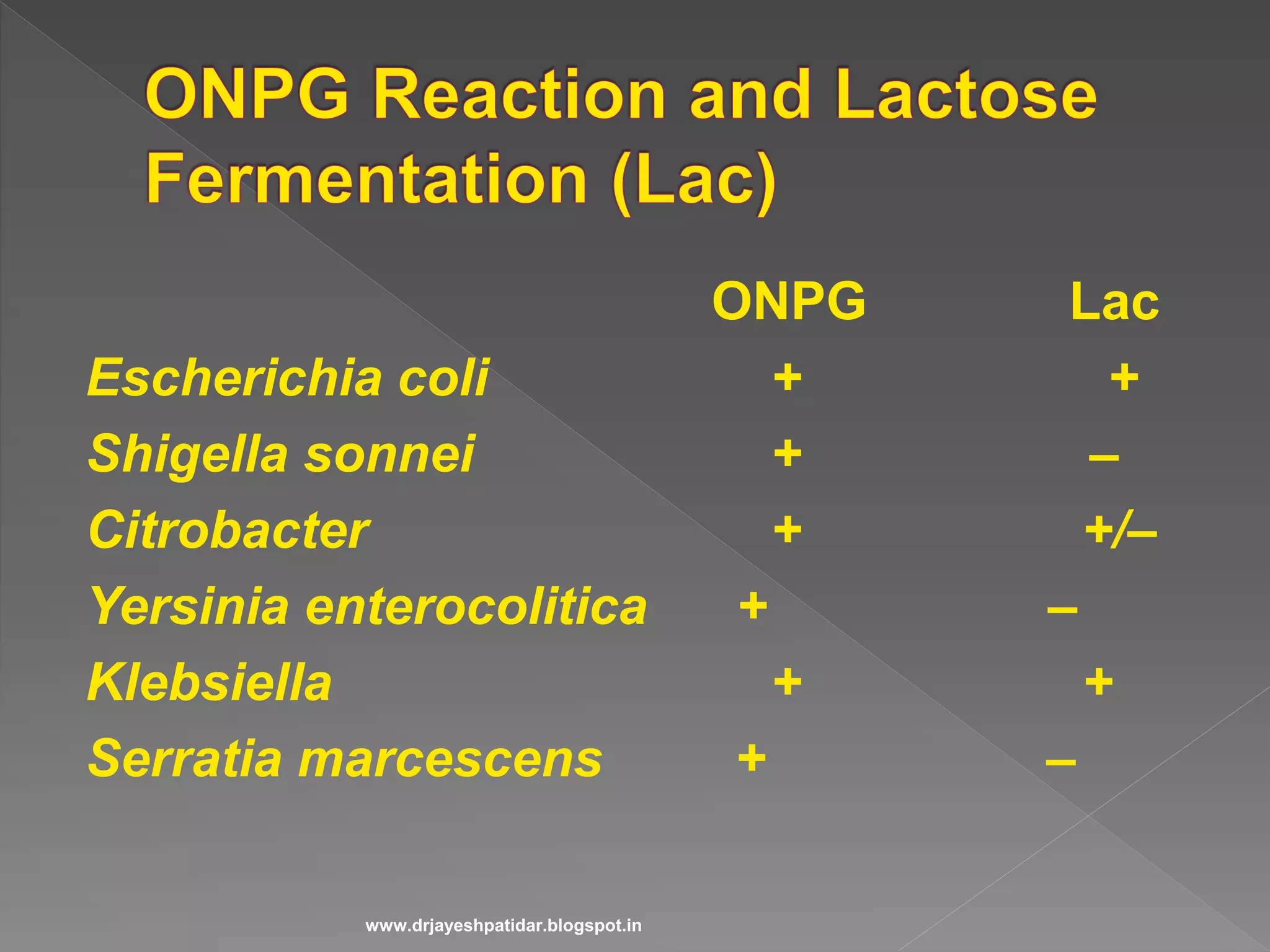
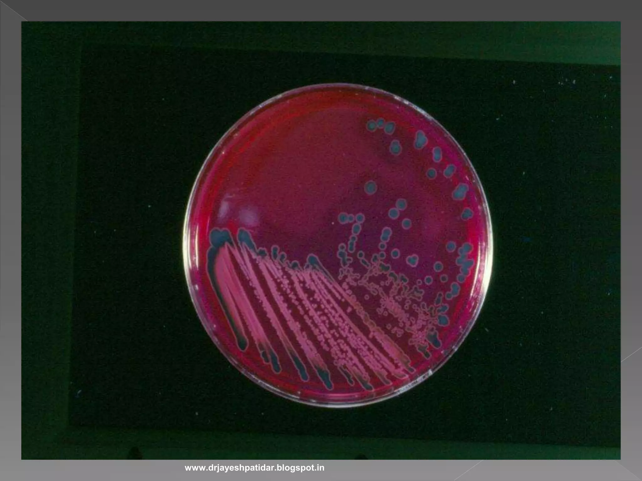
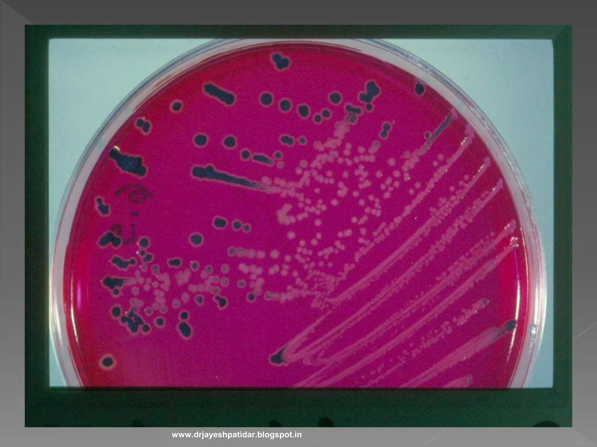
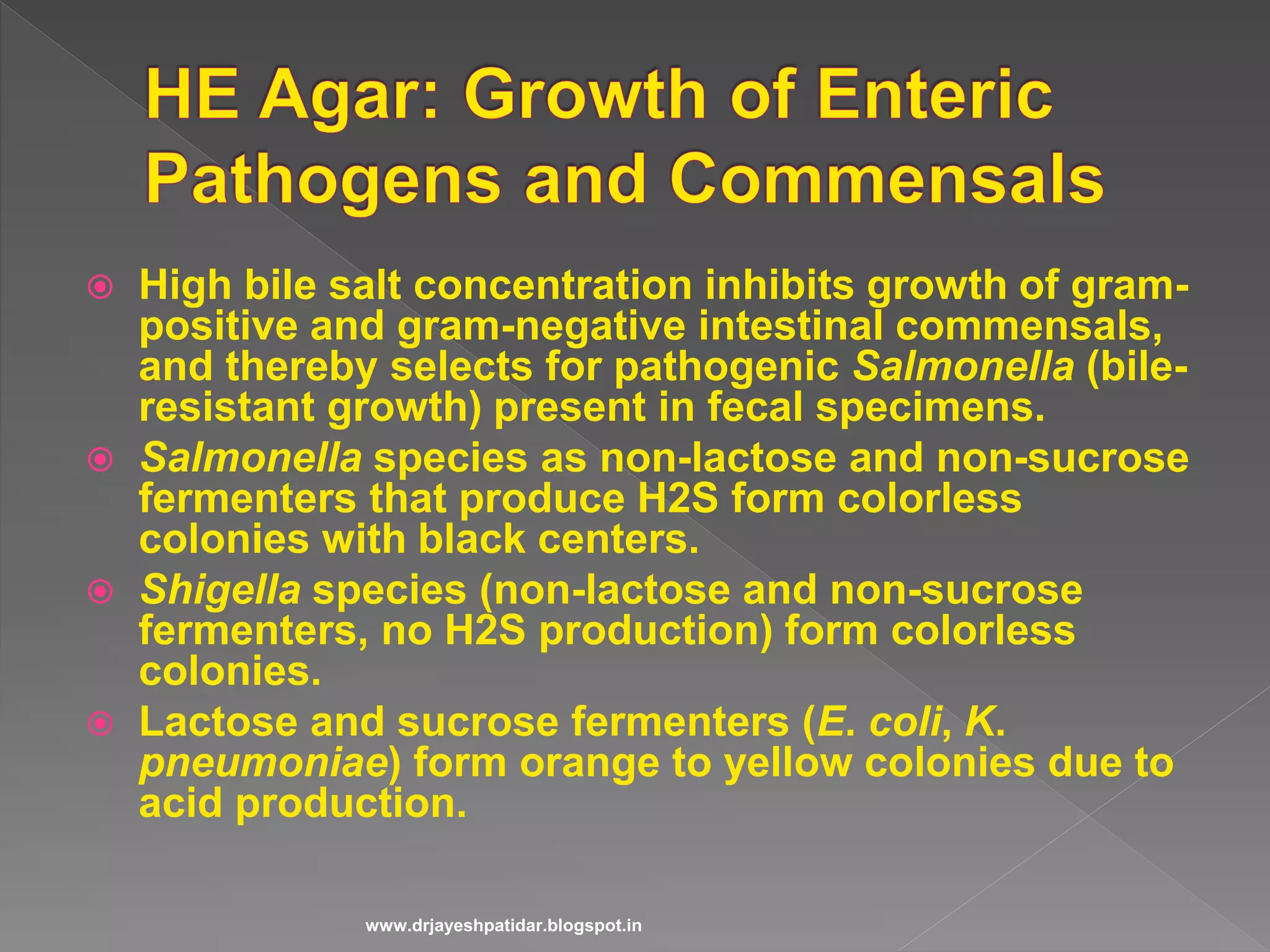
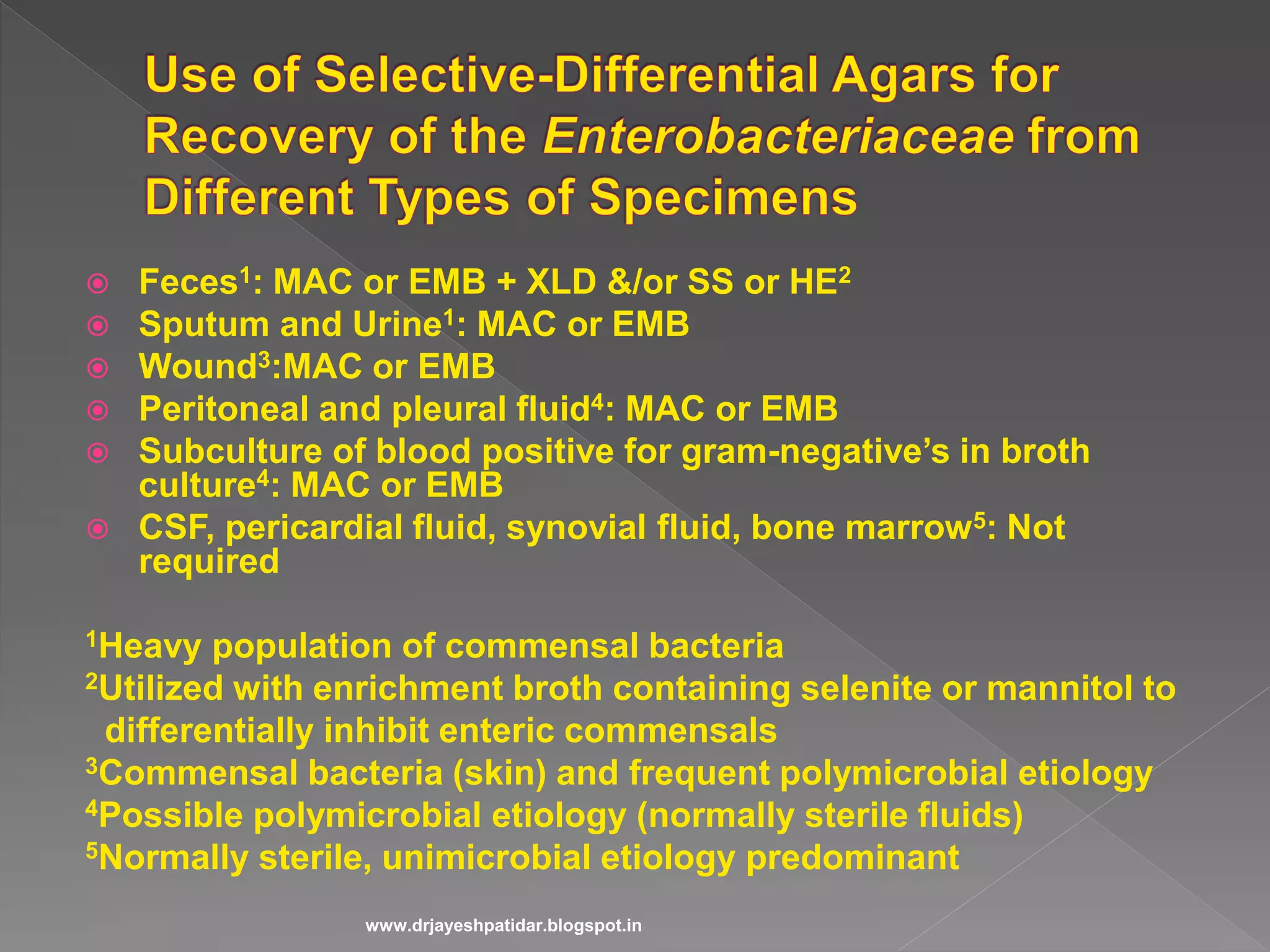
Discusses fermentation test results and identification of various bacteria from the family.
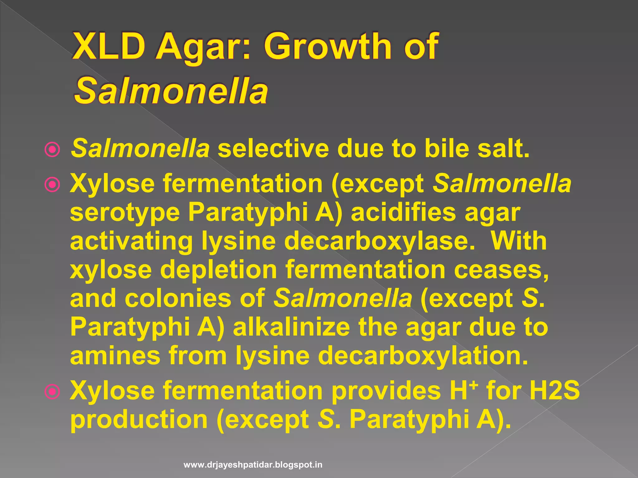
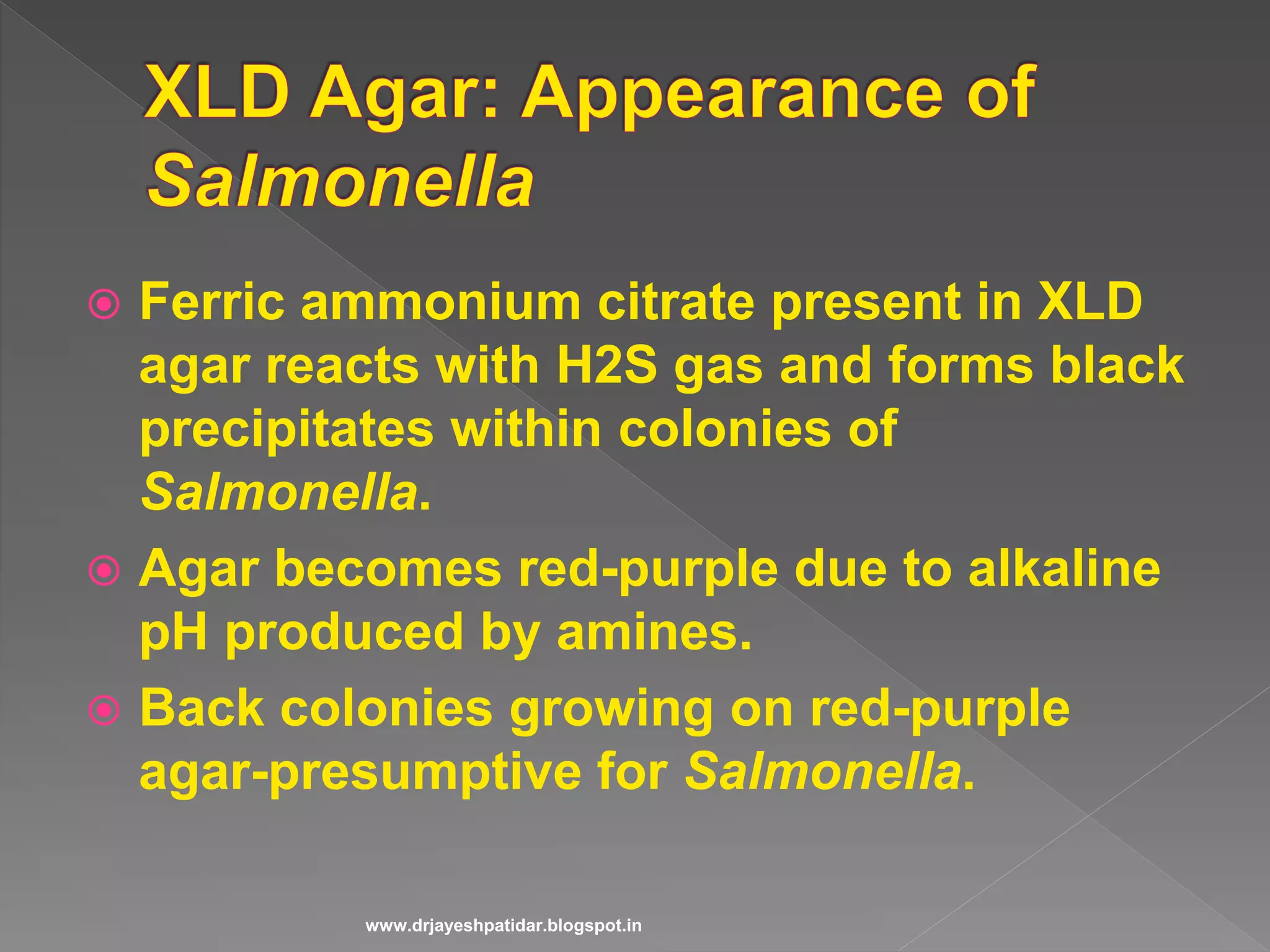
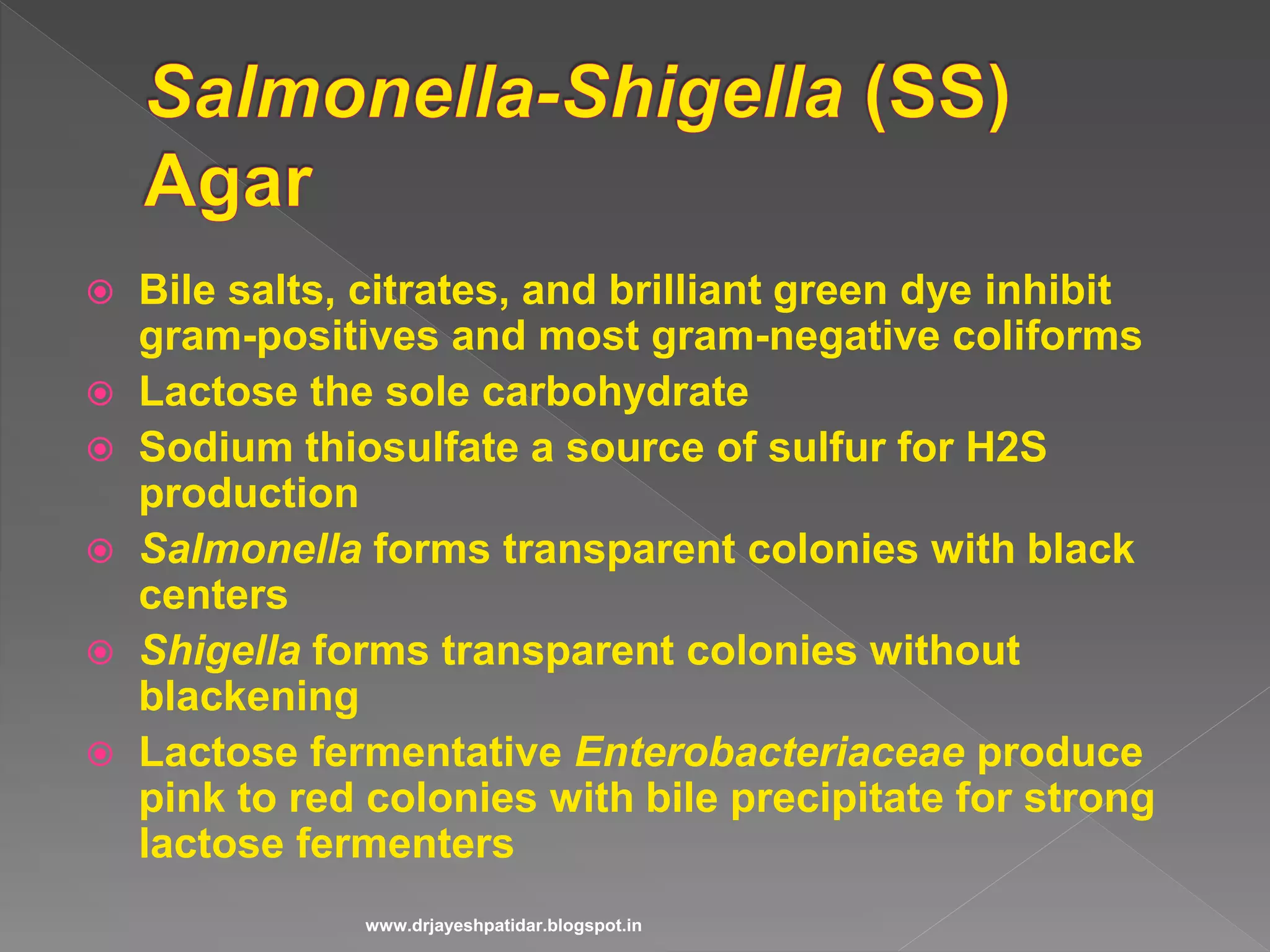
Analyzes media components and their role in distinguishing between Gram-negative bacteria.
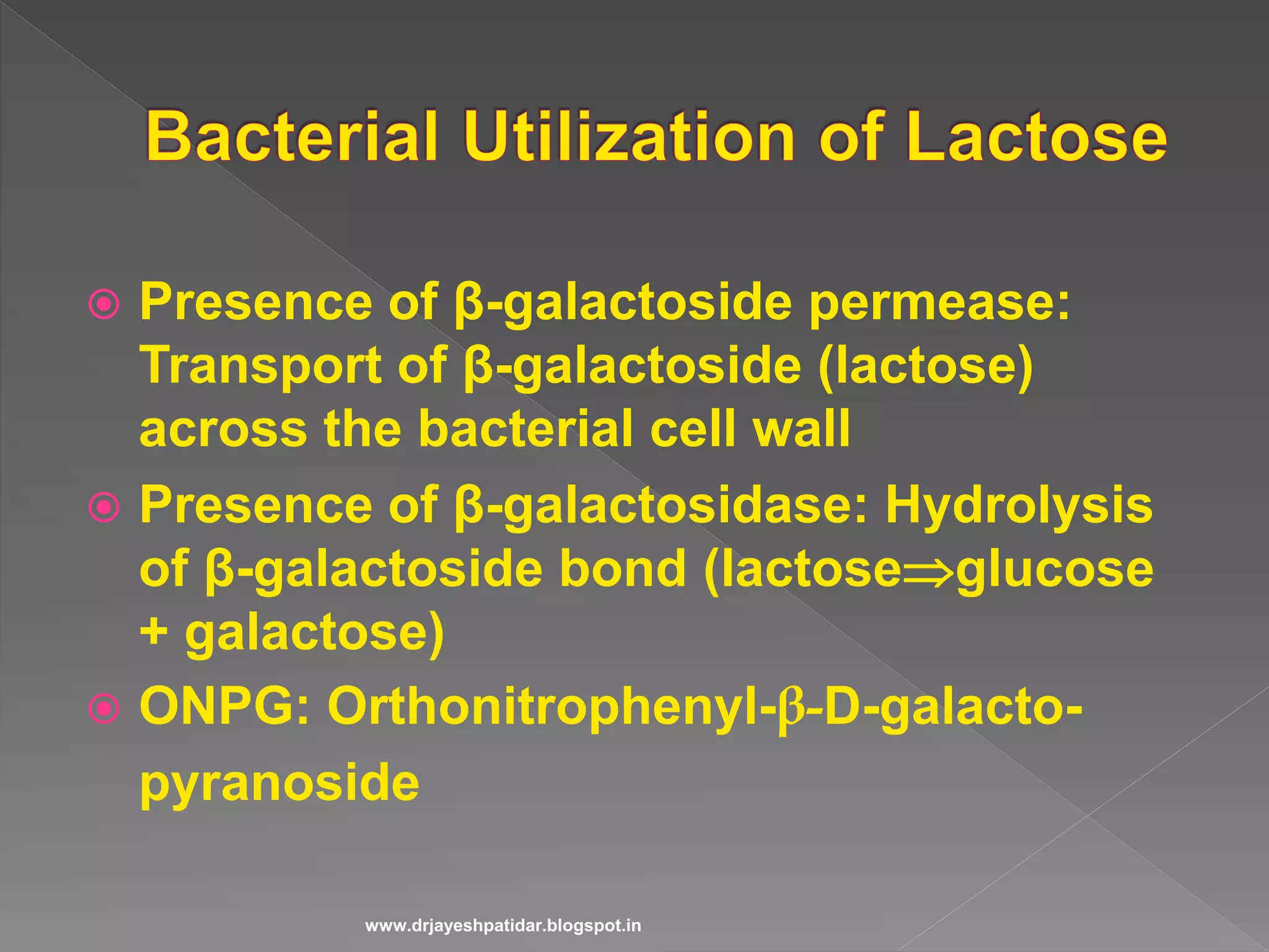
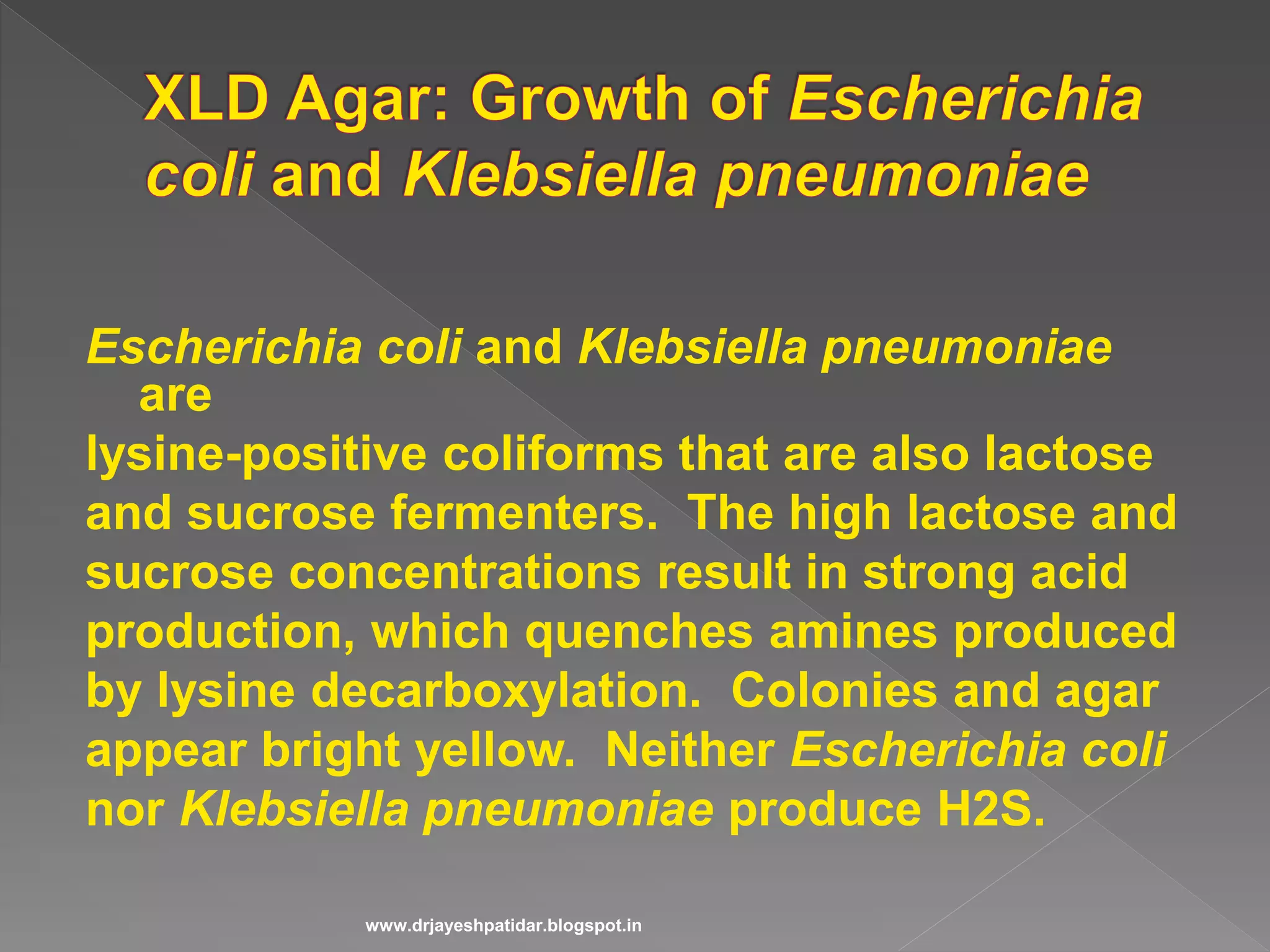
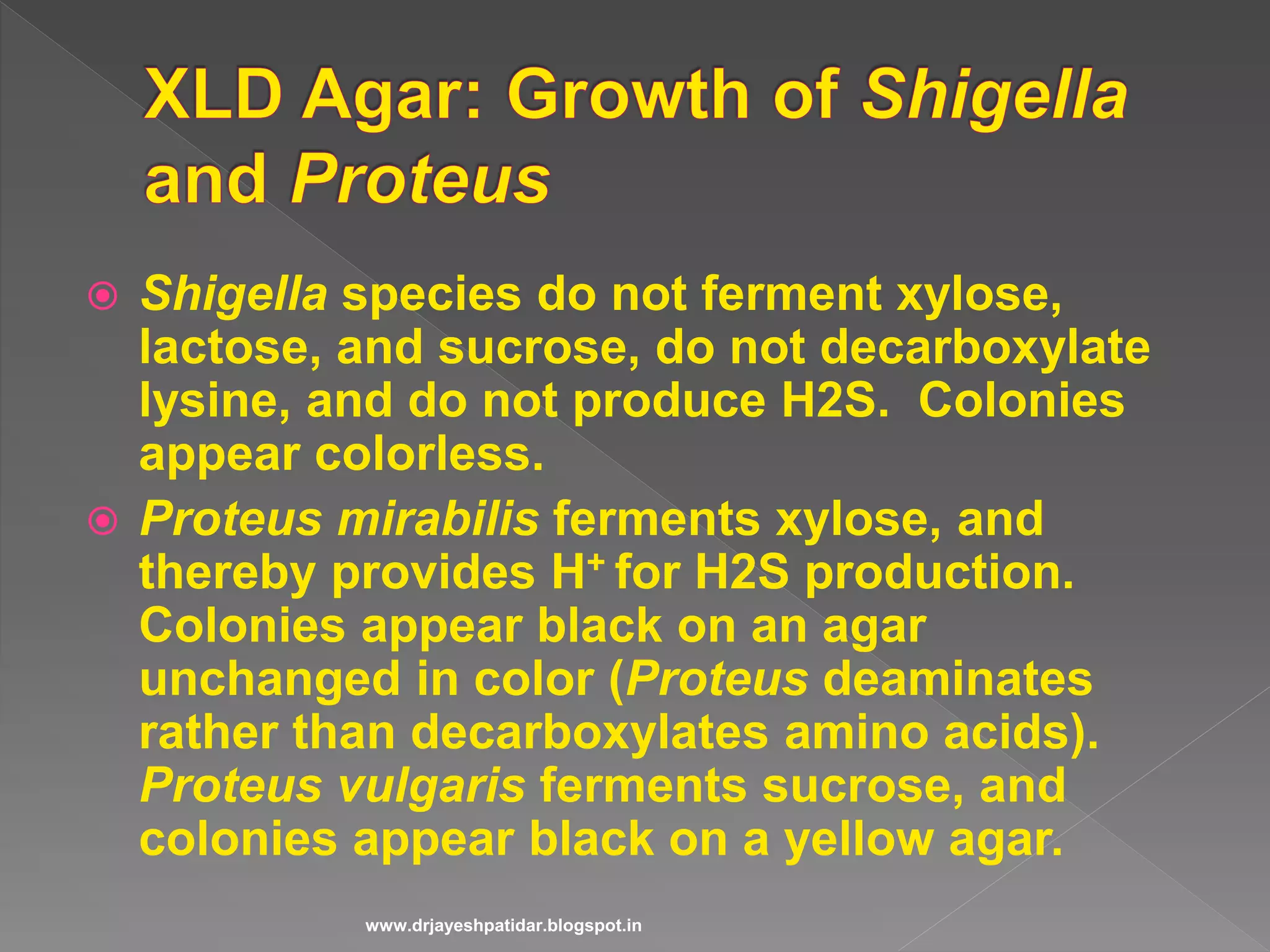
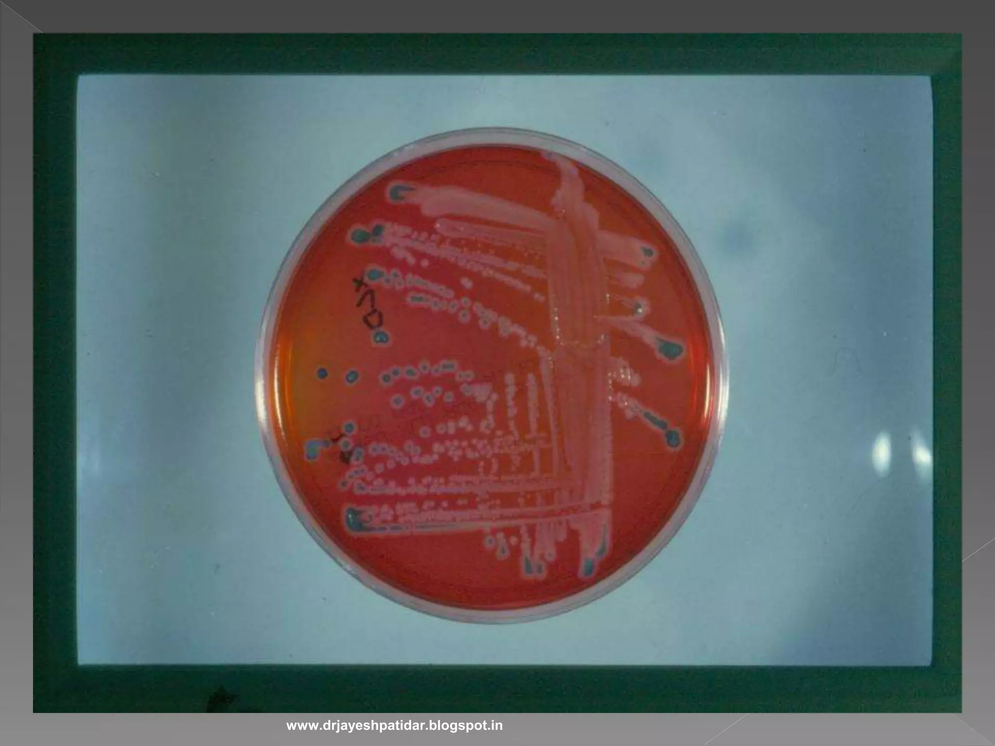
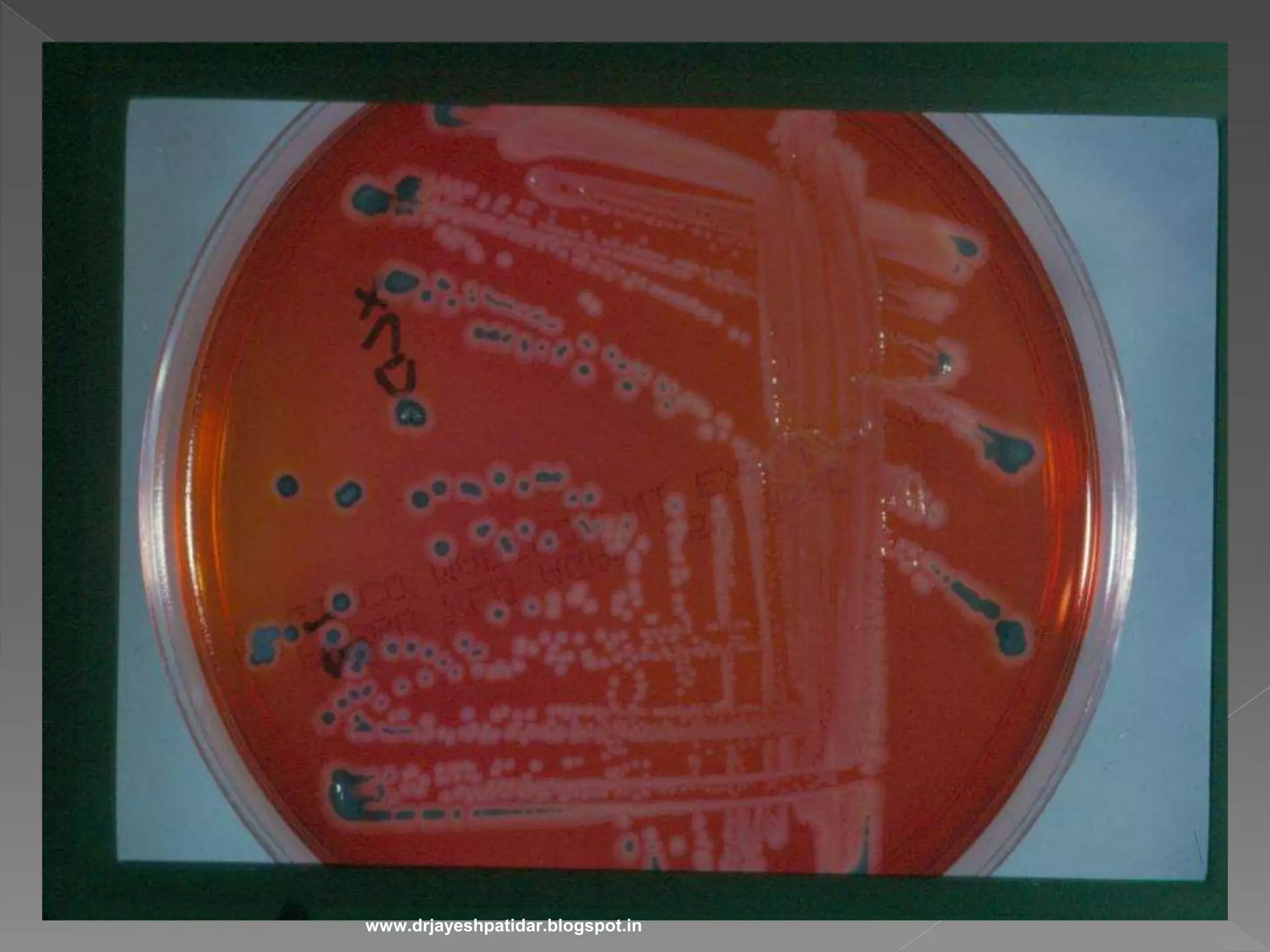
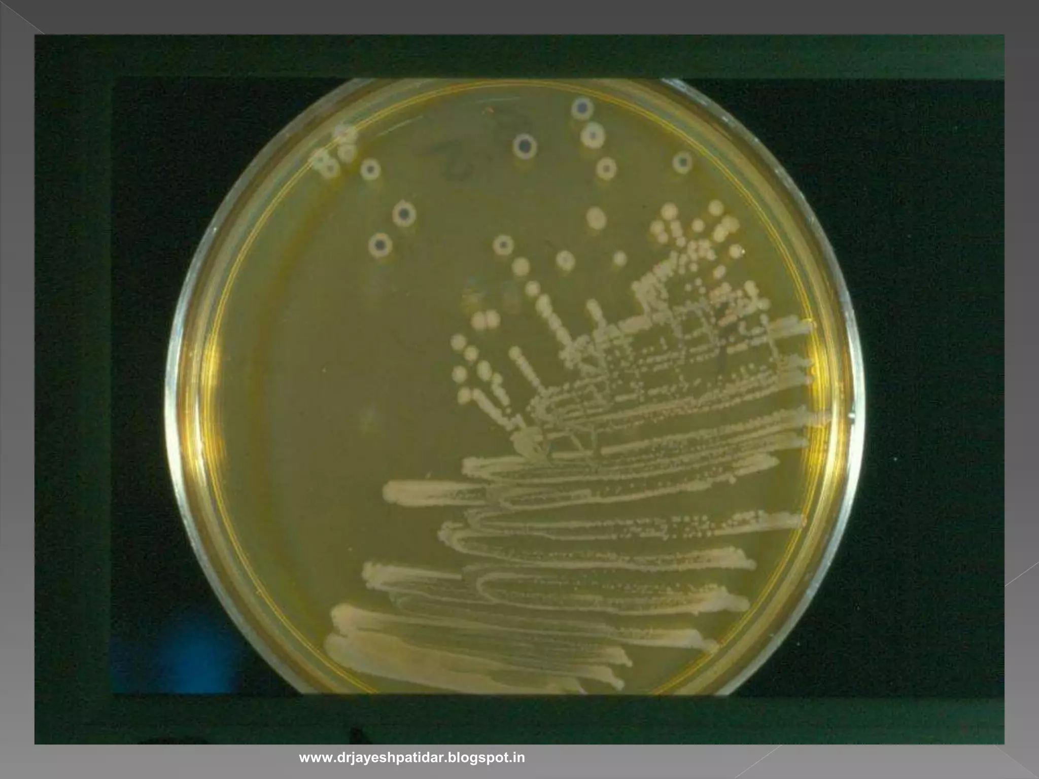
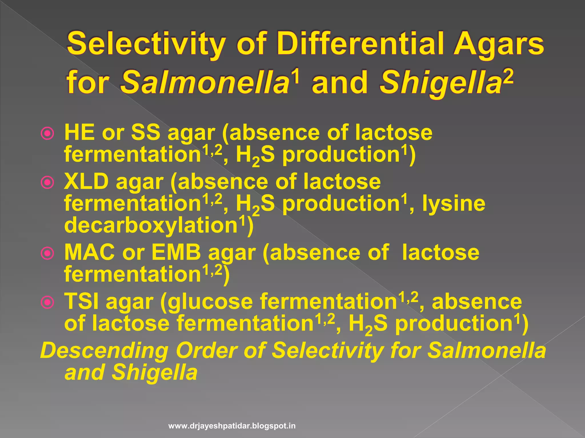
Details the biochemical tests for lactose fermentation and H2S production in various bacteria.
Explains composition and application of specific culture media for isolating Salmonella and Shigella.Lists key references for further reading in medical bacteriology and diagnostic microbiology.