SlideShare a Scribd company logo
RUPTURED ANEURYSMS
OF THE AORTA
VAISHNAVI SURESH NAIR
The aorta is the main trunk of a series of vessels which
convey the oxygenated blood to the tissues of the body
for their nutrition.
It commences at the upper part of the left ventricle,
where it is about 3 cm in diameter, and after ascending
for a short distance, arches backward and to the left side,
over the root of the left lung.
It then descends within the thorax on the left side of the
vertebral column, passes into the abdominal cavity
through the aortic hiatus in the diaphragm, and ends,
considerably diminished in size (about 1.75 cm. in
diameter), opposite the lower border of the fourth lumbar
vertebra, by dividing into the right and left common iliac
arteries.
Branches: The ascending aorta, the arch of the
aorta, and the descending aorta, which is again divided
into the thoracic and abdominal aorta.
THE AORTA
THE AORTIC VALVE
The aortic valve, or aortic semilunar valve, has three leaflets or cusps.
It is located at the base of the aorta. It opens to allow blood to leave
the left ventricle as it contracts. When the ventricular muscles relax,
the valve closes to prevent blood from backing up into the ventricular
chamber.
ASCENDING AORTA (Aorta Ascendens)
 The ascending aorta is about 5 cm. in length.
 It commences at the upper part of the base of the left ventricle, on a level with the lower border
of the third costal cartilage behind the left half of the sternum; it passes obliquely upward,
forward, and to the right, in the direction of the heart’s axis, as high as the upper border of the
second right costal cartilage, describing a slight curve in its course, and being situated, about 6
cm. behind the posterior surface of the sternum.
 At its origin it presents, opposite the segments of the aortic valve, three small dilatations called
the aortic sinuses.
 At the union of the ascending aorta with the aortic arch the caliber of the vessel is increased, owing
to a bulging of its right wall. This dilatation is termed the bulb of the aorta, and on transverse
section presents a somewhat oval figure.
 The ascending aorta is contained within the pericardium, and is enclosed in a tube of the serous
pericardium, common to it and the pulmonary artery
Branches: The only branches of the ascending aorta are the two coronary arteries (Right &
Left) which supply the heart; they arise near the commencement of the aorta immediately above the
attached margins of the semilunar valves.
ARCH OF AORTA (Arcus aorta/transverse aorta)
The arch of the aorta is the second major anatomical region of
the aorta; it curves above the heart between the ascending and
descending aorta. All of the blood delivered from the heart to the
systemic tissues of the body passes through the aorta, making it
the largest artery in the human body. As the aorta extends from
the heart, it begins as the ascending aorta before turning 180
degrees towards the body’s left side in the aortic arch. From the
arch the aorta passes posterior to the heart and descends
through the thorax and abdomen as the descending aorta.
Branches: Three major arteries branch off from the superior
arterial wall of the aortic arch to supply blood to the tissues of the
superior regions of the body: the brachiocephalic trunk, left
common carotid artery, and left subclavian artery.
The brachiocephalic trunk is the first artery to arise from the
aortic arch, carrying blood to the right arm and the right side of
the head and neck. Next to branch from the aorta is the left
common carotid artery that supplies blood to the left side of the
head and neck. Finally, the left subclavian artery arises from the
aortic arch and supplies blood to the left arm.
DESCENDING AORTA
Although the descending aorta is positioned to the
left of the body's midline, it gradually descends to
directly in front of the vertebral column at the left
of the 12th thoracic vertebra.
The portion of the descending aorta above the
diaphragm is called the thoracic aorta, and gives
off branches into the thoracic wall.
Branches of thoracic aorta: The Bronchial
arteries, Mediastinal arteries, Esophageal arteries,
Pericardial arteries, Superior phrenic arteries &
supply blood to the organs for which they were
named.
Below the diaphragm, the descending aorta
become the abdominal aorta and stems off into
branches that reach the abdominal wall and
various tissues and organs of the abdomen.
Branches of abdominal aorta:
Epidemiology
 Ruptured abdominal aortic aneurysms (AAAs) cause 12,000 deaths per year, 8,000 of these are infra-renal.
 Women are much less frequently affected.
 Ruptured abdominal aortic aneurysm (AAA) is one of the most fatal surgical emergencies, with an overall
mortality rate of 90%.
 Rupture of a thoracic aneurysm has a greater than 97% fatality rate.
Risk factors
 The presence of an aneurysm is a risk for rupture.
 The larger the lesion, the more likely it is to bleed; aneurysms over 6 cm have a 25% annual risk of rupture.
 Smoking and hypertension are additional risks.
A ruptured aneurysm should be considered whenever a man aged over 55 or a
woman aged over 70 presents with circulatory collapse.
PATHOPHYSIOLOGY
 Aorta consists of 3 layers- Intima, Media & Adventitia.
 Certain diseases causes weakening of the aortic walls , reduces its elasticity.
When blood is pumped through these weakened areas, it bulges out, which are
called aneurysms.
 As the flow and thus the pressure through this area increases, it causes the
rupture of the aneurysm of the aortic wall.
Clinical Presentation
A ruptured aneurysm usually presents with pain.
Ruptured Thoracic aortic aneurysm (RTAA)
 It will cause chest pain , Ripping sensation in chest, Severe back pain, between shoulder
blades
Haemoptysis (erodes to trachea), Hematemesis (erodes into esophagus) can occur.
 Dizziness, Hypotension, Syncope
 Difficulty in walking or speaking
 If bleeding occurs into the mediastinum, it can cause cardiac tamponade and rapidly be fatal.
The patient will probably never reach hospital alive and the diagnosis is made post-mortem.
Ruptured Abdominal aortic aneurysm (RAAA)
 Ruptured AAA presents with a classical triad of pain in the flank or back, hypotension and a
pulsatile abdominal mass; however, only about half have the full triad. Tachycardia
develops. Shock may occur.
 The patient will complain of the pain and may feel cold, sweaty and faint on standing.
 The following symptoms are listed with approximate frequency of presentation
• Abdominal pain (60%)
• Back pain (70%)
• Syncope (30%)
• Vomiting (20%)
RUPTURED ABDOMINAL AORTIC ANEURYSM
Abdominal aortic aneurysm (AAA) - sites of rupture and their
incidence
•Intraperitoneal rupture (20%)
•Retroperitoneal rupture (80%)
•Aortocaval fistula (3–4%)
•Primary aortoduodenal fistula (<1%)
•Rarely, Into the abdominal veins or
the bowel.
Pressure builds
within aneurysm
Rupture of aortic
aneurysm
Normal abdominal aorta Aneurysm
Anterior intraperitoneal rupture
A tear in the anterior wall of the aneurysm results in sudden severe abdominal or back pain and collapse.
The resultant bleeding into the peritoneal cavity is so rapid that exsanguination and death usually occur before
the patient reaches the hospital.
Posterior retroperitoneal rupture
Classical clinical picture
Rupture into the retroperitoneal cavity is the most common site of ruptured AAA .
A tear in the posterolateral aneurysm wall leads to retroperitoneal bleeding which manifests clinically as back pain
with or without abdominal pain and hypotension.
This tear is often sealed for a few hours, which allows time for the transfer of the patient to the hospital,
diagnosis, and treatment.
On examination, a pulsatile epigastric mass is often palpable, particularly in a thin patient. But this mass may not
be palpable in obese or distended patients or in those with severe hypovolaemia.
Unusual presentations of ruptured abdominal aortic aneurysm
The temporary sealing of the tear in the aneurysm wall may rarely extend beyond a few hours. This results in a variety
of misleading symptoms and signs because of the extension of the retroperitoneal haematoma and its compressive
effects.
In ruptured AAA the duration of symptoms may be unusually long, and that an abdominal pulsatile mass may be
absent.
 Transient lower limb paralysis
 Right hypochondrial pain
 Nephro ureterolithiasis
 Groin pain
 Testicular pain
 Testicular ecchymosis (blue scrotum sign of Bryant)
 Iliofemoral venous thrombosis
 Inguinoscrotal mass mimicking a hernia
The three most frequent emergency presentations of ruptured AAA and their immediate management are:
•A patient known to have an AAA presents with a sudden onset of abdominal or back pain and
hypotension
•A patient presents with the classical triad of pain, hypotension, and a pulsatile mass.
In the two previous scenarios, a haemodynamically unstable patient should be immediately transferred to the
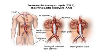
operating theatre for emergency open repair. Recently, endovascular aneurysm repair (EVAR) has been successfully
used to treat ruptured AAA.
In hospitals with capabilities for EVAR, a haemodynamically stable patient can undergo a preoperative computed
tomography (CT) scan, and if the anatomy of the aneurysm is suitable, the patient can undergo endovascular repair
for his/her ruptured aneurysm.
•A patient is suspected of having a ruptured AAA, regardless of the symptoms and signs, and is
haemodynamically stable.
This patient should undergo a CT scan to confirm the diagnosis and assess his/her suitability for EVAR.
Emergency presentations of ruptured AAA
Chronic contained rupture of AAA
Although most patients with ruptured AAA have an acute presentation, some patients may escape detection for
weeks or months after the aneurysm ruptures. This usually occurs when a retroperitoneal rupture leads to slow
progressive bleeding which forms a large haematoma that is contained by the resistance of the periaortic tissues.
Approximately 4% of ruptured AAA are contained ruptures. They are also known as "sealed" or "spontaneously
healed" aneurysms.
Most patients with a contained rupture are haemodynamically stable with no manifestations of acute blood
loss. But a contained rupture is a ruptured aneurysm in a stable patient that may progress to free rupture at any
time. Thus, urgent surgical treatment within 24 h, preferably after admission to an intensive care unit, is
necessary.
Most patients with a contained rupture present with chronic back pain that may radiate to the groin. Other
reported presentations includes lumbar vertebral erosion, lumbar spondylitis-like symptoms, left lower limb
weakness or neuropathy, crural neuropathy, left psoas muscle haematoma, and obstructive jaundice.
Rupture into the abdominal veins
Rarely, AAA ruptures into the inferior vena cava or the left renal vein. This results in an aortocaval fistula or an
aorta–left renal vein fistula, respectively.
Aortocaval fistula
 A spontaneous aortocaval fistula most commonly occurs when an AAA erodes (ruptures) into the inferior
vena cava
 Approximately 3–4% of patients with ruptured AAA have an aortocaval fistula.
 In these patients, the manifestations of rupture usually dominate the clinical picture and significantly
diminish the chance of preoperative diagnosis.
 Aortocaval fistulae are probably missed in 50% of patients and are discovered accidentally during elective
repair of AAA.
 Trauma and surgery of the lumbar spine are other known causes of aortocaval fistulae.
 The manifestations of an aortocaval fistula are variable because they depend on the size of the communication
between the aorta and the inferior vena cava.
 Thus, temporary or permanent closure of this communication by an aortic mural thrombus or by a
compressing aneurysm will change the clinical picture.
 The discovery of an aortocaval fistula during surgery is associated with major blood loss and the possibility of
pulmonary embolization with thrombus material from the aneurysm sac. Thus, unless the patient presents in
extremis due to the rupture, every effort should be made to detect the fistula preoperatively.
 Abdominal CT scan is the definitive imaging test for the evaluation of AAA, and all patients suspected of
having an aortocaval fistula should undergo a contrast CT whenever possible.
 Characteristic CT findings are loss of the fat plane between the aorta and inferior vena cava, vena caval
effacement, and direct inflow of contrast from the aorta to the inferior vena cava.
 Clinically, a patient with an aortocaval fistula presents with a classical triad of abdominal or back pain, a
pulsatile abdominal mass, and a continuous bruit on abdominal auscultation.
 This triad is reported in 50–90% of patients.
 Patients with an aortocaval fistula may also present with manifestations of high output heart failure such as
dyspnoea, tachycardia, wide pulse pressure, cyanosis, and lower limb oedema.
 Additional symptoms and signs include angina, palpitations hypotension, fever, oliguria,
haematuria, pulsatile peripheral veins, and diminished lower limb pulses.
Aorta–left renal vein fistula
 It most commonly occurs when the wall of an infrarenal AAA erodes into the left renal vein.
 The left renal vein normally crosses in front of the abdominal aorta on its way to the inferior vena cava. In 1–
2.4% of people, however, the vein crosses behind the aorta.
 A retro aortic left renal vein is involved in more than 90% of cases of aorta–left renal vein fistula.
 The "abdominal pain, haematuria, silent left kidney" syndrome described by Mansour et al summarises
the clinical features of aorta–left renal vein fistula.
 Haematuria is the most important clinical feature in this condition, followed by pain which is usually felt in the
left flank and radiates to the groin, mimicking ureteric colic.
 A pulsatile abdominal mass and a left sided continuous bruit are detected in approximately 60% and
70% of patients, respectively.
 Renal dysfunction is usually seen in 85% of patients.
 High output heart failure, similar to that seen in an aortocaval fistula, can also be seen in patients with an
aorta–left renal vein fistula. The degree of heart failure depends mostly on the size of the fistula.
 Although extremely rare, an aorta–left renal vein fistula should be ruled out if a patient with an AAA develops
haematuria, left loin pain, or manifestations of renal dysfunction.
 A contrast CT scan of the aorta can visualise the fistula, which should be looked for if a retro aortic left renal vein
is seen.
 Preoperative diagnosis can avoid unnecessary blood loss when the aneurysm is opened during surgery. Intra
operatively, a palpable thrill over the aneurysm, or an absent left renal vein anterior to the aorta, should raise
suspicion of an aorta–left renal vein fistula.
 An extremely rare cause of haematuria is an aortoureteric fistula. Here, the communication is usually between the
ureter and a previously inserted aortic graft, but it can also occur with an aortoiliac aneurysm.
 The triad of unilateral hydronephrosis, intermittent haematuria, and previous aortic surgery should alert the
clinician to this life threatening but potentially curable disease.
Rupture into the bowel (aorto enteric fistula)
 An aorto enteric fistula is an abnormal communication between the abdominal aorta and the bowel; it may be
primary or secondary.
 A primary aorto enteric fistula connects an infra renal AAA to the bowel, most commonly the duodenum
(aortoduodenal fistula). This condition is often fatal but fortunately rare, with an estimated incidence at autopsy of
0.04–0.07%.
 A secondary aorto enteric fistula is a late postoperative complication due to erosion of a prosthetic aortic
graft into the duodenum. This condition is more common, occurring in 0.5–2.3% of patients after aortic surgery.
 The third and fourth parts of the duodenum are most commonly involved in an aorto enteric fistula because this
duodenal segment is closely applied to the anterior wall of the aorta, being fixed posteriorly by the ligament of
Treitz. However, communications with other parts of the gastrointestinal tract have also been reported.
 Patients with a primary aortoduodenal fistula commonly present with upper gastrointestinal haemorrhage
(hematemesis, melena, haematochezia). Abdominal pain and a pulsatile abdominal mass may also be
present; however, patients rarely have all three findings.
 Gastrointestinal haemorrhage was the most common presentation in patients with primary aortoduodenal fistula,
occurring in 96% of patients. Massive haemorrhage is uncommon initially; patients usually experience an episode
of small brisk bleeding which stops spontaneously. This "herald bleed" is characteristic of an aortoduodenal
fistula
 Oesophagogastroduodenoscopy (OGD) is likely to be the first diagnostic test done in patients with upper
gastrointestinal haemorrhage, even if the cause of the bleeding is an aortoduodenal fistula.
 OGD is useful to rule out the much more common causes of bleeding such as gastroduodenal ulcers and
oesophageal varices. If bleeding is due to an aortoduodenal fistula, OGD may not be precise in visualising the
fistula because of failure to pass the endoscope into the third or fourth parts of the duodenum.
 Consequently, in patients with known or previously treated AAApresenting with unexplained upper
gastrointestinal haemorrhage, CT has emerged as the most important initial diagnostic test to rule out an
aortoduodenal fistula. Highly suggestive findings on CT include loss of the fat plane between the aorta and
duodenum and the presence of air in the retroperitoneal cavity.
 Unless a primary gastrointestinal source of bleeding has been unequivocally identified in a bleeding patient
with AAA, an aortoduodenal fistula should always be ruled out.
 Currently, the preoperative diagnosis of aortoduodenal fistulae is reached in only 50% of patients.
RUPTURED AAA IN CHILDREN
• Aortic aneurysms are extremely rare in children, and their aetiology is different from those in adults.
• In children, aortic wall infection, vasculitis, and connective tissue disorders are important causative
factors for AAA.
• Umbilical vein catheterisation is also a well recognised cause of childhood AAA, possibly
through infection.
• Most AAAs in children present as painless pulsatile masses; But a few alarming cases of rupture
have been reported.
• Ruptured AAA is not often suspected in children; But its fatal & should immediately rule out ruptured
AAA in children if it is suspected.
Physical Examination
 A patient with a ruptured aneurysm at any level is likely to look pale and unwell and to be cold and sweaty.
 The pulse will be rapid, weak and thready. Hypotension is common.
 With a ruptured AAA there may well be a pulsatile mass in the vicinity of the bifurcation of the aorta. This is a few
centimetres above the umbilicus and a little to the left.
 It may be tender and a bruit may be audible. Bleeding causes peritoneal irritation and it may appear as an acute
abdomen.
 The following findings are listed with approximate frequency:
 Palpable mass (90%).
 Tenderness (80%).
 Systolic blood pressure (BP) below 80 mm Hg (40%).
 NB: presentation can be atypical, eg: intestinal obstruction from haematoma or an apparent irreducible inguinal
hernia.
 Rare presentations are:
 Severe hematemesis from an aorto -duodenal fistula.
 A fistula into the inferior vena cava, producing lower limb oedema and high-output cardiac failure.
Differential diagnosis
 The differential diagnosis for a ruptured TAA is that of chest pain, especially MI with cardiogenic shock but
also massive pulmonary embolism.
 The differential diagnosis for ruptured AAA involves other causes of abdominal pain, including acute abdomen.
Investigations
If an aneurysm is ruptured, investigations need to be swift and pertinent.
Laboratory studies
 CBC: NB: if there has not been time for haemodilution then haemoglobin will be normal. Anaemia is present in
less than half of patients. Around 80% have a white cell count of 10 x 109/L or more.
 CMP
 Group and rapid cross-match: whilst arranging surgery.
 Baseline biochemistry of U&Es: should be performed.
Radiology
 CXR: for a TAA the CXR may well show an enlarged base of aorta.
 Plain abdominal X-ray: for an AAA this will show the lesion in about 75%, as it is often calcified.
 Portable ultrasound: this examination may be helpful but there is not time for detailed assessment. If there is
strong suspicion of a ruptured aneurysm then immediate surgery may be the investigation of choice.
 Other investigations: CT angiography will confirm the diagnosis. MRI and angiography are an alternative
but, practically, more time-consuming so probably only suitable for the stable patient.
ECG
 ECG: is important In patients presenting with chest pain.
TREATMENT
Abdominal aortic aneurysms (AAAs) are typically repaired by an operative intervention.
The possible approaches are the traditional open laparotomy, newer minimally invasive methodologies, or by
the placement of endovascular stents.
INDICATIONS:
Open repair:
Diameter of the aneurysm greater than 2 inches
Abdominal Pain
Abdominal Pulsation
Endovascular repair:
Severe Heart diseases
Age risks
Other underlying medical conditions
PRE-OPERATIVE MEASURES
•Type and cross match blood
•IV line (fluids, antibiotics, anesthesia)
•Administer prophylactic antibiotics (cefazolin, 1 g intravenous piggyback)
•Insert a Foley catheter
•Monitor central venous pressure or establish Swan-Ganz catheterization (if
indicated)
•Prepare the skin from the nipples to the mid thigh
•Administer general anesthesia (open), general/regional anesthesia (endovascular)
•Insert a nasogastric tube
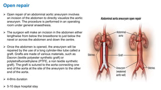
 Open repair of an abdominal aortic aneurysm involves
an incision of the abdomen to directly visualize the aortic
aneurysm. The procedure is performed in an operating
room under general anaesthesia.
 The surgeon will make an incision in the abdomen either
lengthwise from below the breastbone to just below the
navel or across the abdomen and down the centre.
 Once the abdomen is opened, the aneurysm will be
repaired by the use of a long cylinder-like tube called a
graft. Grafts are made of various materials, such as
Dacron (textile polyester synthetic graft) or
polytetrafluoroethylene (PTFE, a non textile synthetic
graft). The graft is sutured to the aorta connecting one
end of the aorta at the site of the aneurysm to the other
end of the aorta.
 4-6hrs duration
 5-10 days hospital stay
Open repair
Endo Vascular Aneurysm Repair (EVAR)
 EVAR is a minimally-invasive (without a large abdominal incision)
procedure performed to repair an abdominal aortic aneurysm. EVAR
may be performed in an operating room, radiology department, or a
catheterization laboratory. Surgeon may use general anesthesia or
regional anesthesia (epidural or spinal anesthesia). The surgeon will
make a small incision in each groin to visualize the femoral arteries in
each leg. With the use of special endovascular instruments, along with
X-ray images for guidance, a stent-graft will be inserted through the
femoral artery and advanced up into the aorta to the site of the
aneurysm. A stent-graft is a long cylinder-like tube made of a thin
metal framework (stent), while the graft portion is made of various
materials such as Dacron or polytetrafluoroethylene (PTFE) and may
cover the stent. The stent helps to hold the graft in place. The stent-
graft is inserted into the aorta in a collapsed position and placed at the
aneurysm site. Once in place, the stent-graft will be expanded (in a
spring-like fashion), attaching to the wall of the aorta to support the
wall of the aorta. The aneurysm will eventually shrink down onto the
stent-graft.
 2-3hrs duration
 2-3 days hospital stay
Postoperative Details
 Fluid shifts are common following aortic surgery. Fluid requirements may be high in the first 12 hours,
depending on the amount of blood loss and fluid resuscitation in the operating room.
 Monitor the patient in the surgical intensive care unit for hemodynamic stability, bleeding, urine output, and
peripheral pulses.
 A postoperative electrocardiogram and chest radiograph are needed.
 Prophylactic antibiotics (eg, cefazolin at 1 g) are administered for 24 hours.
 The patient is seen in 1-2 weeks for suture or skin staple removal, then yearly thereafter.
Complications of the procedure
Open repair
• Myocardial infarction (heart attack)
• Irregular heart rhythms (arrhythmias)
• Bleeding during or after surgery
• Injury to the bowel (intestines)
• Limb ischemia (loss of blood flow to legs/ feet)
• Embolus (clot) to other parts of the body
• Infection of the graft
• Lung problems
• Kidney damage
• Spinal cord injury
EVAR
• Damage to surrounding blood vessels, organs, or
other structures by instruments
• Kidney damage
• Limb ischemia (loss of blood flow to leg/feet) from
clots
• Groin wound infection
• Groin hematoma (large blood-filled bruise)
• Bleeding
• Endo leak (continual leaking of blood out of the
graft and into the aneurysm sac with potential
rupture)
• Spinal cord injury
Prognosis
No more than 1 in 3 patients with a ruptured aortic aneurysm will reach hospital alive, and 20% of those who do, fail
to reach theatre.
Delay in diagnosis is a major risk factor. Elective repair of AAA has a mortality of around 5% compared with 60-80%
for emergency repair.
The following factors are associated with a mortality rate in excess of 80%:
 Age over 80.
 Presentation in shock with free intraperitoneal rupture.
 Failure of BP to rise, despite attempts at resuscitation.
 Haematocrit below 25% on admission.
 Preoperative cardiac arrest.
Ruptured aortic aneurysms

More Related Content

What's hot

Mesenteric ischemia
Mesenteric ischemiaMesenteric ischemia
Mesenteric ischemia
krisshk1989
 
Abdominal aortic aneurysm
Abdominal aortic aneurysmAbdominal aortic aneurysm
Abdominal aortic aneurysm
ArthurMpower
 
ACUTE MESENTERIC ISCHAEMIA
ACUTE MESENTERIC ISCHAEMIAACUTE MESENTERIC ISCHAEMIA
ACUTE MESENTERIC ISCHAEMIA
Arkaprovo Roy
 
Blunt abdominal trauma
Blunt abdominal traumaBlunt abdominal trauma
Blunt abdominal trauma
Anne Odaro
 
Aortic disection
Aortic disectionAortic disection
Aortic disection
Monika Devi NR
 
Abdominal Aortic Aneurysm
Abdominal Aortic AneurysmAbdominal Aortic Aneurysm
Abdominal Aortic Aneurysm
Dr Shibu Chacko MBE
 
Aortic anurysm
Aortic anurysmAortic anurysm
Aortic anurysm
Monika Devi NR
 
Pericardial diseases
Pericardial  diseasesPericardial  diseases
Pericardial diseases
ikramdr01
 
Penetrating chest injury
Penetrating chest injuryPenetrating chest injury
Penetrating chest injuryNote Noteenote
 
Splenic Trauma by Doctor Saleem
Splenic Trauma by Doctor Saleem Splenic Trauma by Doctor Saleem
Splenic Trauma by Doctor Saleem
Muhammad Saleem
 
Mesenteric ishemia ankur
Mesenteric ishemia ankurMesenteric ishemia ankur
Mesenteric ishemia ankur
Burdwan Medical College and Hospital
 
Abdominal Aortic Aneurysms
Abdominal Aortic AneurysmsAbdominal Aortic Aneurysms
Abdominal Aortic Aneurysms
amit jha
 
LOWER GI BLEEDING
LOWER GI BLEEDINGLOWER GI BLEEDING
LOWER GI BLEEDING
Arkaprovo Roy
 
GI Bleeding (Upper and Lower GIB)
GI Bleeding (Upper and Lower GIB)GI Bleeding (Upper and Lower GIB)
GI Bleeding (Upper and Lower GIB)
Chy Yong
 
Acute intestinal obstruction
Acute intestinal obstructionAcute intestinal obstruction
Acute intestinal obstruction
Shambhavi Sharma
 
Aortic abdominal aneurism
Aortic abdominal aneurismAortic abdominal aneurism
Aortic abdominal aneurismTroy Pennington
 
CAROTID ARTERY STENOSIS
CAROTID ARTERY STENOSISCAROTID ARTERY STENOSIS
CAROTID ARTERY STENOSIS
Srirama Anjaneyulu
 

What's hot (20)

Mesenteric ischemia
Mesenteric ischemiaMesenteric ischemia
Mesenteric ischemia
 
Abdominal aortic aneurysm
Abdominal aortic aneurysmAbdominal aortic aneurysm
Abdominal aortic aneurysm
 
Aortic Aneurysms
Aortic AneurysmsAortic Aneurysms
Aortic Aneurysms
 
ACUTE MESENTERIC ISCHAEMIA
ACUTE MESENTERIC ISCHAEMIAACUTE MESENTERIC ISCHAEMIA
ACUTE MESENTERIC ISCHAEMIA
 
Blunt abdominal trauma
Blunt abdominal traumaBlunt abdominal trauma
Blunt abdominal trauma
 
Aortic disection
Aortic disectionAortic disection
Aortic disection
 
Aortic arneurysm
Aortic arneurysmAortic arneurysm
Aortic arneurysm
 
Abdominal Aortic Aneurysm
Abdominal Aortic AneurysmAbdominal Aortic Aneurysm
Abdominal Aortic Aneurysm
 
Aortic anurysm
Aortic anurysmAortic anurysm
Aortic anurysm
 
Pericardial diseases
Pericardial  diseasesPericardial  diseases
Pericardial diseases
 
Penetrating chest injury
Penetrating chest injuryPenetrating chest injury
Penetrating chest injury
 
Splenic Trauma by Doctor Saleem
Splenic Trauma by Doctor Saleem Splenic Trauma by Doctor Saleem
Splenic Trauma by Doctor Saleem
 
Mesenteric ishemia ankur
Mesenteric ishemia ankurMesenteric ishemia ankur
Mesenteric ishemia ankur
 
Abdominal Aortic Aneurysms
Abdominal Aortic AneurysmsAbdominal Aortic Aneurysms
Abdominal Aortic Aneurysms
 
LOWER GI BLEEDING
LOWER GI BLEEDINGLOWER GI BLEEDING
LOWER GI BLEEDING
 
Shock
Shock Shock
Shock
 
GI Bleeding (Upper and Lower GIB)
GI Bleeding (Upper and Lower GIB)GI Bleeding (Upper and Lower GIB)
GI Bleeding (Upper and Lower GIB)
 
Acute intestinal obstruction
Acute intestinal obstructionAcute intestinal obstruction
Acute intestinal obstruction
 
Aortic abdominal aneurism
Aortic abdominal aneurismAortic abdominal aneurism
Aortic abdominal aneurism
 
CAROTID ARTERY STENOSIS
CAROTID ARTERY STENOSISCAROTID ARTERY STENOSIS
CAROTID ARTERY STENOSIS
 

Viewers also liked

Abdominal Aortic Aneurysms
Abdominal Aortic AneurysmsAbdominal Aortic Aneurysms
Abdominal Aortic Aneurysmstgraphos
 
Abdominal aortic aneurysm
Abdominal aortic aneurysm Abdominal aortic aneurysm
Abdominal aortic aneurysm
LadyKFI
 
Aortic aneurysm imaging
Aortic aneurysm imagingAortic aneurysm imaging
Aortic aneurysm imaging
Sanal Kumar
 
Abdominal Aortic Aneurysm Nursing Care Plan and Management
Abdominal Aortic Aneurysm Nursing Care Plan and ManagementAbdominal Aortic Aneurysm Nursing Care Plan and Management
Abdominal Aortic Aneurysm Nursing Care Plan and Management
Nursing Path
 
Thoracic aortic aneurysm
Thoracic aortic aneurysmThoracic aortic aneurysm
Thoracic aortic aneurysmAhmed Almumtin
 
Anaesthetic management of Abdominal aortic aneurysms
Anaesthetic management of Abdominal aortic aneurysmsAnaesthetic management of Abdominal aortic aneurysms
Anaesthetic management of Abdominal aortic aneurysmsAbhijit Nair
 
Aortic aneurysms and dissection 2016
Aortic aneurysms and dissection 2016Aortic aneurysms and dissection 2016
Aortic aneurysms and dissection 2016
Ashraf Banoub
 
Hodgkin lymphoma
Hodgkin lymphomaHodgkin lymphoma
Hodgkin lymphoma
tashagarwal
 
Heart failure
Heart failureHeart failure
Heart failure
snich
 
Damage control-surgery
Damage control-surgeryDamage control-surgery
Damage control-surgery
TunO pulciņš
 
Pancreatic neoplasms
Pancreatic neoplasmsPancreatic neoplasms
Pancreatic neoplasms
drksreenath
 
Pancreatic cystic neoplasm - Dr Dheeraj Yadav
Pancreatic cystic neoplasm   - Dr Dheeraj YadavPancreatic cystic neoplasm   - Dr Dheeraj Yadav
Pancreatic cystic neoplasm - Dr Dheeraj Yadav
dheeraj_maddoc
 
Survue daw 2016
Survue daw 2016Survue daw 2016
Survue daw 2016
Dicky A Wartono
 
Spectrum Of Ct Findings In Rupture And Impendinging Rupture Of AAA
Spectrum Of Ct Findings In Rupture And Impendinging Rupture Of AAASpectrum Of Ct Findings In Rupture And Impendinging Rupture Of AAA
Spectrum Of Ct Findings In Rupture And Impendinging Rupture Of AAA
Xiu Srithammasit
 
Emergencies in vascular surgery
Emergencies in vascular  surgery Emergencies in vascular  surgery
Emergencies in vascular surgery
Joel Arudchelvam MBBS, MD, MRCS, FCSSL
 
Vascular Emergencies
Vascular EmergenciesVascular Emergencies
Vascular Emergencies
Steve Henao MD FACS FACC
 
Aortic dissection Nightmare
Aortic dissection NightmareAortic dissection Nightmare

Viewers also liked (20)

Abdominal Aortic Aneurysms
Abdominal Aortic AneurysmsAbdominal Aortic Aneurysms
Abdominal Aortic Aneurysms
 
Aortic aneurysm imaging
Aortic aneurysm imagingAortic aneurysm imaging
Aortic aneurysm imaging
 
Abdominal aortic aneurysm
Abdominal aortic aneurysm Abdominal aortic aneurysm
Abdominal aortic aneurysm
 
Aortic aneurysm imaging
Aortic aneurysm imagingAortic aneurysm imaging
Aortic aneurysm imaging
 
Abdominal Aortic Aneurysm Nursing Care Plan and Management
Abdominal Aortic Aneurysm Nursing Care Plan and ManagementAbdominal Aortic Aneurysm Nursing Care Plan and Management
Abdominal Aortic Aneurysm Nursing Care Plan and Management
 
Thoracic aortic aneurysm
Thoracic aortic aneurysmThoracic aortic aneurysm
Thoracic aortic aneurysm
 
Anaesthetic management of Abdominal aortic aneurysms
Anaesthetic management of Abdominal aortic aneurysmsAnaesthetic management of Abdominal aortic aneurysms
Anaesthetic management of Abdominal aortic aneurysms
 
Aortic aneurysms and dissection 2016
Aortic aneurysms and dissection 2016Aortic aneurysms and dissection 2016
Aortic aneurysms and dissection 2016
 
Hodgkin lymphoma
Hodgkin lymphomaHodgkin lymphoma
Hodgkin lymphoma
 
Heart failure
Heart failureHeart failure
Heart failure
 
Aneurysms & dissection 7
Aneurysms & dissection 7Aneurysms & dissection 7
Aneurysms & dissection 7
 
Damage control-surgery
Damage control-surgeryDamage control-surgery
Damage control-surgery
 
Pancreatic neoplasms
Pancreatic neoplasmsPancreatic neoplasms
Pancreatic neoplasms
 
Ecg
EcgEcg
Ecg
 
Pancreatic cystic neoplasm - Dr Dheeraj Yadav
Pancreatic cystic neoplasm   - Dr Dheeraj YadavPancreatic cystic neoplasm   - Dr Dheeraj Yadav
Pancreatic cystic neoplasm - Dr Dheeraj Yadav
 
Survue daw 2016
Survue daw 2016Survue daw 2016
Survue daw 2016
 
Spectrum Of Ct Findings In Rupture And Impendinging Rupture Of AAA
Spectrum Of Ct Findings In Rupture And Impendinging Rupture Of AAASpectrum Of Ct Findings In Rupture And Impendinging Rupture Of AAA
Spectrum Of Ct Findings In Rupture And Impendinging Rupture Of AAA
 
Emergencies in vascular surgery
Emergencies in vascular  surgery Emergencies in vascular  surgery
Emergencies in vascular surgery
 
Vascular Emergencies
Vascular EmergenciesVascular Emergencies
Vascular Emergencies
 
Aortic dissection Nightmare
Aortic dissection NightmareAortic dissection Nightmare
Aortic dissection Nightmare
 

Similar to Ruptured aortic aneurysms

Aorta, SVC, PULMONARY TRUNK.pptx
Aorta, SVC, PULMONARY TRUNK.pptxAorta, SVC, PULMONARY TRUNK.pptx
Aorta, SVC, PULMONARY TRUNK.pptx
MovieDownload83
 
USMLE CVS 005 Blood vessels – Arteries and veins.pdf
USMLE   CVS  005 Blood vessels – Arteries and veins.pdfUSMLE   CVS  005 Blood vessels – Arteries and veins.pdf
USMLE CVS 005 Blood vessels – Arteries and veins.pdf
AHMED ASHOUR
 
USMLE CVS 001 Mediastinum anatomy medical chest .pdf
USMLE   CVS  001 Mediastinum anatomy medical chest .pdfUSMLE   CVS  001 Mediastinum anatomy medical chest .pdf
USMLE CVS 001 Mediastinum anatomy medical chest .pdf
AHMED ASHOUR
 
tro-200922025135 2.pdf
tro-200922025135 2.pdftro-200922025135 2.pdf
tro-200922025135 2.pdf
monicaaneesha
 
Triple Rule Out Computed Tomography
Triple Rule Out Computed TomographyTriple Rule Out Computed Tomography
Triple Rule Out Computed Tomography
Daniel Steven
 
USMLE CVS 004 Coronary circulation and venous drainage heart.pdf
USMLE   CVS  004 Coronary circulation and venous drainage heart.pdfUSMLE   CVS  004 Coronary circulation and venous drainage heart.pdf
USMLE CVS 004 Coronary circulation and venous drainage heart.pdf
AHMED ASHOUR
 
Aorta
AortaAorta
Cynotic heart disease plane radiography
Cynotic heart disease plane radiography Cynotic heart disease plane radiography
Cynotic heart disease plane radiography
Dr-Girish Gunari
 
268099 human-heart
268099 human-heart268099 human-heart
268099 human-heartabctutor
 
Human Heart
Human HeartHuman Heart
Human Heart
Examville.com LLC
 
268099 human-heart
268099 human-heart268099 human-heart
268099 human-heartYoAmoNYC
 
Disorders of the aorta OM VERMA 2022...pdf
Disorders of the aorta OM VERMA 2022...pdfDisorders of the aorta OM VERMA 2022...pdf
Disorders of the aorta OM VERMA 2022...pdf
OM VERMA
 
4.abdominal aorta and ivc
4.abdominal aorta and ivc4.abdominal aorta and ivc
4.abdominal aorta and ivc
JbRahimi
 
Aorta&its branches . pptx.pptx
Aorta&its branches . pptx.pptxAorta&its branches . pptx.pptx
Aorta&its branches . pptx.pptx
KannurLion
 
Anatomy of heart by dr rajesh-1.pptx
Anatomy of heart by dr rajesh-1.pptxAnatomy of heart by dr rajesh-1.pptx
Anatomy of heart by dr rajesh-1.pptx
Rajesh Munigial
 
4. BLOOD SUPPLY OF HEART 1.pdf
4. BLOOD SUPPLY OF HEART 1.pdf4. BLOOD SUPPLY OF HEART 1.pdf
4. BLOOD SUPPLY OF HEART 1.pdf
sajadkhan30
 
Do you Know your Heart?
Do you Know your Heart?Do you Know your Heart?
Do you Know your Heart?
HBT Media Management Ltd
 
Surgery 5th year, 9th, 10th &11th lectures (Dr. Ahmed Al-Azzawi)
Surgery 5th year, 9th, 10th &11th lectures (Dr. Ahmed Al-Azzawi)Surgery 5th year, 9th, 10th &11th lectures (Dr. Ahmed Al-Azzawi)
Surgery 5th year, 9th, 10th &11th lectures (Dr. Ahmed Al-Azzawi)
College of Medicine, Sulaymaniyah
 

Similar to Ruptured aortic aneurysms (20)

Aorta, SVC, PULMONARY TRUNK.pptx
Aorta, SVC, PULMONARY TRUNK.pptxAorta, SVC, PULMONARY TRUNK.pptx
Aorta, SVC, PULMONARY TRUNK.pptx
 
USMLE CVS 005 Blood vessels – Arteries and veins.pdf
USMLE   CVS  005 Blood vessels – Arteries and veins.pdfUSMLE   CVS  005 Blood vessels – Arteries and veins.pdf
USMLE CVS 005 Blood vessels – Arteries and veins.pdf
 
USMLE CVS 001 Mediastinum anatomy medical chest .pdf
USMLE   CVS  001 Mediastinum anatomy medical chest .pdfUSMLE   CVS  001 Mediastinum anatomy medical chest .pdf
USMLE CVS 001 Mediastinum anatomy medical chest .pdf
 
tro-200922025135 2.pdf
tro-200922025135 2.pdftro-200922025135 2.pdf
tro-200922025135 2.pdf
 
Triple Rule Out Computed Tomography
Triple Rule Out Computed TomographyTriple Rule Out Computed Tomography
Triple Rule Out Computed Tomography
 
USMLE CVS 004 Coronary circulation and venous drainage heart.pdf
USMLE   CVS  004 Coronary circulation and venous drainage heart.pdfUSMLE   CVS  004 Coronary circulation and venous drainage heart.pdf
USMLE CVS 004 Coronary circulation and venous drainage heart.pdf
 
Aorta
AortaAorta
Aorta
 
Cynotic heart disease plane radiography
Cynotic heart disease plane radiography Cynotic heart disease plane radiography
Cynotic heart disease plane radiography
 
268099 human-heart
268099 human-heart268099 human-heart
268099 human-heart
 
Human Heart
Human HeartHuman Heart
Human Heart
 
268099 human-heart
268099 human-heart268099 human-heart
268099 human-heart
 
Disorders of the aorta OM VERMA 2022...pdf
Disorders of the aorta OM VERMA 2022...pdfDisorders of the aorta OM VERMA 2022...pdf
Disorders of the aorta OM VERMA 2022...pdf
 
4.abdominal aorta and ivc
4.abdominal aorta and ivc4.abdominal aorta and ivc
4.abdominal aorta and ivc
 
Aorta&its branches . pptx.pptx
Aorta&its branches . pptx.pptxAorta&its branches . pptx.pptx
Aorta&its branches . pptx.pptx
 
Anatomy of heart by dr rajesh-1.pptx
Anatomy of heart by dr rajesh-1.pptxAnatomy of heart by dr rajesh-1.pptx
Anatomy of heart by dr rajesh-1.pptx
 
17 Heart
17 Heart17 Heart
17 Heart
 
Heart
HeartHeart
Heart
 
4. BLOOD SUPPLY OF HEART 1.pdf
4. BLOOD SUPPLY OF HEART 1.pdf4. BLOOD SUPPLY OF HEART 1.pdf
4. BLOOD SUPPLY OF HEART 1.pdf
 
Do you Know your Heart?
Do you Know your Heart?Do you Know your Heart?
Do you Know your Heart?
 
Surgery 5th year, 9th, 10th &11th lectures (Dr. Ahmed Al-Azzawi)
Surgery 5th year, 9th, 10th &11th lectures (Dr. Ahmed Al-Azzawi)Surgery 5th year, 9th, 10th &11th lectures (Dr. Ahmed Al-Azzawi)
Surgery 5th year, 9th, 10th &11th lectures (Dr. Ahmed Al-Azzawi)
 

More from Vaishnavi S Nair

Critical congenital heart diseases
Critical congenital heart diseases  Critical congenital heart diseases
Critical congenital heart diseases
Vaishnavi S Nair
 
Manic depressive psychosis (bipolar disease)
Manic depressive psychosis (bipolar disease)Manic depressive psychosis (bipolar disease)
Manic depressive psychosis (bipolar disease)Vaishnavi S Nair
 
Complications of total knee replacement
Complications of total knee replacementComplications of total knee replacement
Complications of total knee replacementVaishnavi S Nair
 
Key principles of newborn care at birth
Key principles of newborn care at birthKey principles of newborn care at birth
Key principles of newborn care at birthVaishnavi S Nair
 

More from Vaishnavi S Nair (10)

Critical congenital heart diseases
Critical congenital heart diseases  Critical congenital heart diseases
Critical congenital heart diseases
 
Manic depressive psychosis (bipolar disease)
Manic depressive psychosis (bipolar disease)Manic depressive psychosis (bipolar disease)
Manic depressive psychosis (bipolar disease)
 
Tb hiv-coinfection
Tb hiv-coinfectionTb hiv-coinfection
Tb hiv-coinfection
 
Dic syndrome
Dic syndromeDic syndrome
Dic syndrome
 
Melanoma
MelanomaMelanoma
Melanoma
 
Complications of total knee replacement
Complications of total knee replacementComplications of total knee replacement
Complications of total knee replacement
 
Acute MI - NSTEMI
Acute MI - NSTEMIAcute MI - NSTEMI
Acute MI - NSTEMI
 
Key principles of newborn care at birth
Key principles of newborn care at birthKey principles of newborn care at birth
Key principles of newborn care at birth
 
Dementia
DementiaDementia
Dementia
 
Lymphangioleiomyomatosis
LymphangioleiomyomatosisLymphangioleiomyomatosis
Lymphangioleiomyomatosis
 

Recently uploaded

Ophthalmology Clinical Tests for OSCE exam
Ophthalmology Clinical Tests for OSCE examOphthalmology Clinical Tests for OSCE exam
Ophthalmology Clinical Tests for OSCE exam
KafrELShiekh University
 
Physiology of Special Chemical Sensation of Taste
Physiology of Special Chemical Sensation of TastePhysiology of Special Chemical Sensation of Taste
Physiology of Special Chemical Sensation of Taste
MedicoseAcademics
 
For Better Surat #ℂall #Girl Service ❤85270-49040❤ Surat #ℂall #Girls
For Better Surat #ℂall #Girl Service ❤85270-49040❤ Surat #ℂall #GirlsFor Better Surat #ℂall #Girl Service ❤85270-49040❤ Surat #ℂall #Girls
For Better Surat #ℂall #Girl Service ❤85270-49040❤ Surat #ℂall #Girls
Savita Shen $i11
 
Prix Galien International 2024 Forum Program
Prix Galien International 2024 Forum ProgramPrix Galien International 2024 Forum Program
Prix Galien International 2024 Forum Program
Levi Shapiro
 
basicmodesofventilation2022-220313203758.pdf
basicmodesofventilation2022-220313203758.pdfbasicmodesofventilation2022-220313203758.pdf
basicmodesofventilation2022-220313203758.pdf
aljamhori teaching hospital
 
ACUTE SCROTUM.....pdf. ACUTE SCROTAL CONDITIOND
ACUTE SCROTUM.....pdf. ACUTE SCROTAL CONDITIONDACUTE SCROTUM.....pdf. ACUTE SCROTAL CONDITIOND
ACUTE SCROTUM.....pdf. ACUTE SCROTAL CONDITIOND
DR SETH JOTHAM
 
Couples presenting to the infertility clinic- Do they really have infertility...
Couples presenting to the infertility clinic- Do they really have infertility...Couples presenting to the infertility clinic- Do they really have infertility...
Couples presenting to the infertility clinic- Do they really have infertility...
Sujoy Dasgupta
 
New Directions in Targeted Therapeutic Approaches for Older Adults With Mantl...
New Directions in Targeted Therapeutic Approaches for Older Adults With Mantl...New Directions in Targeted Therapeutic Approaches for Older Adults With Mantl...
New Directions in Targeted Therapeutic Approaches for Older Adults With Mantl...
i3 Health
 
Evaluation of antidepressant activity of clitoris ternatea in animals
Evaluation of antidepressant activity of clitoris ternatea in animalsEvaluation of antidepressant activity of clitoris ternatea in animals
Evaluation of antidepressant activity of clitoris ternatea in animals
Shweta
 
Knee anatomy and clinical tests 2024.pdf
Knee anatomy and clinical tests 2024.pdfKnee anatomy and clinical tests 2024.pdf
Knee anatomy and clinical tests 2024.pdf
vimalpl1234
 
micro teaching on communication m.sc nursing.pdf
micro teaching on communication m.sc nursing.pdfmicro teaching on communication m.sc nursing.pdf
micro teaching on communication m.sc nursing.pdf
Anurag Sharma
 
BENIGN PROSTATIC HYPERPLASIA.BPH. BPHpdf
BENIGN PROSTATIC HYPERPLASIA.BPH. BPHpdfBENIGN PROSTATIC HYPERPLASIA.BPH. BPHpdf
BENIGN PROSTATIC HYPERPLASIA.BPH. BPHpdf
DR SETH JOTHAM
 
New Drug Discovery and Development .....
New Drug Discovery and Development .....New Drug Discovery and Development .....
New Drug Discovery and Development .....
NEHA GUPTA
 
Are There Any Natural Remedies To Treat Syphilis.pdf
Are There Any Natural Remedies To Treat Syphilis.pdfAre There Any Natural Remedies To Treat Syphilis.pdf
Are There Any Natural Remedies To Treat Syphilis.pdf
Little Cross Family Clinic
 
Hemodialysis: Chapter 3, Dialysis Water Unit - Dr.Gawad
Hemodialysis: Chapter 3, Dialysis Water Unit - Dr.GawadHemodialysis: Chapter 3, Dialysis Water Unit - Dr.Gawad
Hemodialysis: Chapter 3, Dialysis Water Unit - Dr.Gawad
NephroTube - Dr.Gawad
 
Cervical & Brachial Plexus By Dr. RIG.pptx
Cervical & Brachial Plexus By Dr. RIG.pptxCervical & Brachial Plexus By Dr. RIG.pptx
Cervical & Brachial Plexus By Dr. RIG.pptx
Dr. Rabia Inam Gandapore
 
Ozempic: Preoperative Management of Patients on GLP-1 Receptor Agonists
Ozempic: Preoperative Management of Patients on GLP-1 Receptor Agonists  Ozempic: Preoperative Management of Patients on GLP-1 Receptor Agonists
Ozempic: Preoperative Management of Patients on GLP-1 Receptor Agonists
Saeid Safari
 
Novas diretrizes da OMS para os cuidados perinatais de mais qualidade
Novas diretrizes da OMS para os cuidados perinatais de mais qualidadeNovas diretrizes da OMS para os cuidados perinatais de mais qualidade
Novas diretrizes da OMS para os cuidados perinatais de mais qualidade
Prof. Marcus Renato de Carvalho
 
ARTIFICIAL INTELLIGENCE IN HEALTHCARE.pdf
ARTIFICIAL INTELLIGENCE IN  HEALTHCARE.pdfARTIFICIAL INTELLIGENCE IN  HEALTHCARE.pdf
ARTIFICIAL INTELLIGENCE IN HEALTHCARE.pdf
Anujkumaranit
 
Alcohol_Dr. Jeenal Mistry MD Pharmacology.pdf
Alcohol_Dr. Jeenal Mistry MD Pharmacology.pdfAlcohol_Dr. Jeenal Mistry MD Pharmacology.pdf
Alcohol_Dr. Jeenal Mistry MD Pharmacology.pdf
Dr Jeenal Mistry
 

Recently uploaded (20)

Ophthalmology Clinical Tests for OSCE exam
Ophthalmology Clinical Tests for OSCE examOphthalmology Clinical Tests for OSCE exam
Ophthalmology Clinical Tests for OSCE exam
 
Physiology of Special Chemical Sensation of Taste
Physiology of Special Chemical Sensation of TastePhysiology of Special Chemical Sensation of Taste
Physiology of Special Chemical Sensation of Taste
 
For Better Surat #ℂall #Girl Service ❤85270-49040❤ Surat #ℂall #Girls
For Better Surat #ℂall #Girl Service ❤85270-49040❤ Surat #ℂall #GirlsFor Better Surat #ℂall #Girl Service ❤85270-49040❤ Surat #ℂall #Girls
For Better Surat #ℂall #Girl Service ❤85270-49040❤ Surat #ℂall #Girls
 
Prix Galien International 2024 Forum Program
Prix Galien International 2024 Forum ProgramPrix Galien International 2024 Forum Program
Prix Galien International 2024 Forum Program
 
basicmodesofventilation2022-220313203758.pdf
basicmodesofventilation2022-220313203758.pdfbasicmodesofventilation2022-220313203758.pdf
basicmodesofventilation2022-220313203758.pdf
 
ACUTE SCROTUM.....pdf. ACUTE SCROTAL CONDITIOND
ACUTE SCROTUM.....pdf. ACUTE SCROTAL CONDITIONDACUTE SCROTUM.....pdf. ACUTE SCROTAL CONDITIOND
ACUTE SCROTUM.....pdf. ACUTE SCROTAL CONDITIOND
 
Couples presenting to the infertility clinic- Do they really have infertility...
Couples presenting to the infertility clinic- Do they really have infertility...Couples presenting to the infertility clinic- Do they really have infertility...
Couples presenting to the infertility clinic- Do they really have infertility...
 
New Directions in Targeted Therapeutic Approaches for Older Adults With Mantl...
New Directions in Targeted Therapeutic Approaches for Older Adults With Mantl...New Directions in Targeted Therapeutic Approaches for Older Adults With Mantl...
New Directions in Targeted Therapeutic Approaches for Older Adults With Mantl...
 
Evaluation of antidepressant activity of clitoris ternatea in animals
Evaluation of antidepressant activity of clitoris ternatea in animalsEvaluation of antidepressant activity of clitoris ternatea in animals
Evaluation of antidepressant activity of clitoris ternatea in animals
 
Knee anatomy and clinical tests 2024.pdf
Knee anatomy and clinical tests 2024.pdfKnee anatomy and clinical tests 2024.pdf
Knee anatomy and clinical tests 2024.pdf
 
micro teaching on communication m.sc nursing.pdf
micro teaching on communication m.sc nursing.pdfmicro teaching on communication m.sc nursing.pdf
micro teaching on communication m.sc nursing.pdf
 
BENIGN PROSTATIC HYPERPLASIA.BPH. BPHpdf
BENIGN PROSTATIC HYPERPLASIA.BPH. BPHpdfBENIGN PROSTATIC HYPERPLASIA.BPH. BPHpdf
BENIGN PROSTATIC HYPERPLASIA.BPH. BPHpdf
 
New Drug Discovery and Development .....
New Drug Discovery and Development .....New Drug Discovery and Development .....
New Drug Discovery and Development .....
 
Are There Any Natural Remedies To Treat Syphilis.pdf
Are There Any Natural Remedies To Treat Syphilis.pdfAre There Any Natural Remedies To Treat Syphilis.pdf
Are There Any Natural Remedies To Treat Syphilis.pdf
 
Hemodialysis: Chapter 3, Dialysis Water Unit - Dr.Gawad
Hemodialysis: Chapter 3, Dialysis Water Unit - Dr.GawadHemodialysis: Chapter 3, Dialysis Water Unit - Dr.Gawad
Hemodialysis: Chapter 3, Dialysis Water Unit - Dr.Gawad
 
Cervical & Brachial Plexus By Dr. RIG.pptx
Cervical & Brachial Plexus By Dr. RIG.pptxCervical & Brachial Plexus By Dr. RIG.pptx
Cervical & Brachial Plexus By Dr. RIG.pptx
 
Ozempic: Preoperative Management of Patients on GLP-1 Receptor Agonists
Ozempic: Preoperative Management of Patients on GLP-1 Receptor Agonists  Ozempic: Preoperative Management of Patients on GLP-1 Receptor Agonists
Ozempic: Preoperative Management of Patients on GLP-1 Receptor Agonists
 
Novas diretrizes da OMS para os cuidados perinatais de mais qualidade
Novas diretrizes da OMS para os cuidados perinatais de mais qualidadeNovas diretrizes da OMS para os cuidados perinatais de mais qualidade
Novas diretrizes da OMS para os cuidados perinatais de mais qualidade
 
ARTIFICIAL INTELLIGENCE IN HEALTHCARE.pdf
ARTIFICIAL INTELLIGENCE IN  HEALTHCARE.pdfARTIFICIAL INTELLIGENCE IN  HEALTHCARE.pdf
ARTIFICIAL INTELLIGENCE IN HEALTHCARE.pdf
 
Alcohol_Dr. Jeenal Mistry MD Pharmacology.pdf
Alcohol_Dr. Jeenal Mistry MD Pharmacology.pdfAlcohol_Dr. Jeenal Mistry MD Pharmacology.pdf
Alcohol_Dr. Jeenal Mistry MD Pharmacology.pdf
 

Ruptured aortic aneurysms

  • 1. RUPTURED ANEURYSMS OF THE AORTA VAISHNAVI SURESH NAIR
  • 2. The aorta is the main trunk of a series of vessels which convey the oxygenated blood to the tissues of the body for their nutrition. It commences at the upper part of the left ventricle, where it is about 3 cm in diameter, and after ascending for a short distance, arches backward and to the left side, over the root of the left lung. It then descends within the thorax on the left side of the vertebral column, passes into the abdominal cavity through the aortic hiatus in the diaphragm, and ends, considerably diminished in size (about 1.75 cm. in diameter), opposite the lower border of the fourth lumbar vertebra, by dividing into the right and left common iliac arteries. Branches: The ascending aorta, the arch of the aorta, and the descending aorta, which is again divided into the thoracic and abdominal aorta. THE AORTA
  • 3.
  • 4. THE AORTIC VALVE The aortic valve, or aortic semilunar valve, has three leaflets or cusps. It is located at the base of the aorta. It opens to allow blood to leave the left ventricle as it contracts. When the ventricular muscles relax, the valve closes to prevent blood from backing up into the ventricular chamber.
  • 5. ASCENDING AORTA (Aorta Ascendens)  The ascending aorta is about 5 cm. in length.  It commences at the upper part of the base of the left ventricle, on a level with the lower border of the third costal cartilage behind the left half of the sternum; it passes obliquely upward, forward, and to the right, in the direction of the heart’s axis, as high as the upper border of the second right costal cartilage, describing a slight curve in its course, and being situated, about 6 cm. behind the posterior surface of the sternum.  At its origin it presents, opposite the segments of the aortic valve, three small dilatations called the aortic sinuses.  At the union of the ascending aorta with the aortic arch the caliber of the vessel is increased, owing to a bulging of its right wall. This dilatation is termed the bulb of the aorta, and on transverse section presents a somewhat oval figure.  The ascending aorta is contained within the pericardium, and is enclosed in a tube of the serous pericardium, common to it and the pulmonary artery Branches: The only branches of the ascending aorta are the two coronary arteries (Right & Left) which supply the heart; they arise near the commencement of the aorta immediately above the attached margins of the semilunar valves.
  • 6. ARCH OF AORTA (Arcus aorta/transverse aorta) The arch of the aorta is the second major anatomical region of the aorta; it curves above the heart between the ascending and descending aorta. All of the blood delivered from the heart to the systemic tissues of the body passes through the aorta, making it the largest artery in the human body. As the aorta extends from the heart, it begins as the ascending aorta before turning 180 degrees towards the body’s left side in the aortic arch. From the arch the aorta passes posterior to the heart and descends through the thorax and abdomen as the descending aorta. Branches: Three major arteries branch off from the superior arterial wall of the aortic arch to supply blood to the tissues of the superior regions of the body: the brachiocephalic trunk, left common carotid artery, and left subclavian artery. The brachiocephalic trunk is the first artery to arise from the aortic arch, carrying blood to the right arm and the right side of the head and neck. Next to branch from the aorta is the left common carotid artery that supplies blood to the left side of the head and neck. Finally, the left subclavian artery arises from the aortic arch and supplies blood to the left arm.
  • 7. DESCENDING AORTA Although the descending aorta is positioned to the left of the body's midline, it gradually descends to directly in front of the vertebral column at the left of the 12th thoracic vertebra. The portion of the descending aorta above the diaphragm is called the thoracic aorta, and gives off branches into the thoracic wall. Branches of thoracic aorta: The Bronchial arteries, Mediastinal arteries, Esophageal arteries, Pericardial arteries, Superior phrenic arteries & supply blood to the organs for which they were named. Below the diaphragm, the descending aorta become the abdominal aorta and stems off into branches that reach the abdominal wall and various tissues and organs of the abdomen.
  • 9. Epidemiology  Ruptured abdominal aortic aneurysms (AAAs) cause 12,000 deaths per year, 8,000 of these are infra-renal.  Women are much less frequently affected.  Ruptured abdominal aortic aneurysm (AAA) is one of the most fatal surgical emergencies, with an overall mortality rate of 90%.  Rupture of a thoracic aneurysm has a greater than 97% fatality rate. Risk factors  The presence of an aneurysm is a risk for rupture.  The larger the lesion, the more likely it is to bleed; aneurysms over 6 cm have a 25% annual risk of rupture.  Smoking and hypertension are additional risks. A ruptured aneurysm should be considered whenever a man aged over 55 or a woman aged over 70 presents with circulatory collapse.
  • 10. PATHOPHYSIOLOGY  Aorta consists of 3 layers- Intima, Media & Adventitia.  Certain diseases causes weakening of the aortic walls , reduces its elasticity. When blood is pumped through these weakened areas, it bulges out, which are called aneurysms.  As the flow and thus the pressure through this area increases, it causes the rupture of the aneurysm of the aortic wall.
  • 11. Clinical Presentation A ruptured aneurysm usually presents with pain. Ruptured Thoracic aortic aneurysm (RTAA)  It will cause chest pain , Ripping sensation in chest, Severe back pain, between shoulder blades Haemoptysis (erodes to trachea), Hematemesis (erodes into esophagus) can occur.  Dizziness, Hypotension, Syncope  Difficulty in walking or speaking  If bleeding occurs into the mediastinum, it can cause cardiac tamponade and rapidly be fatal. The patient will probably never reach hospital alive and the diagnosis is made post-mortem.
  • 12. Ruptured Abdominal aortic aneurysm (RAAA)  Ruptured AAA presents with a classical triad of pain in the flank or back, hypotension and a pulsatile abdominal mass; however, only about half have the full triad. Tachycardia develops. Shock may occur.  The patient will complain of the pain and may feel cold, sweaty and faint on standing.  The following symptoms are listed with approximate frequency of presentation • Abdominal pain (60%) • Back pain (70%) • Syncope (30%) • Vomiting (20%)
  • 14. RUPTURED ABDOMINAL AORTIC ANEURYSM Abdominal aortic aneurysm (AAA) - sites of rupture and their incidence •Intraperitoneal rupture (20%) •Retroperitoneal rupture (80%) •Aortocaval fistula (3–4%) •Primary aortoduodenal fistula (<1%) •Rarely, Into the abdominal veins or the bowel.
  • 15. Pressure builds within aneurysm Rupture of aortic aneurysm Normal abdominal aorta Aneurysm
  • 16. Anterior intraperitoneal rupture A tear in the anterior wall of the aneurysm results in sudden severe abdominal or back pain and collapse. The resultant bleeding into the peritoneal cavity is so rapid that exsanguination and death usually occur before the patient reaches the hospital. Posterior retroperitoneal rupture Classical clinical picture Rupture into the retroperitoneal cavity is the most common site of ruptured AAA . A tear in the posterolateral aneurysm wall leads to retroperitoneal bleeding which manifests clinically as back pain with or without abdominal pain and hypotension. This tear is often sealed for a few hours, which allows time for the transfer of the patient to the hospital, diagnosis, and treatment. On examination, a pulsatile epigastric mass is often palpable, particularly in a thin patient. But this mass may not be palpable in obese or distended patients or in those with severe hypovolaemia.
  • 17. Unusual presentations of ruptured abdominal aortic aneurysm The temporary sealing of the tear in the aneurysm wall may rarely extend beyond a few hours. This results in a variety of misleading symptoms and signs because of the extension of the retroperitoneal haematoma and its compressive effects. In ruptured AAA the duration of symptoms may be unusually long, and that an abdominal pulsatile mass may be absent.  Transient lower limb paralysis  Right hypochondrial pain  Nephro ureterolithiasis  Groin pain  Testicular pain  Testicular ecchymosis (blue scrotum sign of Bryant)  Iliofemoral venous thrombosis  Inguinoscrotal mass mimicking a hernia
  • 18. The three most frequent emergency presentations of ruptured AAA and their immediate management are: •A patient known to have an AAA presents with a sudden onset of abdominal or back pain and hypotension •A patient presents with the classical triad of pain, hypotension, and a pulsatile mass. In the two previous scenarios, a haemodynamically unstable patient should be immediately transferred to the operating theatre for emergency open repair. Recently, endovascular aneurysm repair (EVAR) has been successfully used to treat ruptured AAA. In hospitals with capabilities for EVAR, a haemodynamically stable patient can undergo a preoperative computed tomography (CT) scan, and if the anatomy of the aneurysm is suitable, the patient can undergo endovascular repair for his/her ruptured aneurysm. •A patient is suspected of having a ruptured AAA, regardless of the symptoms and signs, and is haemodynamically stable. This patient should undergo a CT scan to confirm the diagnosis and assess his/her suitability for EVAR. Emergency presentations of ruptured AAA
  • 19. Chronic contained rupture of AAA Although most patients with ruptured AAA have an acute presentation, some patients may escape detection for weeks or months after the aneurysm ruptures. This usually occurs when a retroperitoneal rupture leads to slow progressive bleeding which forms a large haematoma that is contained by the resistance of the periaortic tissues. Approximately 4% of ruptured AAA are contained ruptures. They are also known as "sealed" or "spontaneously healed" aneurysms. Most patients with a contained rupture are haemodynamically stable with no manifestations of acute blood loss. But a contained rupture is a ruptured aneurysm in a stable patient that may progress to free rupture at any time. Thus, urgent surgical treatment within 24 h, preferably after admission to an intensive care unit, is necessary. Most patients with a contained rupture present with chronic back pain that may radiate to the groin. Other reported presentations includes lumbar vertebral erosion, lumbar spondylitis-like symptoms, left lower limb weakness or neuropathy, crural neuropathy, left psoas muscle haematoma, and obstructive jaundice. Rupture into the abdominal veins Rarely, AAA ruptures into the inferior vena cava or the left renal vein. This results in an aortocaval fistula or an aorta–left renal vein fistula, respectively.
  • 20. Aortocaval fistula  A spontaneous aortocaval fistula most commonly occurs when an AAA erodes (ruptures) into the inferior vena cava  Approximately 3–4% of patients with ruptured AAA have an aortocaval fistula.  In these patients, the manifestations of rupture usually dominate the clinical picture and significantly diminish the chance of preoperative diagnosis.  Aortocaval fistulae are probably missed in 50% of patients and are discovered accidentally during elective repair of AAA.  Trauma and surgery of the lumbar spine are other known causes of aortocaval fistulae.  The manifestations of an aortocaval fistula are variable because they depend on the size of the communication between the aorta and the inferior vena cava.  Thus, temporary or permanent closure of this communication by an aortic mural thrombus or by a compressing aneurysm will change the clinical picture.
  • 21.  The discovery of an aortocaval fistula during surgery is associated with major blood loss and the possibility of pulmonary embolization with thrombus material from the aneurysm sac. Thus, unless the patient presents in extremis due to the rupture, every effort should be made to detect the fistula preoperatively.  Abdominal CT scan is the definitive imaging test for the evaluation of AAA, and all patients suspected of having an aortocaval fistula should undergo a contrast CT whenever possible.  Characteristic CT findings are loss of the fat plane between the aorta and inferior vena cava, vena caval effacement, and direct inflow of contrast from the aorta to the inferior vena cava.  Clinically, a patient with an aortocaval fistula presents with a classical triad of abdominal or back pain, a pulsatile abdominal mass, and a continuous bruit on abdominal auscultation.  This triad is reported in 50–90% of patients.  Patients with an aortocaval fistula may also present with manifestations of high output heart failure such as dyspnoea, tachycardia, wide pulse pressure, cyanosis, and lower limb oedema.  Additional symptoms and signs include angina, palpitations hypotension, fever, oliguria, haematuria, pulsatile peripheral veins, and diminished lower limb pulses.
  • 22. Aorta–left renal vein fistula  It most commonly occurs when the wall of an infrarenal AAA erodes into the left renal vein.  The left renal vein normally crosses in front of the abdominal aorta on its way to the inferior vena cava. In 1– 2.4% of people, however, the vein crosses behind the aorta.  A retro aortic left renal vein is involved in more than 90% of cases of aorta–left renal vein fistula.  The "abdominal pain, haematuria, silent left kidney" syndrome described by Mansour et al summarises the clinical features of aorta–left renal vein fistula.  Haematuria is the most important clinical feature in this condition, followed by pain which is usually felt in the left flank and radiates to the groin, mimicking ureteric colic.  A pulsatile abdominal mass and a left sided continuous bruit are detected in approximately 60% and 70% of patients, respectively.  Renal dysfunction is usually seen in 85% of patients.  High output heart failure, similar to that seen in an aortocaval fistula, can also be seen in patients with an aorta–left renal vein fistula. The degree of heart failure depends mostly on the size of the fistula.  Although extremely rare, an aorta–left renal vein fistula should be ruled out if a patient with an AAA develops haematuria, left loin pain, or manifestations of renal dysfunction.
  • 23.  A contrast CT scan of the aorta can visualise the fistula, which should be looked for if a retro aortic left renal vein is seen.  Preoperative diagnosis can avoid unnecessary blood loss when the aneurysm is opened during surgery. Intra operatively, a palpable thrill over the aneurysm, or an absent left renal vein anterior to the aorta, should raise suspicion of an aorta–left renal vein fistula.  An extremely rare cause of haematuria is an aortoureteric fistula. Here, the communication is usually between the ureter and a previously inserted aortic graft, but it can also occur with an aortoiliac aneurysm.  The triad of unilateral hydronephrosis, intermittent haematuria, and previous aortic surgery should alert the clinician to this life threatening but potentially curable disease.
  • 24. Rupture into the bowel (aorto enteric fistula)  An aorto enteric fistula is an abnormal communication between the abdominal aorta and the bowel; it may be primary or secondary.  A primary aorto enteric fistula connects an infra renal AAA to the bowel, most commonly the duodenum (aortoduodenal fistula). This condition is often fatal but fortunately rare, with an estimated incidence at autopsy of 0.04–0.07%.  A secondary aorto enteric fistula is a late postoperative complication due to erosion of a prosthetic aortic graft into the duodenum. This condition is more common, occurring in 0.5–2.3% of patients after aortic surgery.  The third and fourth parts of the duodenum are most commonly involved in an aorto enteric fistula because this duodenal segment is closely applied to the anterior wall of the aorta, being fixed posteriorly by the ligament of Treitz. However, communications with other parts of the gastrointestinal tract have also been reported.  Patients with a primary aortoduodenal fistula commonly present with upper gastrointestinal haemorrhage (hematemesis, melena, haematochezia). Abdominal pain and a pulsatile abdominal mass may also be present; however, patients rarely have all three findings.  Gastrointestinal haemorrhage was the most common presentation in patients with primary aortoduodenal fistula, occurring in 96% of patients. Massive haemorrhage is uncommon initially; patients usually experience an episode of small brisk bleeding which stops spontaneously. This "herald bleed" is characteristic of an aortoduodenal fistula
  • 25.  Oesophagogastroduodenoscopy (OGD) is likely to be the first diagnostic test done in patients with upper gastrointestinal haemorrhage, even if the cause of the bleeding is an aortoduodenal fistula.  OGD is useful to rule out the much more common causes of bleeding such as gastroduodenal ulcers and oesophageal varices. If bleeding is due to an aortoduodenal fistula, OGD may not be precise in visualising the fistula because of failure to pass the endoscope into the third or fourth parts of the duodenum.  Consequently, in patients with known or previously treated AAApresenting with unexplained upper gastrointestinal haemorrhage, CT has emerged as the most important initial diagnostic test to rule out an aortoduodenal fistula. Highly suggestive findings on CT include loss of the fat plane between the aorta and duodenum and the presence of air in the retroperitoneal cavity.  Unless a primary gastrointestinal source of bleeding has been unequivocally identified in a bleeding patient with AAA, an aortoduodenal fistula should always be ruled out.  Currently, the preoperative diagnosis of aortoduodenal fistulae is reached in only 50% of patients.
  • 26. RUPTURED AAA IN CHILDREN • Aortic aneurysms are extremely rare in children, and their aetiology is different from those in adults. • In children, aortic wall infection, vasculitis, and connective tissue disorders are important causative factors for AAA. • Umbilical vein catheterisation is also a well recognised cause of childhood AAA, possibly through infection. • Most AAAs in children present as painless pulsatile masses; But a few alarming cases of rupture have been reported. • Ruptured AAA is not often suspected in children; But its fatal & should immediately rule out ruptured AAA in children if it is suspected.
  • 27. Physical Examination  A patient with a ruptured aneurysm at any level is likely to look pale and unwell and to be cold and sweaty.  The pulse will be rapid, weak and thready. Hypotension is common.  With a ruptured AAA there may well be a pulsatile mass in the vicinity of the bifurcation of the aorta. This is a few centimetres above the umbilicus and a little to the left.  It may be tender and a bruit may be audible. Bleeding causes peritoneal irritation and it may appear as an acute abdomen.  The following findings are listed with approximate frequency:  Palpable mass (90%).  Tenderness (80%).  Systolic blood pressure (BP) below 80 mm Hg (40%).  NB: presentation can be atypical, eg: intestinal obstruction from haematoma or an apparent irreducible inguinal hernia.  Rare presentations are:  Severe hematemesis from an aorto -duodenal fistula.  A fistula into the inferior vena cava, producing lower limb oedema and high-output cardiac failure.
  • 28. Differential diagnosis  The differential diagnosis for a ruptured TAA is that of chest pain, especially MI with cardiogenic shock but also massive pulmonary embolism.  The differential diagnosis for ruptured AAA involves other causes of abdominal pain, including acute abdomen. Investigations If an aneurysm is ruptured, investigations need to be swift and pertinent. Laboratory studies  CBC: NB: if there has not been time for haemodilution then haemoglobin will be normal. Anaemia is present in less than half of patients. Around 80% have a white cell count of 10 x 109/L or more.  CMP  Group and rapid cross-match: whilst arranging surgery.  Baseline biochemistry of U&Es: should be performed.
  • 29. Radiology  CXR: for a TAA the CXR may well show an enlarged base of aorta.  Plain abdominal X-ray: for an AAA this will show the lesion in about 75%, as it is often calcified.  Portable ultrasound: this examination may be helpful but there is not time for detailed assessment. If there is strong suspicion of a ruptured aneurysm then immediate surgery may be the investigation of choice.  Other investigations: CT angiography will confirm the diagnosis. MRI and angiography are an alternative but, practically, more time-consuming so probably only suitable for the stable patient. ECG  ECG: is important In patients presenting with chest pain.
  • 30. TREATMENT Abdominal aortic aneurysms (AAAs) are typically repaired by an operative intervention. The possible approaches are the traditional open laparotomy, newer minimally invasive methodologies, or by the placement of endovascular stents. INDICATIONS: Open repair: Diameter of the aneurysm greater than 2 inches Abdominal Pain Abdominal Pulsation Endovascular repair: Severe Heart diseases Age risks Other underlying medical conditions
  • 31. PRE-OPERATIVE MEASURES •Type and cross match blood •IV line (fluids, antibiotics, anesthesia) •Administer prophylactic antibiotics (cefazolin, 1 g intravenous piggyback) •Insert a Foley catheter •Monitor central venous pressure or establish Swan-Ganz catheterization (if indicated) •Prepare the skin from the nipples to the mid thigh •Administer general anesthesia (open), general/regional anesthesia (endovascular) •Insert a nasogastric tube
  • 32.  Open repair of an abdominal aortic aneurysm involves an incision of the abdomen to directly visualize the aortic aneurysm. The procedure is performed in an operating room under general anaesthesia.  The surgeon will make an incision in the abdomen either lengthwise from below the breastbone to just below the navel or across the abdomen and down the centre.  Once the abdomen is opened, the aneurysm will be repaired by the use of a long cylinder-like tube called a graft. Grafts are made of various materials, such as Dacron (textile polyester synthetic graft) or polytetrafluoroethylene (PTFE, a non textile synthetic graft). The graft is sutured to the aorta connecting one end of the aorta at the site of the aneurysm to the other end of the aorta.  4-6hrs duration  5-10 days hospital stay Open repair
  • 33.
  • 34. Endo Vascular Aneurysm Repair (EVAR)  EVAR is a minimally-invasive (without a large abdominal incision) procedure performed to repair an abdominal aortic aneurysm. EVAR may be performed in an operating room, radiology department, or a catheterization laboratory. Surgeon may use general anesthesia or regional anesthesia (epidural or spinal anesthesia). The surgeon will make a small incision in each groin to visualize the femoral arteries in each leg. With the use of special endovascular instruments, along with X-ray images for guidance, a stent-graft will be inserted through the femoral artery and advanced up into the aorta to the site of the aneurysm. A stent-graft is a long cylinder-like tube made of a thin metal framework (stent), while the graft portion is made of various materials such as Dacron or polytetrafluoroethylene (PTFE) and may cover the stent. The stent helps to hold the graft in place. The stent- graft is inserted into the aorta in a collapsed position and placed at the aneurysm site. Once in place, the stent-graft will be expanded (in a spring-like fashion), attaching to the wall of the aorta to support the wall of the aorta. The aneurysm will eventually shrink down onto the stent-graft.  2-3hrs duration  2-3 days hospital stay
  • 35.
  • 36. Postoperative Details  Fluid shifts are common following aortic surgery. Fluid requirements may be high in the first 12 hours, depending on the amount of blood loss and fluid resuscitation in the operating room.  Monitor the patient in the surgical intensive care unit for hemodynamic stability, bleeding, urine output, and peripheral pulses.  A postoperative electrocardiogram and chest radiograph are needed.  Prophylactic antibiotics (eg, cefazolin at 1 g) are administered for 24 hours.  The patient is seen in 1-2 weeks for suture or skin staple removal, then yearly thereafter.
  • 37. Complications of the procedure Open repair • Myocardial infarction (heart attack) • Irregular heart rhythms (arrhythmias) • Bleeding during or after surgery • Injury to the bowel (intestines) • Limb ischemia (loss of blood flow to legs/ feet) • Embolus (clot) to other parts of the body • Infection of the graft • Lung problems • Kidney damage • Spinal cord injury EVAR • Damage to surrounding blood vessels, organs, or other structures by instruments • Kidney damage • Limb ischemia (loss of blood flow to leg/feet) from clots • Groin wound infection • Groin hematoma (large blood-filled bruise) • Bleeding • Endo leak (continual leaking of blood out of the graft and into the aneurysm sac with potential rupture) • Spinal cord injury
  • 38. Prognosis No more than 1 in 3 patients with a ruptured aortic aneurysm will reach hospital alive, and 20% of those who do, fail to reach theatre. Delay in diagnosis is a major risk factor. Elective repair of AAA has a mortality of around 5% compared with 60-80% for emergency repair. The following factors are associated with a mortality rate in excess of 80%:  Age over 80.  Presentation in shock with free intraperitoneal rupture.  Failure of BP to rise, despite attempts at resuscitation.  Haematocrit below 25% on admission.  Preoperative cardiac arrest.