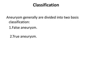
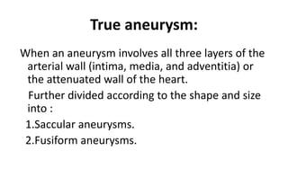
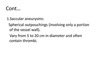
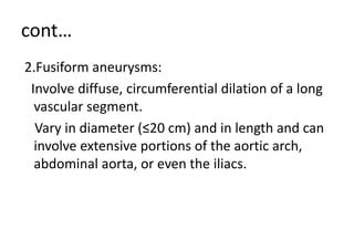
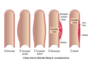
AORTIC ANEURYSMS
Presented by
Monika Devi
Msc. (N)
HCN, SRHU
ANEURYSMS
An aneurysm is a localized sac or dilation formed at a weak
point in the wall of the aorta.
Aortic aneurysm may involve the aortic arch, thoracic aorta
and or abdominal aorta.
most aneurysms however are found in the abdominal
aorta below the level of the renal arteries.
Causes
• Congenital:- primary connective tissue disorder (Marfan’s,
syndrome, Ehlers- Danlos Syndrome) and other diseases (focal
medial agenesis, tuberious scierosis,Turner’s Syndrome
Menkes’syndrome)
• Mechanical (hemodynamic):-Poststenotic and Arteriovenous
fistula and amputation related.
• Traumatic (pseudoaneurysms): Penetrating arterial injuries blunt
arterial injuries pseudoaneurysms
Causes
• Inflammatory (noninfectionus ): Associated with arteritist (Takayasu’s
diseases giant cell arteritis ,systemic lupus erythematosus ,Behcet’s
syndrome, Kawasaki’s disease) and periarterial inflammation (i.e
percreatitis).
• Infectious (mycotic ): Bacterial, fungal, spirochetal infections.
• Pregnancy –related degenerative: Nonspecific inflammatory variant .
• Anastomotic (post-arteriotomy) and graft aneurysms: Infection,
arterial wall failure, suture failure, graft failure.
Risk factors
• Hypertension
• Diabetes
• Atherosclerosis
• Smoking
• Family history
Classification
Aneurysm generally are divided into two basis
classification:
1.False aneurysm.
2.True aneurysm.
True aneurysm:
When an aneurysm involves all three layers of the
arterial wall (intima, media, and adventitia) or
the attenuated wall of the heart.
Further divided according to the shape and size
into :
1.Saccular aneurysms.
2.Fusiform aneurysms.
Cont…
1.Saccular aneurysms:
Spherical outpouchings (involving only a portion
of the vessel wall).
Vary from 5 to 20 cm in diameter and often
contain thrombi.
cont…
2.Fusiform aneurysms:
Involve diffuse, circumferential dilation of a long
vascular segment.
Vary in diameter (≤20 cm) and in length and can
involve extensive portions of the aortic arch,
abdominal aorta, or even the iliacs.
Cont…
• A false aneurysm or pseudoaneurysm, is not an aneusysm but a
disruption of all layers of the arterial wall resulting in bleeding that is
contained by surrounding structure. false aneurysms may result from
trauma or infection or occur after peripherial arteries bypass graft
surgery at the site of the graft to artery anastomosis they also may
result from arterial leakage after removal of cannulae such as upper
or lower extremity arterial catheters and intra aortic balloon pump
devices.
Etiologic types :
1. Atherosclerotic aneurysm.
2. Syphilitic aneurysm.
3. Congenital aneurysms (e.g., berry aneurysms).
4. Ventricular aneurysms that follow transmural
myocardial infarctions.
Cont…
5. Mycotic aneurysms can originate:
(a) From embolization of a septic thrombus, usually as a
complication of infective endocarditis.
(b) As an extension of an adjacent suppurative process.
(c) By circulating organisms directly infecting the arterial
wall.
Abdominal Aortic Aneurysm:
Atherosclerosis, the most common cause.
It causes thinning and weakening of the media secondary to
intimal plaques.
Such plaques compress the underlying media.
The media consequently undergoes degeneration and
necrosis.
Most frequently in the abdominal aorta (abdominal aortic
aneurysm AAA)
Clinical Course
The clinical consequences of AAA include:
1. Rupture into the peritoneal cavity or retroperitoneal
tissues with massive, potentially fatal hemorrhage
2.Obstruction of a branch vessel resulting in downstream
tissue ischemic injury.
Cont…
The risk of rupture is directly related to the size of the
aneurysm.
Timely surgery is critical; operative mortality for
unruptured aneurysms is approximately 5%,
whereas emergency surgery after rupture carries a
mortality rate of more than 50%.
Thoracic aortic aneurysms
(1) Encroachment on mediastinal structures.
(2) Respiratory difficulties caused by encroachment on the lungs and
airways.
(3) Difficulty in swallowing caused by compression of the esophagus
(4) Persistent cough from irritation of the recurrent laryngeal nerves.
(5) Pain caused by erosion of bone (i.e., ribs and vertebral bodies).
(6) Cardiac disease due to valvular insufficiency.
(7) Aortic rupture.
Diagnostic studies
• Chest X-Ray are useful in demonstrating the mediastinal
silhouette and any abnormal widening of the thoracic aorta.
• Plain X-Ray of the abdomen may show calcification within the
wall of abdominal aorta aneurysms
• Electrocardiogram (ECG)may be performed to rule out evidence
of myocardial infarction (MI) because some person with thoracic
aneurysms have symptoms suggestive of angina.
Echocardiography assists in the diagnosis of aortic valve
insufficiency related to ascending aortic dilation.
Cont…
• Ultrasonography is useful in screening for aneurysms and to
serially monitor aneurysm size.
• CT scan is the most accurate test to determine the anterior
to posterior length the cross sectional diameter and the
presence of thrombus in the aneurysm.
• Magnetic resonance imaging (MRI) also may be used to
diagnose and assess the location and severity of aneurysms.
Management
The goal of management is to prevent the aneurysm from
rupturing. therefore early detection and prompt treatment is
imperative.
• once an aneurysm is suspected studies are performed to
determine its exact size and location a careful review of all
body system is necessary to identify any coexisting disorder
especially of the lungs heart or kidney because they may
influence the patient’s surgical risk if carotid and /or coronary
artery obstructions are present they may need to be
corrected before the aneurysm is repaired for individual with
small aneurysms(<4cm).
Cont…
• conservative therapy typically is initiated which consist
of risk factor modification, decreasing blood pressure
(BP) and monitoring aneurysm size every 6 month using
ultrasound MRI or CT scan if the aneurysm size more
than 5.5cm or the increase in aneurysm size more than
0.5cm in 6 month should be repaired surgically.
Surgical management
The surgical technique involve
• Incising the deased segment of the aorta ;
• Removing intraluminal thrombus or plaque;
• Inserting synthetic graft dacron or polytetrafluoroethylene
(ptfe),which is sutured to the normal aorta oroximal and
distal to the aneurysm
Conventional surgical repair
• Suturing the native aortic wall around the graft so that it will
act a protective cover. if the iliac arteries also are aneurysmal,
the entire diseased segment is replaced with a bifurcation
graft. With saccular aneurysms. It may be possible to excise
only the bulbous lesion, repairing the artery by primary
closure (suturing the artery together) or by application of an
autogenous or synthetic patch graft over the arterial defect.
Cont…
• All aaa resections require cross clamping of the aorta
proximal and distal to the aneurysm.
• Most resections can be completed in 30 to 45 minutes, after
which time the clamps are removed and blood flow to the
lower extremities is restored.
• If the aaa extends above the renal arteries or if the cross
clamp must be applied above the renal arteries, adequate
renal perfusion
Complications
The most serous complication related to an untreated
aneurysm is rupture if rupture occure posteriorly into the
retroperitoneal space bleeding may be temponaded by
surrounding structure preventing exsanguinations and death
in this case the patient often sever back pain and may or
may not have back or flank ecchymosis (Grey turner’ssign)
Complications
• If rupture occurs anteriorly into the abdominal cavity most patient do
not servive long enough to get to the hospital they die from massive
hemorrhage if the patient does reach the hospital, he or she is in
hypovolemic shock with tachycardia hypotension pale clammy skin
decreased urine output altered level of consciousness and abdominal
tenderness on palpation in this situation simultaneous resuscitation
and immediate surgical repair are necessary.
THANK YOU

Aortic anurysm

  • 1.
    AORTIC ANEURYSMS Presented by MonikaDevi Msc. (N) HCN, SRHU
  • 2.
    ANEURYSMS An aneurysm isa localized sac or dilation formed at a weak point in the wall of the aorta. Aortic aneurysm may involve the aortic arch, thoracic aorta and or abdominal aorta. most aneurysms however are found in the abdominal aorta below the level of the renal arteries.
  • 4.
    Causes • Congenital:- primaryconnective tissue disorder (Marfan’s, syndrome, Ehlers- Danlos Syndrome) and other diseases (focal medial agenesis, tuberious scierosis,Turner’s Syndrome Menkes’syndrome) • Mechanical (hemodynamic):-Poststenotic and Arteriovenous fistula and amputation related. • Traumatic (pseudoaneurysms): Penetrating arterial injuries blunt arterial injuries pseudoaneurysms
  • 5.
    Causes • Inflammatory (noninfectionus): Associated with arteritist (Takayasu’s diseases giant cell arteritis ,systemic lupus erythematosus ,Behcet’s syndrome, Kawasaki’s disease) and periarterial inflammation (i.e percreatitis). • Infectious (mycotic ): Bacterial, fungal, spirochetal infections. • Pregnancy –related degenerative: Nonspecific inflammatory variant . • Anastomotic (post-arteriotomy) and graft aneurysms: Infection, arterial wall failure, suture failure, graft failure.
  • 6.
    Risk factors • Hypertension •Diabetes • Atherosclerosis • Smoking • Family history
  • 7.
    Classification Aneurysm generally aredivided into two basis classification: 1.False aneurysm. 2.True aneurysm.
  • 8.
    True aneurysm: When ananeurysm involves all three layers of the arterial wall (intima, media, and adventitia) or the attenuated wall of the heart. Further divided according to the shape and size into : 1.Saccular aneurysms. 2.Fusiform aneurysms.
  • 9.
    Cont… 1.Saccular aneurysms: Spherical outpouchings(involving only a portion of the vessel wall). Vary from 5 to 20 cm in diameter and often contain thrombi.
  • 10.
    cont… 2.Fusiform aneurysms: Involve diffuse,circumferential dilation of a long vascular segment. Vary in diameter (≤20 cm) and in length and can involve extensive portions of the aortic arch, abdominal aorta, or even the iliacs.
  • 12.
    Cont… • A falseaneurysm or pseudoaneurysm, is not an aneusysm but a disruption of all layers of the arterial wall resulting in bleeding that is contained by surrounding structure. false aneurysms may result from trauma or infection or occur after peripherial arteries bypass graft surgery at the site of the graft to artery anastomosis they also may result from arterial leakage after removal of cannulae such as upper or lower extremity arterial catheters and intra aortic balloon pump devices.
  • 13.
    Etiologic types : 1.Atherosclerotic aneurysm. 2. Syphilitic aneurysm. 3. Congenital aneurysms (e.g., berry aneurysms). 4. Ventricular aneurysms that follow transmural myocardial infarctions.
  • 14.
    Cont… 5. Mycotic aneurysmscan originate: (a) From embolization of a septic thrombus, usually as a complication of infective endocarditis. (b) As an extension of an adjacent suppurative process. (c) By circulating organisms directly infecting the arterial wall.
  • 15.
    Abdominal Aortic Aneurysm: Atherosclerosis,the most common cause. It causes thinning and weakening of the media secondary to intimal plaques. Such plaques compress the underlying media. The media consequently undergoes degeneration and necrosis. Most frequently in the abdominal aorta (abdominal aortic aneurysm AAA)
  • 16.
    Clinical Course The clinicalconsequences of AAA include: 1. Rupture into the peritoneal cavity or retroperitoneal tissues with massive, potentially fatal hemorrhage 2.Obstruction of a branch vessel resulting in downstream tissue ischemic injury.
  • 17.
    Cont… The risk ofrupture is directly related to the size of the aneurysm. Timely surgery is critical; operative mortality for unruptured aneurysms is approximately 5%, whereas emergency surgery after rupture carries a mortality rate of more than 50%.
  • 18.
    Thoracic aortic aneurysms (1)Encroachment on mediastinal structures. (2) Respiratory difficulties caused by encroachment on the lungs and airways. (3) Difficulty in swallowing caused by compression of the esophagus (4) Persistent cough from irritation of the recurrent laryngeal nerves. (5) Pain caused by erosion of bone (i.e., ribs and vertebral bodies). (6) Cardiac disease due to valvular insufficiency. (7) Aortic rupture.
  • 19.
    Diagnostic studies • ChestX-Ray are useful in demonstrating the mediastinal silhouette and any abnormal widening of the thoracic aorta. • Plain X-Ray of the abdomen may show calcification within the wall of abdominal aorta aneurysms • Electrocardiogram (ECG)may be performed to rule out evidence of myocardial infarction (MI) because some person with thoracic aneurysms have symptoms suggestive of angina. Echocardiography assists in the diagnosis of aortic valve insufficiency related to ascending aortic dilation.
  • 20.
    Cont… • Ultrasonography isuseful in screening for aneurysms and to serially monitor aneurysm size. • CT scan is the most accurate test to determine the anterior to posterior length the cross sectional diameter and the presence of thrombus in the aneurysm. • Magnetic resonance imaging (MRI) also may be used to diagnose and assess the location and severity of aneurysms.
  • 21.
    Management The goal ofmanagement is to prevent the aneurysm from rupturing. therefore early detection and prompt treatment is imperative. • once an aneurysm is suspected studies are performed to determine its exact size and location a careful review of all body system is necessary to identify any coexisting disorder especially of the lungs heart or kidney because they may influence the patient’s surgical risk if carotid and /or coronary artery obstructions are present they may need to be corrected before the aneurysm is repaired for individual with small aneurysms(<4cm).
  • 22.
    Cont… • conservative therapytypically is initiated which consist of risk factor modification, decreasing blood pressure (BP) and monitoring aneurysm size every 6 month using ultrasound MRI or CT scan if the aneurysm size more than 5.5cm or the increase in aneurysm size more than 0.5cm in 6 month should be repaired surgically.
  • 23.
    Surgical management The surgicaltechnique involve • Incising the deased segment of the aorta ; • Removing intraluminal thrombus or plaque; • Inserting synthetic graft dacron or polytetrafluoroethylene (ptfe),which is sutured to the normal aorta oroximal and distal to the aneurysm
  • 24.
    Conventional surgical repair •Suturing the native aortic wall around the graft so that it will act a protective cover. if the iliac arteries also are aneurysmal, the entire diseased segment is replaced with a bifurcation graft. With saccular aneurysms. It may be possible to excise only the bulbous lesion, repairing the artery by primary closure (suturing the artery together) or by application of an autogenous or synthetic patch graft over the arterial defect.
  • 25.
    Cont… • All aaaresections require cross clamping of the aorta proximal and distal to the aneurysm. • Most resections can be completed in 30 to 45 minutes, after which time the clamps are removed and blood flow to the lower extremities is restored. • If the aaa extends above the renal arteries or if the cross clamp must be applied above the renal arteries, adequate renal perfusion
  • 26.
    Complications The most serouscomplication related to an untreated aneurysm is rupture if rupture occure posteriorly into the retroperitoneal space bleeding may be temponaded by surrounding structure preventing exsanguinations and death in this case the patient often sever back pain and may or may not have back or flank ecchymosis (Grey turner’ssign)
  • 27.
    Complications • If ruptureoccurs anteriorly into the abdominal cavity most patient do not servive long enough to get to the hospital they die from massive hemorrhage if the patient does reach the hospital, he or she is in hypovolemic shock with tachycardia hypotension pale clammy skin decreased urine output altered level of consciousness and abdominal tenderness on palpation in this situation simultaneous resuscitation and immediate surgical repair are necessary.
  • 28.