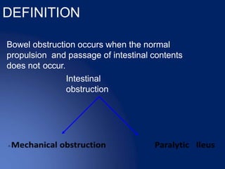
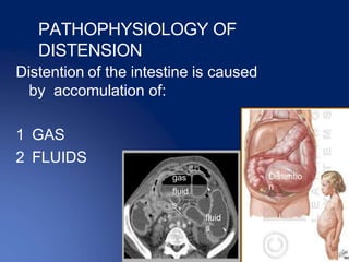
INTESTINAL OBSTRUCTION
DR SHAMBHAVI SHARMA
Bowel obstruction occurs when the normal
propulsion and passage of intestinal contents
does not occur.
Intestinal
obstruction
DEFINITION
CASE 1
65 YEARS HYPERTENSIVE SMOKER MALE PRESENTED WITH
PAIN IN LOWER ABDOMEN A/W VOMITING ,ABDOMINAL
DISTENSION AND ABSOLUTE CONSTIPATION
1.WHAT IS LIKELY DIAGNOSIS?
2.HOW WILL YOU PROCEED ?
CLINICAL FEATURE OF INTESTINAL
OBSTRUCTION
Clinical obstruction of intestinal
obstruction vary according to :
 The location of the obstruction;
 The age of the obstruction;
 The underlying pathology;
 The presence or the absence of the
intestinal obstruction;
Intestinal
obstruction
• small intestine (small bowel obstruction),
• large intestine (large bowel obstruction),
• via systemic alterations, involving both the
small and large intestine (generalized ileus).
PAIN
 Pain is the first symptom
encountered, it occurs
suddenly and is usually
severe..
 It is colicky in nature and
usually centered on the
umbilicus(small bowel) or
lower abdomen (large
bowel).
 The pain coincides with the
increasing peristaltic
activity
VOMITING
 The more distal the obstruction
,the longer the interval between
the onset of symptoms and the
appearance of nausea and
vomiting .
 More proximal the obstruction,
more the frequency.
 The interval ,frequency & nature
of vomitus depends on the site of
obstruction
• In high bowel obstruction :
• the interval is shorter
• Bile stained vomitus
• Vomiting is more frequent and copious ;
• And is relieved by decompressing the
obstructed bowel
In low bowel obstruction :
• the interval is longer may last for a day or
two
• Feculent vomitus
• vomiting is less frequent and does not
cause any relief.
Pyloric obstruction
•Watery and acidic
vomitus Large bowel
obstruction
Uncommon and late symptoms.
Long standing low small bowel
obstruction-
• feculent material.
• Strangulation-blood.
DISTENTION
 In the small bowel, the
degree of distention is
dependent on the site of
obstruction & is greater the
more distal the lesion.
 Central abdomen is
distended in low small bowel
obstruction.
 Distention is much less in
high small bowel obstruction.
CONSTIPATION
• Failure to pass flatus or faeces through
the rectum is important symptom of
bowel obstruction.
• It may be classified as
1. ABSOLUTE
2. RELATIVE
ON THE BASIS OF NATURE
ACUTE
CHRONIC
ACUTE ON
CHRONIC
SUBACUTE
ACUTE
OBSTRUCTION :
 It usually occur in small bowel obstruction with
sudden onset of severe colicky central
abdominal pain,distention and early vomiting
and constipation.
CHRONIC
OBSTRUCTION :
 Usually seen in large bowel obstruction with
lower abdominal colic and absolute
constipation,followed by distention.
ACUTE ON CHRONIC
OBSTRUCTION :
 It starts in large bowel but gradually involves
the small intestine.
 Early symptoms are pain and constipation but
when small intestine is involved it is
characterized by vomiting and general
distention.
CAUSES OF OBSTRUCTION
 DYNAMIC : where peristalsis is working
against a mechanical obstruction
 ADYNAMIC: it may occur in two forms
1. 1st where peristalsis may be absent
(paralytic ileus,)occurring secondarily to
neuromuscular failure in the mesentery.
2. 2nd where peristalsis may be present in
non- propulsive form.(pseudo-
obstruction)
Case
ON PAST HISTORY :
1.He has had undergone exploratory laparotomy for similar
complaints 5 years back
What is your diagnosis ?
2. Swelling in inguinal area which reduced spontaneously in past,
irreducible for last 3 hours .
What is your diagnosis ?
3.He had history of weight loss , alteration in bowel habit and
bleeding per rectum for last 1 month
what is your diagnosis ?
4.He had history of taking warfarin for cardiac disease ?
Will this information help?
5.He has history of hypothyroidism and and passes stool every 3-
4 days for last 3 years
What clue do you get for your diagnosis ?
• Adhesions- 40%
• Tumors -15%
• Inflamatory- 15%
• Obstructed hernia-
12%
• Intraluminal-10%
• Miscellaneous -8%
ETIOLOGY
DYNAMIC(MECHANICAL)FROM
THE WALL
1 TB
2 CROHN’S
3 TUMORS
4 STICTURE
5 CONGENITAL ……… .
ETIOLOGY
MECHANNICAL IN
THE LUMEN
1 GALL STONES
2 F.B
3 BEZOARS
4 WORMS
5 FECES GALL
STONES
BEZOARS
WORMs
FAECES
ETIOLOGY
MECHANICAL
EXTRALUMINAL
1- BANDS
2 ADHESIONS
3 ABSCESS
4 HERNIAS
5 COMPRESSION
6 INTUSSESCEPTION
NDS
ABSCESS
COM
GALLSTO
NES
Duodenal
Artesia
INTUSSUCEPTION
ADHESIVE BANDS AND
CONSTRICTION
Intestinal
tumor
CASE
He says that pain was initially mild however now the pain has
increased in intensity and aggravated by sublte movements as
well
What do you think has happened ?
Presentation of bowel
obstruction
• This mechanical obstruction can be partial ( lumen
narrowed but allow transit some content) or complete (
lumen totally obstruction) this classify to
• simple obstruction (no vascular impairment)
• strangulated obstruction(with vascular impairment
and peritonitis)
• Closed loop obstruction
STARANGULA
TED
OBSTRUCTI
ON
STARANGULATED OBSTRUCTION :
 Strangulating obstruction is obstruction with
compromised blood flow; it occurs in nearly
25% of patients with small-bowel obstruction.
 It is usually associated with hernia, volvulus,
and intussusceptions.
 Strangulating obstruction can progress to
infarction and gangrene in as little as 6 h.
.
• Venous obstruction
occurs first, followed
by arterial occlusion,
resulting in rapid
ischemia of the bowel
wall.
• The ischemic bowel
becomes edematous
and infarcts, leading
to gangrene and
perforation. In large-
bowel obstruction,
strangulation is rare
(except with
volvulus)
CLOSED LOOP
OBSTRUCTION
 Closed loop obstruction is a specific
type of obstruction in which two
points along the course of a bowel
are obstructed at a single location
thus forming a closed loop.
 Usually this is due to adhesions, a
twist of the mesentery or internal
herniation.
• In the large bowel it is known
as a volvulus.
• In the small bowel it is simply known
as small bowel closed loop
obstruction.
• Obstruction to the blood supply
occur either from the same
mechanism which caused
obstruction or by the twist of the
bowel on mesentery.
CASE
1.This patient gives history of cocaine abuse since 10 years
What would be the underlying etiology for obstruction?
2. He has underwent open cholecystectomy with cbd
exploration 2 days back .
D/d?
3.He has history of acute gastroenteritis and has vomited more
than 30 times in past 2 days.
What might be the pathophysiology for obstruction ?
ETIOLOGY
Adynamic Intestinal
Obstruction
1- Peritonitis
2 Electrolytes’ Imbalance
3 Postoperative
4 Ischemia
5 Drugs
6 Retroperitoneal causes
Case
IF he was of younger age group ………
CAUSES ACCORDING TO AGE
BIRTH : Atresia, Meconium, NE, Volvulus,Hirschsprung’s
3 WEEKS : Pyloric stenosis
6-9MONTHS : Intussusception
TEENAGE : Appendicitis , Meckel’s diverticulitis
YOUNG ADULT : Adhesions,Hernia
ADULT : Adhesions ,Hernia, Appendicitis,Crohn’s,
Carcinoma
ELDERLY : Carcinoma, Diverticulitis, Sigmoid Volvulus ,
Feces
How will you examine the patient ?
Examination
findings
Intestinal
obstructio
n
Vomiting
Distention
constipation
Pain
PATHOPHYSIOLOGY
Dehydration caused by :
1. Reduced intake
2. Reduced absorption
3. Increased loss (Vomiting &
sequesration)
Should we examine the vital signs and mental status ??
PATHOPHYSIOLOGY
Systemic Effects of Obstruction :
1. Water and electrolyte losses (lead to
hypovolemia)
2. Toxic materials and toxemia(lead to sepsis)
3. Cardiopulmonary dysfunction(atelectasis)
4. Renal failure
5. Shock and death
PHYSICAL EXAMINATION
Abdominal
examination
Inspection
Palpation
Percussion
Auscultation
Rectal
examination
INSPECTION
INSPECTION
Shape of the abdomen
Movement of the abdomen wall
Umbilicus
Visible loop of bowel/visible
peristalsis
Scar
Striae
Prominent veins
Pubic hair
Hernial orifices
PATHOPHYSIOLOGY OF
DISTENSION
Distention of the intestine is caused
by accomulation of:
1 GAS
2 FLUIDS
fluid
s
Distentio
n
gas
fluid
s
PATHOPYSIOLOGY
SIMPLE OBSTRUCTION :
1 ABOVE THE OBSTRUCTION
OBSTRUCTION  Peristalsis increases  Intstine dilates
 Reduction in peristaltic strength  Flaccidity and
paralysis (protective but late)
2 BELOW THE OBSTRUCTION
NORMAL PERISTALSIS & ABSORBTION  Until it
becomes empty  It contracts & becomes immobile
Pathophysiology
Gas in the intestine is due
to:
1. Swallowed air
2. Bacterial overgrowth
3. Diffusion from blood
PATHOPHYSIOLOGY
Fluids come from :
1. Ingested fluids
2. Saliva
3. Gastric and intestinal
juice
4. Bile & Pancreatic
secretions
VISIBLE
PERISTALSIS
Visible peristalsis may be present if
the abdomen is examined carefully.
Mostly seen in proximal loops.
Borborygmi is quite loud ,does not
require stethoscope to hear it .
In auscultation sound of hyper
peristalsis coinciding with attack of
colic characteristic feature f intestinal
obstruction.
VISIBLE
PERISTALSIS
BLOATING
• The accumulation of chyme and gas gives
rise to a feeling of fullness and causes
bloating. This may also give rise to high-
pitched gurgling sounds from the
abdomen
PALPATION
PALPATION
During colic there may be muscle
guarding.
Slight tenderness may be present
between attacks of pain.
Tenderness and rigidity at the sight of
obstruction usually indicate
strangulation.
All the hernial orifices should be
palpated to exclude the presence of
hernia.
PERCUSSION
PERCUSSION
Percussion to hear any Dullness or
Resonance related to site of obstruction.
Tympanic node will be present.
Tenderness on light percussion
suggest strangulation.
AUSCULTATION
AUSCULTATION
Bowel sounds are Initially Loud and
frequent
Then as bowel distends the sounds
become more resonant and high
pitched
Eventually becoming amphoric.
In strangulation bowel sound is
completely absent.
RECTAL
EXAMINATION
Presence of mass on rectal examination
within or outside the lumen will give a clue
to diagnosis.
Presence or absence of feces in rectum
should be noted.
Absence means obstruction is higher up.
If presence it should be studied for
presence of occult blood which include
mucosal lesion e.g.cancer,Intussuception
or infraction
Revision
what is strangulation ??
PATHOPHYSIOLOGY
Strangulation leads to impaired venous
return
 Increased congestion 
-free peritoneal fluid
-edema of intestinal wall
-blood in the lumen
-impaired arterial blood supply
-ischemia and gangrene
DIAGNOSIS
History
Clinical
examination
Investigations
INVESTIGATI
ONS
• BLOOD EXAMINATION
• RADIOLOGICAL
EXAMINATION
BLOOD
EXAMINATION
• CBC
• Urea &
electrolytes
• Metabolic acidosis
CBC (Complete blood
count)-
A rise white cell count will indicate
an infection.
Normal or slight rise in W.B.C count:
simple mechanical obstruction.
Moderate rise in W.B.C
count(15000-
20000):strangulation.
Very high rise in W.B.C count(30000-
40000):primary mesenteric vascular
occlusion.
Serum Urea &
electrolytes-
Derangement may be seen with
vomiting & diarrhea.
Dehydration will be reflected in raised
serum urea and creatinine.
Metabolic
acidosis
It occurs due to combined effects of
dehydration ketosis and loss of
alkaline secretion.
Very common in distal intestinal
obstruction.
RADIOLOGICAL
EXAMINATION
RADIOLOGICAL
EXAMINATION
Gas fluid levels are the most important
criteria of diagnosis of intestinal
obstruction.
When obstruction occurs, both fluid and
gas collect in the intestine.
They produce a characteristic pattern
called "air-fluid levels"
The air rises above the fluid and there is
a flat surface at the "air-fluid" interface.
RADIOLOGICAL
PICTURE
 Small Bowel Obstruction
- Central distention (GAS)
- Valvulae conniventes
- “Ladder-like dilatation”
- Small diameter
 Large Bowel Obstruction
- Peripheral distention “Picture
frame”
- More gross distention
- Haustral indentation & large
DIAGNOSIS
Small bowel
obstruction
Large bowel
obstruction
In most cases, the abdominal
radiograph will have the following
features:
1. ileated loops of small bowel
proximal to the obstruction
2. predominantly central dilated
loops
3. dilatation of loops over 3cm
4. valvulae conniventes are visible
DIAGNOS ?
Paralytic
Ileus
Volvulus x ray: Sigmoid volvulus -
'coffee bean' sign
The sigmoid colon is very dilated because it is twisted at the root
of its mesentery in the left iliac fossa (LIF)
The twisted loop of sigmoid colon is said to resemble a coffee
bean
Barium
studies
• Are recommended in patient with a history
of recurring obstruction
CT
scan
Useful in patient with a
history of abdominal
malignancy
in postsurgical patients
and in patient who have no
history of abdominal surgery
and present with symptoms
of bowel obstruction.
Ultrasound: small
bowel
obstruction
CASE
YOU ARE ON DUTY INTERN AND CALLED TO MANAGE THIS CASE
WHAT WILL YOU DO ?
DANGEROUS SIGNS
(Red Flags)
Constant pain
Absent bowel sounds
Tenderness with
rigidity
Leukocytosis
Fever and
tachycardia
Shock
Three main
measures-
 GI drainage
 Fluid &Electrolyte replacement
 Relief of obstruction, usually
surgical
Treatme
nt
Conservative:
 Nasogastric aspiration by Ryles tube
 IV fluids- volume varies depending on
dehydration
 NPO
 urinary catheter
 check temp. and pulse 2 hourly
 abdominal examination 8 hourly
 Broad spectrum antibiotics initiated early-
reduce bacterial overgrowth.
Some cases will settle by using this conservative
regimen, other need surgical intervention.
Surgery should be delayed till resuscitation is complete
unless signs of strangulation and evidence of
closed-loop obstruction.
Cases that show reasons for delay should be monitored
continuously for 72 hours in hope of spontaneous
resolution
e.g. adhesions with radiological findings but no pain
or tenderness
Indication for surgery:
- failure of conservative management
- tender, irreducible hernia
- strangulation
- virgin abdomen
• If the site of obstruction is unknown; laparotomy
assessment is directed to-
-The site of obstruction.
-The nature of obstruction.
-The viability of gut.
• The site of obstruction can be determined by caecum
Surgical
treatment
Operative decompression required-if
dilatation of bowel loops prevent exposure,
bowel wall viability is compromised,
or if subsequent closure will be compromised.
Savage’s decompressor used
within seromuscular purse-string
suture.
Or large-bore NG tube maybe used for milking
intestinal contents into stomach.
The type of surgical procedure depend upon the cause of
obstruction viz division of bands,adhesiolysis, excision ,or
bypass
*Once obstruction relieved, the bowel is inspected for
viability, and if non-viable, resection is required.
Indication of non-viability
1.absent peristalsis
2.loss of normal shine
3.loss of pulsation in mesentry
4.green or black color of bowel
5.absent mesentric pulsations
• If in doubt of viability, bowel is wrapped
in hot packs for 10 minutes with
increased oxygen and reassessed for
viability.
• Resection of non viable gut should be
done followed by stoma.
• Sometimes a second look
laprotomy is required in 24-48
hours e.g. multiple ischemic areas.
MANAGEMENT OF ACUTE
CASE (Plan)
I.V Fluids and electrolytes rescusitation for all
N.G tube if repeated vomiting
Antibiotics for all
Hernia  Operation
Adhesions  Conservative first
Obstruction  Remove
Volvulus  Derotate and or Operate
Mesenteric ischemia  Operate
Abscess or Peritonitis  Drain and Treat
Intussusception  Pneumatic or Barium
Reductio
n or Operate
Do not take to OR
if:
• Post-op
• Carcinomatosis
• Recurrent adhesive bowel
obstruction
• Post radiotherapy
MANAGEMENT ACCORDING
TO CAUSE
LETS REVISE THE CAUSES …………………….
THANK YOU

Acute intestinal obstruction

  • 1.
  • 2.
    Bowel obstruction occurswhen the normal propulsion and passage of intestinal contents does not occur. Intestinal obstruction DEFINITION
  • 3.
    CASE 1 65 YEARSHYPERTENSIVE SMOKER MALE PRESENTED WITH PAIN IN LOWER ABDOMEN A/W VOMITING ,ABDOMINAL DISTENSION AND ABSOLUTE CONSTIPATION 1.WHAT IS LIKELY DIAGNOSIS? 2.HOW WILL YOU PROCEED ?
  • 4.
    CLINICAL FEATURE OFINTESTINAL OBSTRUCTION Clinical obstruction of intestinal obstruction vary according to :  The location of the obstruction;  The age of the obstruction;  The underlying pathology;  The presence or the absence of the intestinal obstruction;
  • 5.
    Intestinal obstruction • small intestine(small bowel obstruction), • large intestine (large bowel obstruction), • via systemic alterations, involving both the small and large intestine (generalized ileus).
  • 6.
    PAIN  Pain isthe first symptom encountered, it occurs suddenly and is usually severe..  It is colicky in nature and usually centered on the umbilicus(small bowel) or lower abdomen (large bowel).  The pain coincides with the increasing peristaltic activity
  • 7.
    VOMITING  The moredistal the obstruction ,the longer the interval between the onset of symptoms and the appearance of nausea and vomiting .  More proximal the obstruction, more the frequency.  The interval ,frequency & nature of vomitus depends on the site of obstruction
  • 8.
    • In highbowel obstruction : • the interval is shorter • Bile stained vomitus • Vomiting is more frequent and copious ; • And is relieved by decompressing the obstructed bowel
  • 9.
    In low bowelobstruction : • the interval is longer may last for a day or two • Feculent vomitus • vomiting is less frequent and does not cause any relief.
  • 10.
    Pyloric obstruction •Watery andacidic vomitus Large bowel obstruction Uncommon and late symptoms.
  • 11.
    Long standing lowsmall bowel obstruction- • feculent material. • Strangulation-blood.
  • 12.
    DISTENTION  In thesmall bowel, the degree of distention is dependent on the site of obstruction & is greater the more distal the lesion.  Central abdomen is distended in low small bowel obstruction.  Distention is much less in high small bowel obstruction.
  • 13.
    CONSTIPATION • Failure topass flatus or faeces through the rectum is important symptom of bowel obstruction. • It may be classified as 1. ABSOLUTE 2. RELATIVE
  • 14.
    ON THE BASISOF NATURE ACUTE CHRONIC ACUTE ON CHRONIC SUBACUTE
  • 15.
    ACUTE OBSTRUCTION :  Itusually occur in small bowel obstruction with sudden onset of severe colicky central abdominal pain,distention and early vomiting and constipation.
  • 16.
    CHRONIC OBSTRUCTION :  Usuallyseen in large bowel obstruction with lower abdominal colic and absolute constipation,followed by distention.
  • 17.
    ACUTE ON CHRONIC OBSTRUCTION:  It starts in large bowel but gradually involves the small intestine.  Early symptoms are pain and constipation but when small intestine is involved it is characterized by vomiting and general distention.
  • 18.
    CAUSES OF OBSTRUCTION DYNAMIC : where peristalsis is working against a mechanical obstruction  ADYNAMIC: it may occur in two forms 1. 1st where peristalsis may be absent (paralytic ileus,)occurring secondarily to neuromuscular failure in the mesentery. 2. 2nd where peristalsis may be present in non- propulsive form.(pseudo- obstruction)
  • 19.
    Case ON PAST HISTORY: 1.He has had undergone exploratory laparotomy for similar complaints 5 years back What is your diagnosis ? 2. Swelling in inguinal area which reduced spontaneously in past, irreducible for last 3 hours . What is your diagnosis ?
  • 20.
    3.He had historyof weight loss , alteration in bowel habit and bleeding per rectum for last 1 month what is your diagnosis ? 4.He had history of taking warfarin for cardiac disease ? Will this information help? 5.He has history of hypothyroidism and and passes stool every 3- 4 days for last 3 years What clue do you get for your diagnosis ?
  • 21.
    • Adhesions- 40% •Tumors -15% • Inflamatory- 15% • Obstructed hernia- 12% • Intraluminal-10% • Miscellaneous -8%
  • 22.
    ETIOLOGY DYNAMIC(MECHANICAL)FROM THE WALL 1 TB 2CROHN’S 3 TUMORS 4 STICTURE 5 CONGENITAL ……… .
  • 23.
    ETIOLOGY MECHANNICAL IN THE LUMEN 1GALL STONES 2 F.B 3 BEZOARS 4 WORMS 5 FECES GALL STONES BEZOARS WORMs FAECES
  • 24.
    ETIOLOGY MECHANICAL EXTRALUMINAL 1- BANDS 2 ADHESIONS 3ABSCESS 4 HERNIAS 5 COMPRESSION 6 INTUSSESCEPTION NDS ABSCESS COM
  • 25.
  • 26.
  • 27.
  • 28.
  • 29.
  • 30.
    CASE He says thatpain was initially mild however now the pain has increased in intensity and aggravated by sublte movements as well What do you think has happened ?
  • 31.
    Presentation of bowel obstruction •This mechanical obstruction can be partial ( lumen narrowed but allow transit some content) or complete ( lumen totally obstruction) this classify to • simple obstruction (no vascular impairment) • strangulated obstruction(with vascular impairment and peritonitis) • Closed loop obstruction
  • 32.
  • 33.
    STARANGULATED OBSTRUCTION : Strangulating obstruction is obstruction with compromised blood flow; it occurs in nearly 25% of patients with small-bowel obstruction.  It is usually associated with hernia, volvulus, and intussusceptions.  Strangulating obstruction can progress to infarction and gangrene in as little as 6 h.
  • 34.
    . • Venous obstruction occursfirst, followed by arterial occlusion, resulting in rapid ischemia of the bowel wall. • The ischemic bowel becomes edematous and infarcts, leading to gangrene and perforation. In large- bowel obstruction, strangulation is rare (except with volvulus)
  • 35.
    CLOSED LOOP OBSTRUCTION  Closedloop obstruction is a specific type of obstruction in which two points along the course of a bowel are obstructed at a single location thus forming a closed loop.  Usually this is due to adhesions, a twist of the mesentery or internal herniation.
  • 36.
    • In thelarge bowel it is known as a volvulus. • In the small bowel it is simply known as small bowel closed loop obstruction. • Obstruction to the blood supply occur either from the same mechanism which caused obstruction or by the twist of the bowel on mesentery.
  • 37.
    CASE 1.This patient giveshistory of cocaine abuse since 10 years What would be the underlying etiology for obstruction? 2. He has underwent open cholecystectomy with cbd exploration 2 days back . D/d? 3.He has history of acute gastroenteritis and has vomited more than 30 times in past 2 days. What might be the pathophysiology for obstruction ?
  • 38.
    ETIOLOGY Adynamic Intestinal Obstruction 1- Peritonitis 2Electrolytes’ Imbalance 3 Postoperative 4 Ischemia 5 Drugs 6 Retroperitoneal causes
  • 39.
    Case IF he wasof younger age group ………
  • 40.
    CAUSES ACCORDING TOAGE BIRTH : Atresia, Meconium, NE, Volvulus,Hirschsprung’s 3 WEEKS : Pyloric stenosis 6-9MONTHS : Intussusception TEENAGE : Appendicitis , Meckel’s diverticulitis YOUNG ADULT : Adhesions,Hernia ADULT : Adhesions ,Hernia, Appendicitis,Crohn’s, Carcinoma ELDERLY : Carcinoma, Diverticulitis, Sigmoid Volvulus , Feces
  • 41.
    How will youexamine the patient ?
  • 42.
  • 43.
    PATHOPHYSIOLOGY Dehydration caused by: 1. Reduced intake 2. Reduced absorption 3. Increased loss (Vomiting & sequesration)
  • 44.
    Should we examinethe vital signs and mental status ??
  • 45.
    PATHOPHYSIOLOGY Systemic Effects ofObstruction : 1. Water and electrolyte losses (lead to hypovolemia) 2. Toxic materials and toxemia(lead to sepsis) 3. Cardiopulmonary dysfunction(atelectasis) 4. Renal failure 5. Shock and death
  • 46.
  • 47.
  • 48.
    INSPECTION Shape of theabdomen Movement of the abdomen wall Umbilicus Visible loop of bowel/visible peristalsis Scar Striae Prominent veins Pubic hair Hernial orifices
  • 49.
    PATHOPHYSIOLOGY OF DISTENSION Distention ofthe intestine is caused by accomulation of: 1 GAS 2 FLUIDS fluid s Distentio n gas fluid s
  • 50.
    PATHOPYSIOLOGY SIMPLE OBSTRUCTION : 1ABOVE THE OBSTRUCTION OBSTRUCTION  Peristalsis increases  Intstine dilates  Reduction in peristaltic strength  Flaccidity and paralysis (protective but late) 2 BELOW THE OBSTRUCTION NORMAL PERISTALSIS & ABSORBTION  Until it becomes empty  It contracts & becomes immobile
  • 51.
    Pathophysiology Gas in theintestine is due to: 1. Swallowed air 2. Bacterial overgrowth 3. Diffusion from blood
  • 52.
    PATHOPHYSIOLOGY Fluids come from: 1. Ingested fluids 2. Saliva 3. Gastric and intestinal juice 4. Bile & Pancreatic secretions
  • 53.
    VISIBLE PERISTALSIS Visible peristalsis maybe present if the abdomen is examined carefully. Mostly seen in proximal loops. Borborygmi is quite loud ,does not require stethoscope to hear it . In auscultation sound of hyper peristalsis coinciding with attack of colic characteristic feature f intestinal obstruction.
  • 54.
  • 55.
    BLOATING • The accumulationof chyme and gas gives rise to a feeling of fullness and causes bloating. This may also give rise to high- pitched gurgling sounds from the abdomen
  • 56.
  • 57.
    PALPATION During colic theremay be muscle guarding. Slight tenderness may be present between attacks of pain. Tenderness and rigidity at the sight of obstruction usually indicate strangulation. All the hernial orifices should be palpated to exclude the presence of hernia.
  • 58.
  • 59.
    PERCUSSION Percussion to hearany Dullness or Resonance related to site of obstruction. Tympanic node will be present. Tenderness on light percussion suggest strangulation.
  • 60.
  • 61.
    AUSCULTATION Bowel sounds areInitially Loud and frequent Then as bowel distends the sounds become more resonant and high pitched Eventually becoming amphoric. In strangulation bowel sound is completely absent.
  • 62.
    RECTAL EXAMINATION Presence of masson rectal examination within or outside the lumen will give a clue to diagnosis. Presence or absence of feces in rectum should be noted. Absence means obstruction is higher up. If presence it should be studied for presence of occult blood which include mucosal lesion e.g.cancer,Intussuception or infraction
  • 63.
  • 64.
    PATHOPHYSIOLOGY Strangulation leads toimpaired venous return  Increased congestion  -free peritoneal fluid -edema of intestinal wall -blood in the lumen -impaired arterial blood supply -ischemia and gangrene
  • 65.
  • 66.
  • 67.
    BLOOD EXAMINATION • CBC • Urea& electrolytes • Metabolic acidosis
  • 68.
    CBC (Complete blood count)- Arise white cell count will indicate an infection. Normal or slight rise in W.B.C count: simple mechanical obstruction. Moderate rise in W.B.C count(15000- 20000):strangulation. Very high rise in W.B.C count(30000- 40000):primary mesenteric vascular occlusion.
  • 69.
    Serum Urea & electrolytes- Derangementmay be seen with vomiting & diarrhea. Dehydration will be reflected in raised serum urea and creatinine.
  • 70.
    Metabolic acidosis It occurs dueto combined effects of dehydration ketosis and loss of alkaline secretion. Very common in distal intestinal obstruction.
  • 71.
  • 72.
    RADIOLOGICAL EXAMINATION Gas fluid levelsare the most important criteria of diagnosis of intestinal obstruction. When obstruction occurs, both fluid and gas collect in the intestine. They produce a characteristic pattern called "air-fluid levels" The air rises above the fluid and there is a flat surface at the "air-fluid" interface.
  • 73.
    RADIOLOGICAL PICTURE  Small BowelObstruction - Central distention (GAS) - Valvulae conniventes - “Ladder-like dilatation” - Small diameter  Large Bowel Obstruction - Peripheral distention “Picture frame” - More gross distention - Haustral indentation & large
  • 74.
  • 75.
    In most cases,the abdominal radiograph will have the following features: 1. ileated loops of small bowel proximal to the obstruction 2. predominantly central dilated loops 3. dilatation of loops over 3cm 4. valvulae conniventes are visible
  • 79.
  • 80.
    Volvulus x ray:Sigmoid volvulus - 'coffee bean' sign The sigmoid colon is very dilated because it is twisted at the root of its mesentery in the left iliac fossa (LIF) The twisted loop of sigmoid colon is said to resemble a coffee bean
  • 81.
    Barium studies • Are recommendedin patient with a history of recurring obstruction
  • 82.
    CT scan Useful in patientwith a history of abdominal malignancy in postsurgical patients and in patient who have no history of abdominal surgery and present with symptoms of bowel obstruction.
  • 83.
  • 85.
    CASE YOU ARE ONDUTY INTERN AND CALLED TO MANAGE THIS CASE WHAT WILL YOU DO ?
  • 87.
    DANGEROUS SIGNS (Red Flags) Constantpain Absent bowel sounds Tenderness with rigidity Leukocytosis Fever and tachycardia Shock
  • 88.
    Three main measures-  GIdrainage  Fluid &Electrolyte replacement  Relief of obstruction, usually surgical
  • 89.
    Treatme nt Conservative:  Nasogastric aspirationby Ryles tube  IV fluids- volume varies depending on dehydration  NPO  urinary catheter  check temp. and pulse 2 hourly  abdominal examination 8 hourly  Broad spectrum antibiotics initiated early- reduce bacterial overgrowth.
  • 90.
    Some cases willsettle by using this conservative regimen, other need surgical intervention. Surgery should be delayed till resuscitation is complete unless signs of strangulation and evidence of closed-loop obstruction. Cases that show reasons for delay should be monitored continuously for 72 hours in hope of spontaneous resolution e.g. adhesions with radiological findings but no pain or tenderness
  • 91.
    Indication for surgery: -failure of conservative management - tender, irreducible hernia - strangulation - virgin abdomen • If the site of obstruction is unknown; laparotomy assessment is directed to- -The site of obstruction. -The nature of obstruction. -The viability of gut. • The site of obstruction can be determined by caecum
  • 92.
    Surgical treatment Operative decompression required-if dilatationof bowel loops prevent exposure, bowel wall viability is compromised, or if subsequent closure will be compromised. Savage’s decompressor used within seromuscular purse-string suture. Or large-bore NG tube maybe used for milking intestinal contents into stomach.
  • 93.
    The type ofsurgical procedure depend upon the cause of obstruction viz division of bands,adhesiolysis, excision ,or bypass *Once obstruction relieved, the bowel is inspected for viability, and if non-viable, resection is required. Indication of non-viability 1.absent peristalsis 2.loss of normal shine 3.loss of pulsation in mesentry 4.green or black color of bowel 5.absent mesentric pulsations
  • 95.
    • If indoubt of viability, bowel is wrapped in hot packs for 10 minutes with increased oxygen and reassessed for viability. • Resection of non viable gut should be done followed by stoma. • Sometimes a second look laprotomy is required in 24-48 hours e.g. multiple ischemic areas.
  • 96.
    MANAGEMENT OF ACUTE CASE(Plan) I.V Fluids and electrolytes rescusitation for all N.G tube if repeated vomiting Antibiotics for all Hernia  Operation Adhesions  Conservative first Obstruction  Remove Volvulus  Derotate and or Operate Mesenteric ischemia  Operate Abscess or Peritonitis  Drain and Treat Intussusception  Pneumatic or Barium Reductio n or Operate
  • 97.
    Do not taketo OR if: • Post-op • Carcinomatosis • Recurrent adhesive bowel obstruction • Post radiotherapy
  • 98.
    MANAGEMENT ACCORDING TO CAUSE LETSREVISE THE CAUSES …………………….
  • 99.