Downloaded 251 times

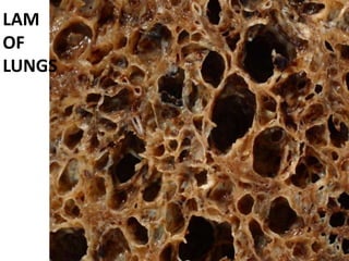
![Lymphangioleiomyomatosis (LAM) is a rare disorder resulting
from proliferation in the lung, kidney, and axial lymphatics of
leiomyoma (LAM cells) that exhibit features of
neoplasia. Cystic destruction of the lung with progressive
pulmonary dysfunction(pneumothorax , dyspnea), and the
presence of abdominal tumors (eg, angiomyolipomas
[AML], lymphangioleiomyomas) characterize the disease.](https://image.slidesharecdn.com/lymphangioleiomyomatosis-130627155218-phpapp02/85/Lymphangioleiomyomatosis-2-320.jpg)
![Proliferation of lymphangioleiomyomatosis (LAM) cells
may obstruct bronchioles,possibly leading to airflow
obstruction, air trapping, formation of bullae, and
pneumothoraces. Obstruction of lymphatics may result in
lymphangioleiomyomas, chylothorax, and chylous ascites.
Obstruction of venules may result in hemosiderosis and
hemoptysis. Excessive proteolytic activity, which relates
to an imbalance of the elastase/alpha1-antitrypsin system
or metalloprotease (MMPs) and their inhibitors (tissue
inhibitors of metalloproteases [TIMPs]), may be
important in lung destruction and formation of cysts.
PATHOLOGY](https://image.slidesharecdn.com/lymphangioleiomyomatosis-130627155218-phpapp02/85/Lymphangioleiomyomatosis-5-320.jpg)
![TREATMENT
General care for patients with lymphangioleiomyomatosis (LAM) addresses
the following findings:
Pleural effusions - Consider chemical pleurodesis; surgical obliteration of
the pleural space; medium-chain triglyceride (MCT [not a component of
chyle]), lipid-free diet to reduce chyle flow (utility unknown)
Ascites - Paracentesis, MCT diet (utility unknown)
Airways disease and hypoxemia - Bronchodilators may be of benefit[25] ;
supplemental oxygen, pulmonary rehabilitation, smoking cessation
Standard vaccination for respiratory infections
Osteoporosis - Standard surveillance and treatment; avoid exogenous
estrogens
Lung transplantation](https://image.slidesharecdn.com/lymphangioleiomyomatosis-130627155218-phpapp02/85/Lymphangioleiomyomatosis-25-320.jpg)


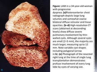
Lymphangioleiomyomatosis (LAM) is a rare lung disease that affects premenopausal women. It involves the proliferation of abnormal smooth muscle-like cells (LAM cells) in the lungs, lymphatic system, and kidneys. This leads to cyst formation in the lungs and lymphatic obstruction, causing symptoms like shortness of breath, pneumothorax, chylous ascites, and lymphangioleiomyomas. The disease has links to tuberous sclerosis complex and is exacerbated by estrogen. Diagnosis involves imaging and biopsy to identify LAM cells. Treatment focuses on managing symptoms, with lung transplantation as a last resort.


