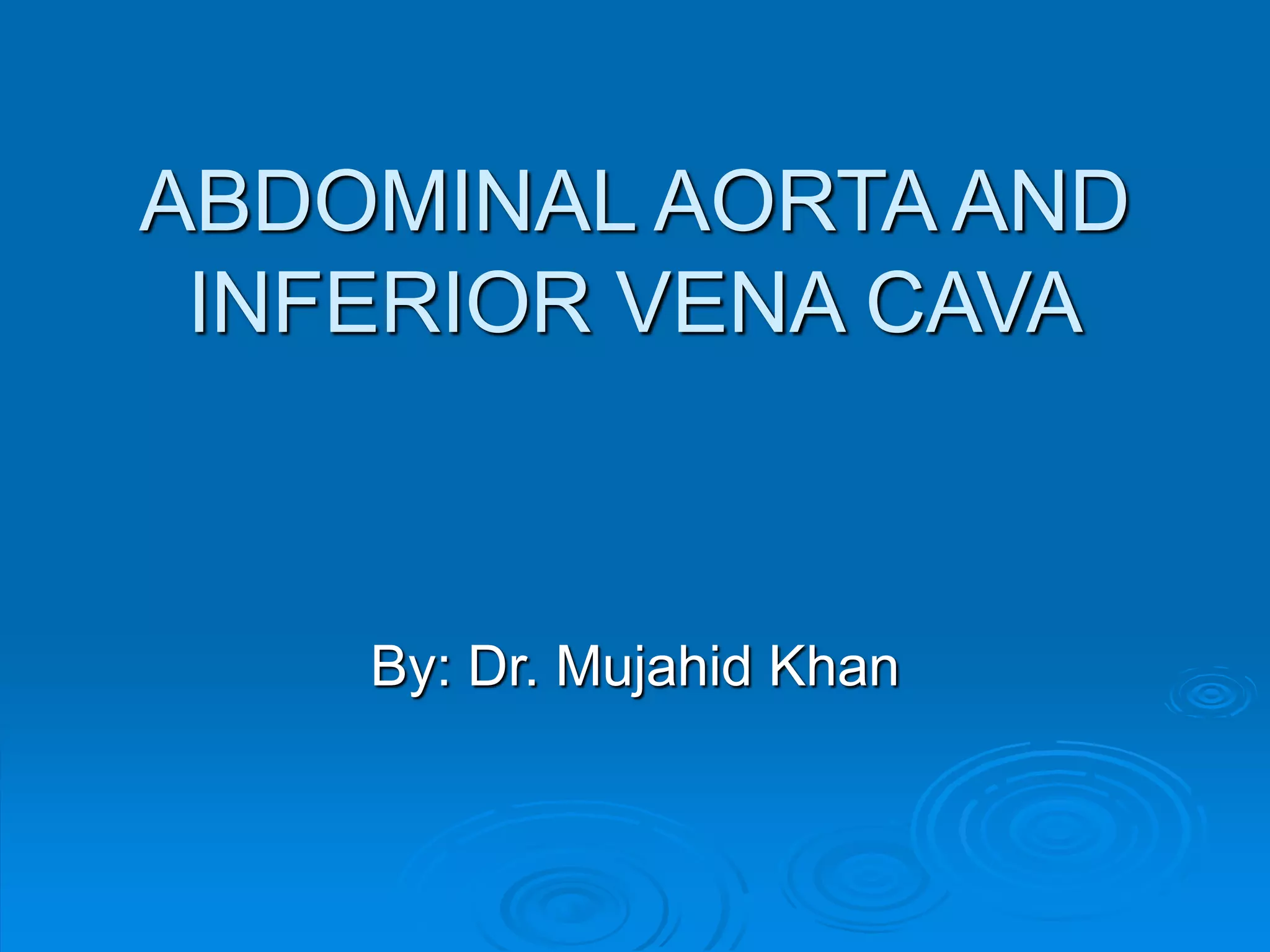
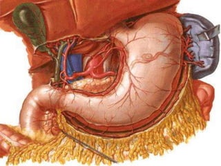
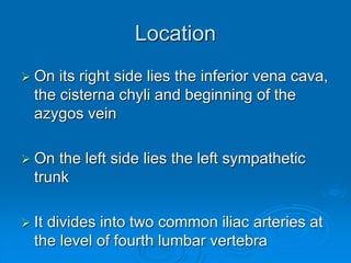
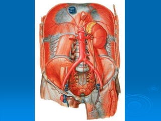
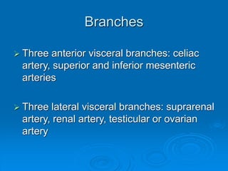
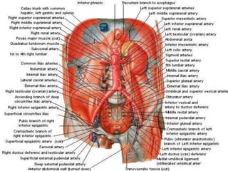
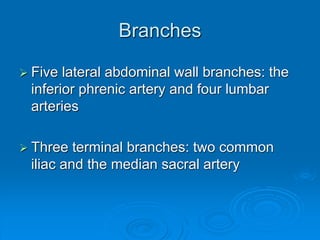
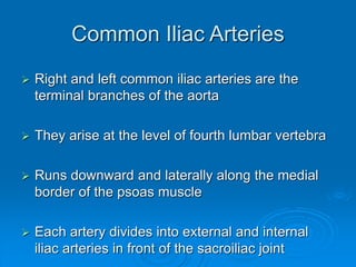
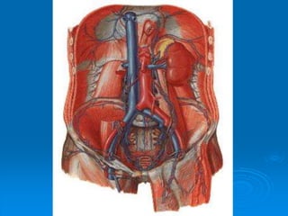
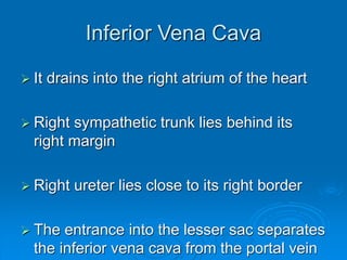
ABDOMINAL AORTA AND
INFERIOR VENA CAVA
By: Dr. Mujahid Khan
Location
 Aorta enters the abdomen through the
aortic opening of the diaphragm
 The opening lies in front of twelfth thoracic
vertebra
 It descends behind the peritoneum on the
anterior surface of the bodies of the
lumbar vertebrae
Location
 On its right side lies the inferior vena cava,
the cisterna chyli and beginning of the
azygos vein
 On the left side lies the left sympathetic
trunk
 It divides into two common iliac arteries at
the level of fourth lumbar vertebra
Branches
 Three anterior visceral branches: celiac
artery, superior and inferior mesenteric
arteries
 Three lateral visceral branches: suprarenal
artery, renal artery, testicular or ovarian
artery
Branches
 Five lateral abdominal wall branches: the
inferior phrenic artery and four lumbar
arteries
 Three terminal branches: two common
iliac and the median sacral artery
Common Iliac Arteries
 Right and left common iliac arteries are the
terminal branches of the aorta
 They arise at the level of fourth lumbar vertebra
 Runs downward and laterally along the medial
border of the psoas muscle
 Each artery divides into external and internal
iliac arteries in front of the sacroiliac joint
External Iliac Artery
 It runs along the medial border of psoas,
following the pelvic brim
 It gives off the inferior epigastric and deep
circumflex iliac branches
 The artery enters the thigh by passing
under the inguinal ligament to become the
femoral artery
Inferior Epigastric Artery
 The inferior epigastric artery arises just
above the inguinal ligament
 Passes upward and medially along the
medial margin of the deep inguinal ring
 Enters the rectus sheath behind the rectus
abdominis muscle
Deep Circumflex Iliac Artery
 Arises close to the inferior epigastric artery
 Ascends laterally to the anterior superior
iliac spine and the iliac crest
 Supplies the muscles of the anterior
abdominal wall
Internal Iliac Artery
 It passes down into the pelvis in front of
the sacroiliac joint
Aortic Aneurisms
 Localized or diffuse dilatations of the
abdominal part of the aorta usually occur
below the origin of the renal arteries
 Most result from atherosclerosis which
causes weakening of the arterial wall
 Occur most commonly in elderly men
Aortic Aneurisms
 Large aneurysms should be surgically
excised and replaced with a prosthetic
graft
 The bifurcation of the abdominal aorta
where the lumen suddenly narrows may
be a lodging site for an embolus from the
heart
 Severe ischemia of the lower limbs results
Obliteration of Abdominal Aorta and
Iliac Arteries
 Gradual occlusion of the bifurcation of the
abdominal aorta produced by atherosclerosis
causes pain in the legs on walking
 Impotence may occur due to lack of blood in
internal iliac arteries
 Surgical treatment by thromboendarterectomy or
a bypass graft should be considered
Inferior Vena Cava
 It conveys most of the blood from the body
below the diaphragm to the right atrium of the
heart
 It is formed by the union of common iliac veins
behind the right common iliac artery at the level
of fifth lumbar vertebra
 It ascends on the right side of the aorta
 Pierces the central tendon of the diaphragm at
the level of the eighth thoracic vertebra
Inferior Vena Cava
 It drains into the right atrium of the heart
 Right sympathetic trunk lies behind its
right margin
 Right ureter lies close to its right border
 The entrance into the lesser sac separates
the inferior vena cava from the portal vein
Tributaries
 Two anterior visceral tributaries: the hepatic
veins
 Three lateral visceral tributaries: the right
suprarenal vein, renal veins, right testicular or
ovarian vein
 Lateral abdominal wall tributaries: inferior
phrenic vein and four lumbar veins
 Three veins of origin: two common iliac veins
and the median sacral vein
Trauma to IVC
 Injuries to inferior vena cava are
commonly lethal
 The anatomical inaccessibility of the
vessel behind the liver, duodenum and
mesentery of the small intestine and the
blocking presence of the right costal
margin make a surgical approach difficult
Trauma to IVC
 The thin wall of the vena cava makes it
prone to extensive tears
 Due to the multiple anastomoses of the
tributaries of IVC, it is impossible in an
emergency to ligate the vessel
 Most patients have venous congestion of
the lower limbs
Compression of IVC
 It is commonly compressed by the
enlarged uterus during the later stages of
pregnancy
 This produces edema of the ankles and
feet and temporary varicose veins
 Malignant retroperitoneal tumors can
cause severe compression and eventual
blockage of IVC
Compression of IVC
 This results in the dilatation of the
extensive anastomoses of the tributaries
 This alternative pathway for the blood to
return to the right atrium is referred to as
the caval-caval shunt

4.abdominal aorta and ivc

  • 1.
    ABDOMINAL AORTA AND INFERIORVENA CAVA By: Dr. Mujahid Khan
  • 2.
    Location  Aorta entersthe abdomen through the aortic opening of the diaphragm  The opening lies in front of twelfth thoracic vertebra  It descends behind the peritoneum on the anterior surface of the bodies of the lumbar vertebrae
  • 4.
    Location  On itsright side lies the inferior vena cava, the cisterna chyli and beginning of the azygos vein  On the left side lies the left sympathetic trunk  It divides into two common iliac arteries at the level of fourth lumbar vertebra
  • 6.
    Branches  Three anteriorvisceral branches: celiac artery, superior and inferior mesenteric arteries  Three lateral visceral branches: suprarenal artery, renal artery, testicular or ovarian artery
  • 8.
    Branches  Five lateralabdominal wall branches: the inferior phrenic artery and four lumbar arteries  Three terminal branches: two common iliac and the median sacral artery
  • 9.
    Common Iliac Arteries Right and left common iliac arteries are the terminal branches of the aorta  They arise at the level of fourth lumbar vertebra  Runs downward and laterally along the medial border of the psoas muscle  Each artery divides into external and internal iliac arteries in front of the sacroiliac joint
  • 11.
    External Iliac Artery It runs along the medial border of psoas, following the pelvic brim  It gives off the inferior epigastric and deep circumflex iliac branches  The artery enters the thigh by passing under the inguinal ligament to become the femoral artery
  • 12.
    Inferior Epigastric Artery The inferior epigastric artery arises just above the inguinal ligament  Passes upward and medially along the medial margin of the deep inguinal ring  Enters the rectus sheath behind the rectus abdominis muscle
  • 13.
    Deep Circumflex IliacArtery  Arises close to the inferior epigastric artery  Ascends laterally to the anterior superior iliac spine and the iliac crest  Supplies the muscles of the anterior abdominal wall
  • 14.
    Internal Iliac Artery It passes down into the pelvis in front of the sacroiliac joint
  • 15.
    Aortic Aneurisms  Localizedor diffuse dilatations of the abdominal part of the aorta usually occur below the origin of the renal arteries  Most result from atherosclerosis which causes weakening of the arterial wall  Occur most commonly in elderly men
  • 16.
    Aortic Aneurisms  Largeaneurysms should be surgically excised and replaced with a prosthetic graft  The bifurcation of the abdominal aorta where the lumen suddenly narrows may be a lodging site for an embolus from the heart  Severe ischemia of the lower limbs results
  • 17.
    Obliteration of AbdominalAorta and Iliac Arteries  Gradual occlusion of the bifurcation of the abdominal aorta produced by atherosclerosis causes pain in the legs on walking  Impotence may occur due to lack of blood in internal iliac arteries  Surgical treatment by thromboendarterectomy or a bypass graft should be considered
  • 18.
    Inferior Vena Cava It conveys most of the blood from the body below the diaphragm to the right atrium of the heart  It is formed by the union of common iliac veins behind the right common iliac artery at the level of fifth lumbar vertebra  It ascends on the right side of the aorta  Pierces the central tendon of the diaphragm at the level of the eighth thoracic vertebra
  • 20.
    Inferior Vena Cava It drains into the right atrium of the heart  Right sympathetic trunk lies behind its right margin  Right ureter lies close to its right border  The entrance into the lesser sac separates the inferior vena cava from the portal vein
  • 21.
    Tributaries  Two anteriorvisceral tributaries: the hepatic veins  Three lateral visceral tributaries: the right suprarenal vein, renal veins, right testicular or ovarian vein  Lateral abdominal wall tributaries: inferior phrenic vein and four lumbar veins  Three veins of origin: two common iliac veins and the median sacral vein
  • 23.
    Trauma to IVC Injuries to inferior vena cava are commonly lethal  The anatomical inaccessibility of the vessel behind the liver, duodenum and mesentery of the small intestine and the blocking presence of the right costal margin make a surgical approach difficult
  • 24.
    Trauma to IVC The thin wall of the vena cava makes it prone to extensive tears  Due to the multiple anastomoses of the tributaries of IVC, it is impossible in an emergency to ligate the vessel  Most patients have venous congestion of the lower limbs
  • 25.
    Compression of IVC It is commonly compressed by the enlarged uterus during the later stages of pregnancy  This produces edema of the ankles and feet and temporary varicose veins  Malignant retroperitoneal tumors can cause severe compression and eventual blockage of IVC
  • 26.
    Compression of IVC This results in the dilatation of the extensive anastomoses of the tributaries  This alternative pathway for the blood to return to the right atrium is referred to as the caval-caval shunt