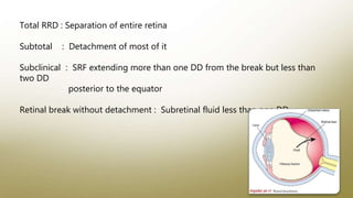
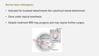
RHEGMATOGENOUS RETINAL
DETACHMENT
Dr.Prakriti Yagnam.K
Moderator : Dr. Swarna Latha
• Retina consists of ten layers which form two distinct functional
components
• The retinal pigment epithelium and sensory retina
• These ten layers from without inward are :
1. Retinal pigment epithelium
2. Layer of rods and cones
3. External limiting membrane
4. Outer nuclear layer
5. Outer plexiform layer
6. Inner nuclear layer
7. Inner plexiform layer
8. Ganglion cell layer
9. Nerve fibre layer
10. Internal limiting membrane
• The potential space between RPE and sensory retina is called
subretinal space
• A separation of RPE and sensory retina is called Retinal detachment
Rhegmatogenous retinal detachment Exudative retinal detachment
Tractional retinal detachment
Combined – Rhegmatogenous plus
tractional
Retinal detachment is
mainly of three types :
Others : Macular hole associated detachment
Factors
causing
retinal
attachment :
An
interphotorecepto
matrix in
space
Oncotic pressure
difference
choroid and
subretinal space
Hydrostatic or
hydraulic forces
related to IOP
Metabolic transfer
of ions and fluid
by RPE
Rhegmatogenous retinal detachment :
• Rhegma means break in Greek
• Separation of neural retina from RPE as a result of vitreous
through retinal defect ( retinal breaks ) into sub retinal space
• It is an important cause of visual disability
• If untreated symptomatic RRD progress to total blindness
Total RRD : Separation of entire retina
Subtotal : Detachment of most of it
Subclinical : SRF extending more than one DD from the break but less than
two DD
posterior to the equator
Retinal break without detachment : Subretinal fluid less than one DD
Epidemiology :
• Incidence is 5.3 – 12.6 cases per 100000
• Peak incidence is between 50 to 79 years
• Males are affected more than female
Pathogenesis :
• The main factors causing RRD are vitreous liquefaction ( synchysis )
and collapse ( syneresis )causing posterior vitreous detachment
synchysis and syneresis
PVD
focal retinal traction
retinal breaks
migration of liquified vitreous into subretinal space
detachment if involves macula
central vision loss
• Vitreous liquefaction occurs due to degenerative changes in
collagen fibril hyaluronan matrix
• Liquefaction cause lacunae which progress to PVD
Conditions which cause liquefaction are :
 Pathological myopia ( > 6 D )
 Surgical / non surgical trauma
 Intraocular inflammation – Viral retinitis
 Congenital – Marfans , Stickler syndromes
 Pseudophakia and aphakia
 Proliferative vitreoretinopathy
Degenerative retinoschisis
Other inherited and acquired ocular disorders
• Common site for retinal break are at peripheral retina that is at
vitreous base
Aggravating factors :
 Rotational and saccadic eye movements play a role
 Young males with posterior capsular rent have high chances of RRD
 Crystalline lens extraction cause early induction of PVD
Rotation of eye
Detached vitreous gel causes it to lag behind rotation of eye and
attached retina
Exerts equal and opposite force on retina
Retinal break
If vitreous remains attached to retina ( horse shoe tear flap at vitreous
base )
Eye movements
Asymmetrical traction at flap holding it open
Liquified vitreous into subretinal space
When movements stop VRT in opposite direction
• When fluid flow into subretinal space overwhelm capacity of RPE to
pump fluid from this space
Other risk factors :
Mainly the peripheral retinal degenerations like
 Lattice degeneration
 Snail track degeneration
 Vitreoretinal tufts
 Meridonial folds
 White with pressure and without pressure
Retinal Breaks :
• They are either retinal tears or retinal holes
• Retinal tears are associated with vitreoretinal
traction while holes are atrophied tissue
• Full thickness retinal tear with persistent VRT
retinal flap tear ( Horse shoe shaped )
leads to RRD
• Dialyses are linear or circumferential retinal breaks along with ora
serrata mostly associated with blunt ocular trauma or
Ocular Manifestations :
• Sudden appearance of floaters – condensation of vitreous gel ,
pigment granules or VH released in process of break formation
• Fleeting photopsias ( flashes ) - due to vitreous traction on retina
resulting in neuronal activity by mechanical manipulation
• More following dark and following rotatory eye movement
Accumulation of SRF – relative scotomata
detachment progresses posterior to equator
symptomatic with loss of peripheral vision
curtain falling over visual field
SRF beneath macula
central vision loss
• Young patients with myopia may have slowly progressive
asymptomatic RD without PVD
• Most common in temporally and inferiorly
• If area of detachment is large there may be afferent pupillary defect
also
Diagnosis :
 Visual acuity
 Visual fields
 IOP
 Slit lamp biomicroscope
 Indirect Ophthalmoscope – binocular stereoscopic examination
entire retina with scleral indentation
 B scan
Visual acuity – spared if macula is spared
Visual field – Depends on location of detachment
IOP – usually is low – due to increased outflow of intraocular fluid
through
subretinal space
- sometimes raised IOP due to photoreceptor outer segment and
other debris clogging the trabecular meshwork – SCHWARTZ –
MATSON SYNDROME
Slit lamp – Mild anterior chamber reaction
- Presence of pigmented cells in anterior vitreous Tobacco
dust / Shafer sign
- Weiss ring
IO – RRD – convex configuration
retina slightly opaque – linear / arc shaped ripples which
hydration lines due to retinal edema
Acute detachment – retina mobile with eye movements
Chronic detachment – decreased mobility of detached retina
• Demarcation lines formed by migration of RPE at border of attached
and detached retina
• Retinal thinning due to atrophy
• Large intraretinal cysts
• Visible preretinal or subretinal membranes ( PVR )
Examination : It includes
1. Identifying the extent of retinal detachment
2. Finding of all retinal breaks
3. Determining whether RRD is macular on or off
4. Checking for associated features
5. Documenting the findings on Amsler Dubey chart
6. B scan ultrasound
1. Extent of detachment :
• Detached area is opaque and corrugated
with undulating retinal folds during
eye movement
• Borders are convex and SRF is clear
and non shifting
2. Finding all retinal breaks :
• Lincoffs rule is useful for identifying precise location of retinal break in cases of
primary RRD
• The number , size and location of the breaks is noted
• If there are multiple breaks highest retinal break is considered as primary hole
Lincoffs rules :
Detachment area Primary break location
1. In shallow RD with SRF slightly higher on temporal
side
Inferiorly on that side
2. Inferior RD with equal fluid levels 6’0 clock
3. In inferior bullous RD Above the horizontal meridian
4. SRF around optic disc and raised on temporal side Upper nasal quadrant
5. Subtotal RD with a superior wedge of attached
retina
Periphery nearest its higher border
6. SRF crosses the vertical midline 12’0 clock corresponding to lower edge of break
Macula on Macula off
B scan :
• Status of macula involved
• Presence of PVD
• Location of break
• Chronicity of RRD
RRD PVD
• High reflectivity
• High spikes on A scan
• Membrane within vitreous
cavity
• Mobility during eye
movements
• Visible with low gain
• OD attachment present
• Posterior hyaloid face
• Low spike on A scan
• High degree of mobility
during eye movements
• Disappears with low gain
• OD attachment present or
absent
Proliferative viteoretinopathy :
• Caused by epiretinal or subretinal
membrane formation leading to
tangential retinal traction
• Complication of RRD or r surgery or
penetrating trauma
Differential
Diagnosis :
TRD – PDR ,
penetrating
trauma
ERD – Inflammatory
disorders , choroidal
neoplasms , Retinal
vascular tumors and
others
Retinoschisis
– Age related
or congenital
Elevated choroidal
lesions – choroidal
detachments or
tumors
Vitreous
hemorrhage
Management :
• Must counter the factors and forces that cause retinal detachment
and
• To reestablish conditions that maintain physiological contact
between neural retina and pigment epithelium
• RRDs with superior breaks that threaten macula require urgent
vitreoretinal intervention
• While awaiting definitive management patients should maintain
posture that prevents SRF from detaching macula
Options :
1. Observation
2. Laser demarcation – Barrier laser retinopexy
3. Pneumatic retinopexy – with drainage of SRF
4. Permanent scleral buckling
5. Temporary scleral buckling – Lincoff balloon / absorbable sutures
6. PPV
7. Combination of PPV and scleral buckle
Barrier laser retinopexy :
• Indicated for localized detachments like subclinical retinal detachment
• Done under topical anesthesia
• Despite treatment RRD may progress and may require further surgery
Pneumatic Retinopexy :
• Creation of chorio retinal adhesion either by cryotherapy or laser
photocoagulation with injection of expansile intraocular gas as
tamponade
• Does not provide release of vitreoretinal traction like PPV or alter
tractional forces like scleral buckling surgery
Indication :
Phakic eye with superior retinal detachment with single retinal break
( located in superior 8 ‘0 clock hours or all breaks confined within 2 ‘0clock hours )
Contraindications :
• Large ( giant ) retinal tears
• Proliferative VitreoRetinopathy ( PVR )
• Advanced glaucoma
• Poor compliance with head posture
• Air travel
• Pseudophakia
Complications :
• Risk of new / mixed retinal breaks
• Gas migration into subretinal space
• CRAO due to raised IOP
• Vitreous incarceration into wound
• Accelerated cataract formation
• Endophthalmitis
Prognosis :
98 % cases – requires more than one procedure
Scleral buckle :
• Exoplant used to create indentation to create buckle effect on sclera
thereby moving the detached retina closer to RPE
• Buckle also alters vector forces and reduces VR traction
Indications :
• RRD in phakic eyes with high myopia and round holes especially
inferior holes without PVD
• RD due to dialyses
Contraindications :
• Giant retinal teras
• PVR
Advantages :
• No or reduced postoperative positioning
• Maintenance of crystalline lens
• Permanent protection of vitreous base
• Help to prevent recurrent retinal detachment
Disadvantages :
• Inability to drain SRF completely
• Risk for SRH in cases of external drainage
• Increased postoperative discomfort and inflammation
Procedure :
360 degree conjunctival peritomy
Recti muscles are slinged
Localising the break with binocular IO
Cryotherapy with or without external drainage of SRF
Inserting segmental and / or encircling or radial scleral buckle
Suturing and tightening of buckle
Check perfusion of CRA to determine need for AC paracentesis
Antibiotic wash around buckle
Conjunctiva is closed
Subconjunctival antibiotic and steroid injection given
Complications :
Intraoperative :
• Scleral perforation
• Recti muscle trauma or slip
• SRF drainage - Suprachoroidal haemorrhage
Hypotony
Retinal incarceration at drainage site
Postoperative :
• PVR formation
• Redetachment
• Buckle migration or exposure
• Buckle related infection
• Refractory changes
• Ocular motility disorders
• Anterior segment ischemia
• Glaucoma ( Due to vortex vein or
ciliary body compression )
Prognosis :
80 – 90 % cases – reattachment possible with single primary scleral
buckle
Study - MUSTARD
Vitrectomy :
Indications :
• Posterior retinal breaks
• Multiple retinal breaks
• Giant retinal tears
• Concurrent PVR
• Dense VH obscuring retinal breaks
Steps :
Three sclerostomy ports ( infusion cannula , light probe , vitrectomy
cutter )
Core vitrectomy and shaving of vitreous base ( relieves vitreous traction over
break )
Perfluorocarbon to flatten retina and displace SRF via original retinal break
Retinopexy around break - laser photocoagulation
Fluid – air exchange Vitreous substitutes ( gas or silicone oil
Postop advice :
• Posturing to allow buoyant vitreous substitutes to tamponade the
break
• For intraocular gas , until the gas bubble has been resolved
Prognosis :
Success rate 64 to 96 %
Complications :
Intraoperative :
• Trauma to iatrogenic structures ( Iaotrgenic RD , cataract , vitreous hemorrhage )
• Vitreous or retinal incarceration at sclerostomy wounds
Postoperative :
• Endophthalmitis
• Sympathetic ophthalmia
• Glaucoma , cataract
Combination of vitrectomy and scleral buckle :
• Inferior retinal breaks ( esp. phakic eyes )
• Multiple retinal breaks
• Extensive lattice degeneration
• Conditions with abnormal vitreoretinal interface ( Stickler syndrome )
• Reoperations
• Poor outcomes of other eye with vitrectomy alone
• Relatively when the encircling band is chosen to protect vitreous base (
360 degree ) prior to PPV
Timing of surgery depends upon certain conditions :
• Status of macula – Macula On – within 24 hours
Macula Off – within a week
• Duration of detachment – Acute – prompt surgery
Chronic – Routine day
• Location of break – RRD with superior breaks progress faster than
detachments with inferior breaks
• Number of days of foveal detachment determines the maximum
number of days that can be awaited for surgery in macula off cases
• Surgery within three days of developing central vision loss has better
visual prognosis
Course and Outcome :
• Approximately 95 % of all RDs can be successfully repaired
• Failure of PPV is due to – Failure to identify all breaks
New retinal breaks
Proliferative vitreoretinopathy
Non compliance of patient to positioning
• Scleral buckle – Inadequate buckle height or position
Inadequate cryotherapy
PVR
Missed breaks
Outcome :
Outcomes are divided mainly into
Anatomical success Functional visual outcome
• Macula detached by SRF some degree of permanent damage to
vision occurs inspite of reattachment
• Macula on detachment – 73% greater than 20/40 vision occurs two
months after surgery
• Macula off Detachment – depends on duration of detachment
Less than 10 days 11 days to 6 weeks > 6 weeks
More than 20/40 vision
71 % 27% 14%
Other reasons for decreased vision are
 Subsequent reattachment surgery
 Progressive ischemic or infectious retinal damage
 Persistent SRF despite closure of all breaks
THANK YOU !!!

Rrd

  • 1.
  • 2.
    • Retina consistsof ten layers which form two distinct functional components • The retinal pigment epithelium and sensory retina • These ten layers from without inward are : 1. Retinal pigment epithelium 2. Layer of rods and cones 3. External limiting membrane 4. Outer nuclear layer 5. Outer plexiform layer 6. Inner nuclear layer 7. Inner plexiform layer 8. Ganglion cell layer
  • 4.
    9. Nerve fibrelayer 10. Internal limiting membrane • The potential space between RPE and sensory retina is called subretinal space • A separation of RPE and sensory retina is called Retinal detachment
  • 5.
    Rhegmatogenous retinal detachmentExudative retinal detachment Tractional retinal detachment Combined – Rhegmatogenous plus tractional Retinal detachment is mainly of three types : Others : Macular hole associated detachment
  • 6.
    Factors causing retinal attachment : An interphotorecepto matrix in space Oncoticpressure difference choroid and subretinal space Hydrostatic or hydraulic forces related to IOP Metabolic transfer of ions and fluid by RPE
  • 7.
    Rhegmatogenous retinal detachment: • Rhegma means break in Greek • Separation of neural retina from RPE as a result of vitreous through retinal defect ( retinal breaks ) into sub retinal space • It is an important cause of visual disability • If untreated symptomatic RRD progress to total blindness
  • 8.
    Total RRD :Separation of entire retina Subtotal : Detachment of most of it Subclinical : SRF extending more than one DD from the break but less than two DD posterior to the equator Retinal break without detachment : Subretinal fluid less than one DD
  • 9.
    Epidemiology : • Incidenceis 5.3 – 12.6 cases per 100000 • Peak incidence is between 50 to 79 years • Males are affected more than female Pathogenesis : • The main factors causing RRD are vitreous liquefaction ( synchysis ) and collapse ( syneresis )causing posterior vitreous detachment
  • 10.
    synchysis and syneresis PVD focalretinal traction retinal breaks migration of liquified vitreous into subretinal space detachment if involves macula central vision loss
  • 12.
    • Vitreous liquefactionoccurs due to degenerative changes in collagen fibril hyaluronan matrix • Liquefaction cause lacunae which progress to PVD Conditions which cause liquefaction are :  Pathological myopia ( > 6 D )  Surgical / non surgical trauma  Intraocular inflammation – Viral retinitis  Congenital – Marfans , Stickler syndromes  Pseudophakia and aphakia  Proliferative vitreoretinopathy
  • 13.
    Degenerative retinoschisis Other inheritedand acquired ocular disorders • Common site for retinal break are at peripheral retina that is at vitreous base Aggravating factors :  Rotational and saccadic eye movements play a role  Young males with posterior capsular rent have high chances of RRD  Crystalline lens extraction cause early induction of PVD
  • 14.
    Rotation of eye Detachedvitreous gel causes it to lag behind rotation of eye and attached retina Exerts equal and opposite force on retina Retinal break If vitreous remains attached to retina ( horse shoe tear flap at vitreous base )
  • 16.
    Eye movements Asymmetrical tractionat flap holding it open Liquified vitreous into subretinal space When movements stop VRT in opposite direction • When fluid flow into subretinal space overwhelm capacity of RPE to pump fluid from this space
  • 17.
    Other risk factors: Mainly the peripheral retinal degenerations like  Lattice degeneration  Snail track degeneration  Vitreoretinal tufts  Meridonial folds  White with pressure and without pressure
  • 19.
    Retinal Breaks : •They are either retinal tears or retinal holes • Retinal tears are associated with vitreoretinal traction while holes are atrophied tissue • Full thickness retinal tear with persistent VRT retinal flap tear ( Horse shoe shaped ) leads to RRD • Dialyses are linear or circumferential retinal breaks along with ora serrata mostly associated with blunt ocular trauma or
  • 20.
    Ocular Manifestations : •Sudden appearance of floaters – condensation of vitreous gel , pigment granules or VH released in process of break formation • Fleeting photopsias ( flashes ) - due to vitreous traction on retina resulting in neuronal activity by mechanical manipulation • More following dark and following rotatory eye movement
  • 21.
    Accumulation of SRF– relative scotomata detachment progresses posterior to equator symptomatic with loss of peripheral vision curtain falling over visual field SRF beneath macula central vision loss
  • 22.
    • Young patientswith myopia may have slowly progressive asymptomatic RD without PVD • Most common in temporally and inferiorly • If area of detachment is large there may be afferent pupillary defect also
  • 23.
    Diagnosis :  Visualacuity  Visual fields  IOP  Slit lamp biomicroscope  Indirect Ophthalmoscope – binocular stereoscopic examination entire retina with scleral indentation  B scan
  • 24.
    Visual acuity –spared if macula is spared Visual field – Depends on location of detachment IOP – usually is low – due to increased outflow of intraocular fluid through subretinal space - sometimes raised IOP due to photoreceptor outer segment and other debris clogging the trabecular meshwork – SCHWARTZ – MATSON SYNDROME Slit lamp – Mild anterior chamber reaction - Presence of pigmented cells in anterior vitreous Tobacco dust / Shafer sign - Weiss ring
  • 25.
    IO – RRD– convex configuration retina slightly opaque – linear / arc shaped ripples which hydration lines due to retinal edema Acute detachment – retina mobile with eye movements Chronic detachment – decreased mobility of detached retina • Demarcation lines formed by migration of RPE at border of attached and detached retina • Retinal thinning due to atrophy • Large intraretinal cysts • Visible preretinal or subretinal membranes ( PVR )
  • 26.
    Examination : Itincludes 1. Identifying the extent of retinal detachment 2. Finding of all retinal breaks 3. Determining whether RRD is macular on or off 4. Checking for associated features 5. Documenting the findings on Amsler Dubey chart 6. B scan ultrasound
  • 27.
    1. Extent ofdetachment : • Detached area is opaque and corrugated with undulating retinal folds during eye movement • Borders are convex and SRF is clear and non shifting
  • 29.
    2. Finding allretinal breaks : • Lincoffs rule is useful for identifying precise location of retinal break in cases of primary RRD • The number , size and location of the breaks is noted • If there are multiple breaks highest retinal break is considered as primary hole
  • 30.
    Lincoffs rules : Detachmentarea Primary break location 1. In shallow RD with SRF slightly higher on temporal side Inferiorly on that side 2. Inferior RD with equal fluid levels 6’0 clock 3. In inferior bullous RD Above the horizontal meridian 4. SRF around optic disc and raised on temporal side Upper nasal quadrant 5. Subtotal RD with a superior wedge of attached retina Periphery nearest its higher border 6. SRF crosses the vertical midline 12’0 clock corresponding to lower edge of break
  • 32.
  • 33.
    B scan : •Status of macula involved • Presence of PVD • Location of break • Chronicity of RRD RRD PVD • High reflectivity • High spikes on A scan • Membrane within vitreous cavity • Mobility during eye movements • Visible with low gain • OD attachment present • Posterior hyaloid face • Low spike on A scan • High degree of mobility during eye movements • Disappears with low gain • OD attachment present or absent
  • 35.
    Proliferative viteoretinopathy : •Caused by epiretinal or subretinal membrane formation leading to tangential retinal traction • Complication of RRD or r surgery or penetrating trauma
  • 37.
    Differential Diagnosis : TRD –PDR , penetrating trauma ERD – Inflammatory disorders , choroidal neoplasms , Retinal vascular tumors and others Retinoschisis – Age related or congenital Elevated choroidal lesions – choroidal detachments or tumors Vitreous hemorrhage
  • 39.
    Management : • Mustcounter the factors and forces that cause retinal detachment and • To reestablish conditions that maintain physiological contact between neural retina and pigment epithelium • RRDs with superior breaks that threaten macula require urgent vitreoretinal intervention • While awaiting definitive management patients should maintain posture that prevents SRF from detaching macula
  • 40.
    Options : 1. Observation 2.Laser demarcation – Barrier laser retinopexy 3. Pneumatic retinopexy – with drainage of SRF 4. Permanent scleral buckling 5. Temporary scleral buckling – Lincoff balloon / absorbable sutures 6. PPV 7. Combination of PPV and scleral buckle
  • 41.
    Barrier laser retinopexy: • Indicated for localized detachments like subclinical retinal detachment • Done under topical anesthesia • Despite treatment RRD may progress and may require further surgery
  • 42.
    Pneumatic Retinopexy : •Creation of chorio retinal adhesion either by cryotherapy or laser photocoagulation with injection of expansile intraocular gas as tamponade • Does not provide release of vitreoretinal traction like PPV or alter tractional forces like scleral buckling surgery
  • 43.
    Indication : Phakic eyewith superior retinal detachment with single retinal break ( located in superior 8 ‘0 clock hours or all breaks confined within 2 ‘0clock hours ) Contraindications : • Large ( giant ) retinal tears • Proliferative VitreoRetinopathy ( PVR ) • Advanced glaucoma • Poor compliance with head posture • Air travel • Pseudophakia
  • 44.
    Complications : • Riskof new / mixed retinal breaks • Gas migration into subretinal space • CRAO due to raised IOP • Vitreous incarceration into wound • Accelerated cataract formation • Endophthalmitis Prognosis : 98 % cases – requires more than one procedure
  • 45.
    Scleral buckle : •Exoplant used to create indentation to create buckle effect on sclera thereby moving the detached retina closer to RPE • Buckle also alters vector forces and reduces VR traction Indications : • RRD in phakic eyes with high myopia and round holes especially inferior holes without PVD • RD due to dialyses Contraindications : • Giant retinal teras • PVR
  • 46.
    Advantages : • Noor reduced postoperative positioning • Maintenance of crystalline lens • Permanent protection of vitreous base • Help to prevent recurrent retinal detachment Disadvantages : • Inability to drain SRF completely • Risk for SRH in cases of external drainage • Increased postoperative discomfort and inflammation
  • 47.
    Procedure : 360 degreeconjunctival peritomy Recti muscles are slinged Localising the break with binocular IO Cryotherapy with or without external drainage of SRF Inserting segmental and / or encircling or radial scleral buckle
  • 48.
    Suturing and tighteningof buckle Check perfusion of CRA to determine need for AC paracentesis Antibiotic wash around buckle Conjunctiva is closed Subconjunctival antibiotic and steroid injection given
  • 49.
    Complications : Intraoperative : •Scleral perforation • Recti muscle trauma or slip • SRF drainage - Suprachoroidal haemorrhage Hypotony Retinal incarceration at drainage site Postoperative : • PVR formation • Redetachment • Buckle migration or exposure
  • 50.
    • Buckle relatedinfection • Refractory changes • Ocular motility disorders • Anterior segment ischemia • Glaucoma ( Due to vortex vein or ciliary body compression ) Prognosis : 80 – 90 % cases – reattachment possible with single primary scleral buckle Study - MUSTARD
  • 51.
    Vitrectomy : Indications : •Posterior retinal breaks • Multiple retinal breaks • Giant retinal tears • Concurrent PVR • Dense VH obscuring retinal breaks
  • 52.
    Steps : Three sclerostomyports ( infusion cannula , light probe , vitrectomy cutter ) Core vitrectomy and shaving of vitreous base ( relieves vitreous traction over break ) Perfluorocarbon to flatten retina and displace SRF via original retinal break Retinopexy around break - laser photocoagulation Fluid – air exchange Vitreous substitutes ( gas or silicone oil
  • 53.
    Postop advice : •Posturing to allow buoyant vitreous substitutes to tamponade the break • For intraocular gas , until the gas bubble has been resolved Prognosis : Success rate 64 to 96 %
  • 54.
    Complications : Intraoperative : •Trauma to iatrogenic structures ( Iaotrgenic RD , cataract , vitreous hemorrhage ) • Vitreous or retinal incarceration at sclerostomy wounds Postoperative : • Endophthalmitis • Sympathetic ophthalmia • Glaucoma , cataract
  • 55.
    Combination of vitrectomyand scleral buckle : • Inferior retinal breaks ( esp. phakic eyes ) • Multiple retinal breaks • Extensive lattice degeneration • Conditions with abnormal vitreoretinal interface ( Stickler syndrome ) • Reoperations • Poor outcomes of other eye with vitrectomy alone • Relatively when the encircling band is chosen to protect vitreous base ( 360 degree ) prior to PPV
  • 56.
    Timing of surgerydepends upon certain conditions : • Status of macula – Macula On – within 24 hours Macula Off – within a week • Duration of detachment – Acute – prompt surgery Chronic – Routine day • Location of break – RRD with superior breaks progress faster than detachments with inferior breaks • Number of days of foveal detachment determines the maximum number of days that can be awaited for surgery in macula off cases • Surgery within three days of developing central vision loss has better visual prognosis
  • 57.
    Course and Outcome: • Approximately 95 % of all RDs can be successfully repaired • Failure of PPV is due to – Failure to identify all breaks New retinal breaks Proliferative vitreoretinopathy Non compliance of patient to positioning • Scleral buckle – Inadequate buckle height or position Inadequate cryotherapy PVR Missed breaks
  • 58.
    Outcome : Outcomes aredivided mainly into Anatomical success Functional visual outcome • Macula detached by SRF some degree of permanent damage to vision occurs inspite of reattachment • Macula on detachment – 73% greater than 20/40 vision occurs two months after surgery
  • 59.
    • Macula offDetachment – depends on duration of detachment Less than 10 days 11 days to 6 weeks > 6 weeks More than 20/40 vision 71 % 27% 14%
  • 60.
    Other reasons fordecreased vision are  Subsequent reattachment surgery  Progressive ischemic or infectious retinal damage  Persistent SRF despite closure of all breaks
  • 61.