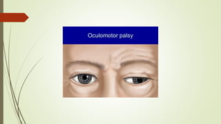
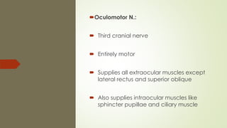
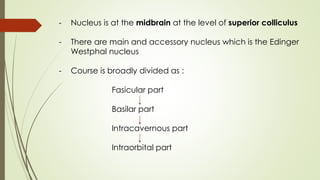
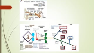
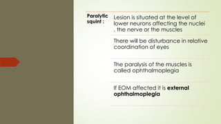
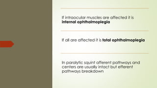
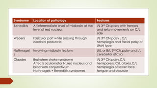
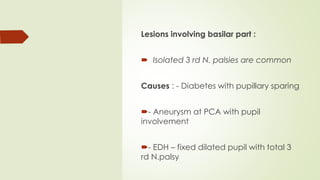
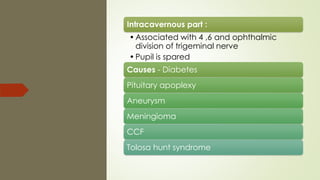
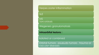
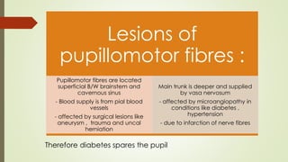
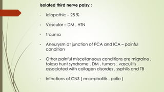
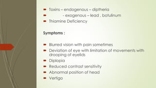
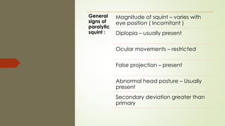
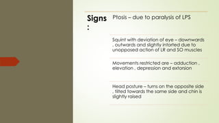
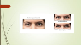
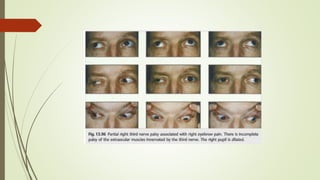
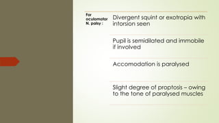
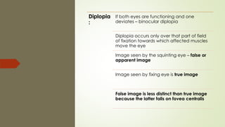
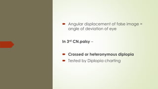
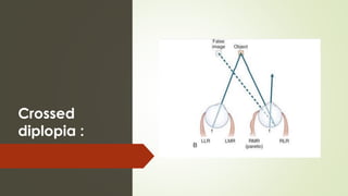
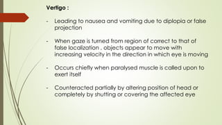
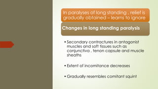
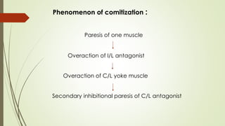
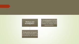
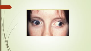
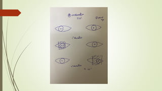
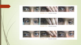
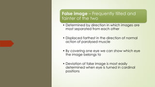
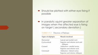
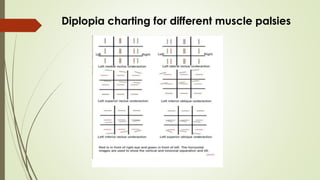
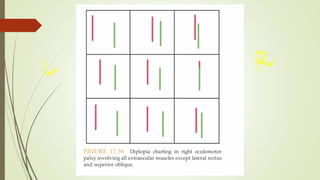
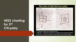
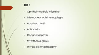
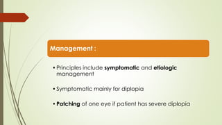
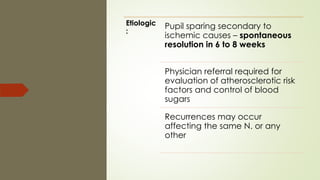
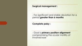
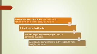
SlideShare a Scribd company logo
PARALYTIC
STRABISMUS(3rd CN )
DR.PRAKRITI YAGNAM.K
Oculomotor N.:
 Third cranial nerve
 Entirely motor
 Supplies all extraocular muscles except
lateral rectus and superior oblique
 Also supplies intraocular muscles like
sphincter pupillae and ciliary muscle
- Nucleus is at the midbrain at the level of superior colliculus
- There are main and accessory nucleus which is the Edinger
Westphal nucleus
- Course is broadly divided as :
Fasicular part
Basilar part
Intracavernous part
Intraorbital part
 There are mainly two divisions – superior
and inferior
 Superior supplies SR and LPS
 Inferior division supplies the remaining
EOM
 The intraocular muscles are supplied by
the parasympathetic root of nerve from
Edinger Westphal nucleus through ciliary
ganglion
Paralytic
squint :
Lesion is situated at the level of
lower neurons affecting the nuclei
, the nerve or the muscles
There will be disturbance in relative
coordination of eyes
The paralysis of the muscles is
called ophthalmoplegia
If EOM affected it is external
ophthalmoplegia
If intraocular muscles are affected it is
internal ophthalmoplegia
If all are affected it is total ophthalmoplegia
In paralytic squint afferent pathways and
centers are usually intact but efferent
pathways breakdown
Etiology :
 Different mechanisms come into play at
different sites of lesion
 Features help to differentiate the location
of the lesion also
Supranuclear :
Lesions of cerebral cortex and supranuclear
pathway
 Conjugate paresis affects both eyes
equally
 Position and movements of eyes are
abnormal but maintain relative
coordination and there will be no
diplopia
Nuclear lesions :
Common causes – Vascular diseases
 Demylienation
 Primary tumors
 Metastasis
 Lesions involving entire nucleus –
Ipsilateral third N. palsy with adjacent
fourth N. palsy
 Ipsilateral sparing and contralateral
weakness of elevator is seen
 Involving paired MR subnuclei – wall eyed
bilateral internuclear ophthalmoplegia (
WEBINO )
 There will be defective convergence and
adduction
Fasicular lesions :
Causes are similar to nuclear
Syndromic associations seen :
1. Benedikts syndrome
2. Weber syndrome
3. Nothnagels syndrome
4. Claudes syndrome
Syndrome Location of pathology Features
Benedikts At intermediate level of midbrain at the
level of red nucleus
I/L 3rd CN.palsy with tremors
and jerky movements on C/L
side
Webers Fasicular part while passing through
cerebral peduncle
I/L 3rd CN.palsy , C/L
hemiplegia and facial palsy of
UMN type
Nothnagel
s
Involving midbrain tectum U/L or B/L 3rd CN.palsy and I/L
cerebellar ataxia
Claudes Brainstem stroke syndrome
Affects oculomotor N.,red nucleus and
brachium conjunctivum
Nothnagels + Benedikts syndromes
I/L 3rd CN.palsy,C/L
hemiparesis,C/L ataxia,C/L
hemiplegia of lower face ,
tongue and shoulder
Lesions involving basilar part :
 Isolated 3 rd N. palsies are common
Causes : - Diabetes with pupillary sparing
- Aneurysm at PCA with pupil
involvement
- EDH – fixed dilated pupil with total 3
rd N.palsy
Intracavernous part :
•Associated with 4 ,6 and ophthalmic
division of trigeminal nerve
•Pupil is spared
Causes - Diabetes
Pituitary apoplexy
Aneurysms
Meningioma
CCF
Tolosa hunt syndrome
Herpes zoster inflammation
SLE
MS
Sarcoidosis
Wegeners granulomatosis
Intraorbital lesions :
- Isolated or combined
- Orbital tumors , psueudo tumors , trauma or
vascular diseases
Lesions of
pupillomotor fibres :
Pupillomotor fibres are located
superficial B/W brainstem and
cavernous sinus
- Blood supply is from pial blood
vessels
- affected by surgical lesions like
aneurysm , trauma and uncal
herniation
Main trunk is deeper and supplied
by vasa nervosum
- affected by microangiopathy in
conditions like diabetes ,
hypertension
- due to infarction of nerve fibres
Therefore diabetes spares the pupil
Isolated third nerve palsy :
- Idiopathic – 25 %
- Vascular – DM , HTN
- Trauma
- Aneurysm at junction of PCA and ICA – painful
condition
- Other painful miscellaneous conditions are migraine ,
tolosa hunt syndrome , DM , tumors , vasculitis
associated with collagen disordes , syphilis and TB
- Infections of CNS ( encephalitis , polio )
 Toxins – endogenous – diptheria
 - exogenous – lead , botulinum
 Thiamine Deficiency
Symptoms :
 Blurred vision with pain sometimes
 Deviation of eye with limitation of movements with
drooping of eyelids
 Diplopia
 Reduced contrast sensitivity
 Abnormal position of head
 Vertigo
General
signs of
paralytic
squint :
Magnitude of squint – varies with
eye position ( Incomitant )
Diplopia – usually present
Ocular movements – restricted
False projection – present
Abnormal head posture – Usually
present
Secondary deviation greater than
primary
Signs
:
Ptosis – due to paralysis of LPS
Squint with deviation of eye – downwards
, outwards and slightly intorted due to
unopposed action of LR and SO muscles
Movements restricted are – adduction ,
elevation , depression and extorsion
Head posture – turns on the opposite side
, tilted towards the same side and chin is
slightly raised
Squint :
- Magnitude of squint or abnormal alignment of eyes is of
variable degree – depends on degree of paralysis and in
which direction patient is looking
- Amount of squint determined by angle of deviation is
more if paralysis is severe
- More if patient is looking in the direction in which
paralysed muscle comes into play
For
oculomotor
N. palsy :
Divergent squint or exotropia with
intorsion seen
Pupil is semidilated and immobile
if involved
Accomodation is paralysed
Slight degree of proptosis – owing
to the tone of paralysed muscles
Diplopia
:
If both eyes are functioning and one
deviates – binocular diplopia
Diplopia occurs only over that part of field
of fixation towards which affected muscles
move the eye
Image seen by the squinting eye – false or
apparent image
Image seen by fixing eye is true image
False image is less distinct than true image
because the latter falls on fovea centralis
 Angular displacement of false image =
angle of deviation of eye
In 3rd CN.palsy –
 Crossed or heteronymous diplopia
 Tested by Diplopia charting
Crossed
diplopia :
False
orientation
:
Necessary accompianment of
binocular diplopia
Same principle as the increase of
secondary deviation for the object
is projected according to the
amount of nervous energy exerted
As this is greater than that exerted
in normal circumstances , object is
projected too far in the direction
of action of paralyzed muscle
Test :
patient fixes with paretic eye to an object
quickly points with index finger
finger moves past the object
- In long standing paralysis may compensate the error
Position of head :
•To eliminate diplopia
•Patient holds his head , so that his face is
turned in the direction of action of
paralyzed muscle so that eyes are
rotated away from field of action
•In defective vertical movements – tilting
of head present
DD : ocular torticollis
Vertigo :
- Leading to nausea and vomiting due to diplopia or false
projection
- When gaze is turned from region of correct to that of
false localization , objects appear to move with
increasing velocity in the direction in which eye is moving
- Occurs chiefly when paralysed muscle is called upon to
exert itself
- Counteracted partially by altering position of head or
completely by shutting or covering the affected eye
In paralyses of long standing , relief is
gradually obtained – learns to ignore
Changes in long standing paralysis :
•Secondary contractures in antagonist
muscles and soft tissues such as
conjunctiva , tenon capsule and muscle
sheaths
•Extent of incomitance decreases
•Gradually resembles comitant squint
Paresis of one muscle
Overaction of I/L antagonist
Overaction of C/L yoke muscle
Secondary inhibitional paresis of C/L antagonist
Phenomenon of comitization :
Workup and
Investigation :
Evaluation of squint
and determination of
involved N. or muscle
2. Investigation to find
out underlying causes
based on history and
examination
History taking :
Ophthalmoplegia – Onset , duration and
progression
 Associated ocular symptoms – loss of
vision diplopia , pain
 eyelid signs like retraction , ptosis ,
edema , fatiguability , proptosis
 Associated systemic symptoms –
headache , generalized asthenia etc…
 Associated systemic comorbities ( DM ,
HTN )
Examination :
 Visual acuity
 Pupillary examination
 Ocular alignment –
 Angle of deviation by Hirschberg test
or synaptophore
 Movements in all directions noted for
overaction and under action
Cover , cover and uncover test and
Alternate cover test done
 To determine relative movements of two
eyes when each eye is used for fixation
 The position of eyes with normal eye
fixing – primary deviation
 Deviation of sound eye when paralyzed
eye fixates the target – secondary
deviation
 In paralytic squint
 secondary deviation > primary
deviation
- Due to overaction of C/L synergist of
palsied muscle
Diplopia charting :
 Dark room – Red googles in front of right
eye
 Green in front of left eye
 Bar of light through stenopaeic slit with a
hand torch moved about in the field of
binocular fixation
 Distance – 120cm. From the patient
Data
derived
:
Area of single vision and diplopia
Distance between two images in
areas of diplopia
Whether images are at same level
or not
One image in inclined or both are
erect
Diplopia is homonymous or
crossed
False image – Frequently tilted and
fainter of the two
• Determined by direction in which images are
most separated from each other
• Displaced farthest in the direction of normal
action of paralysed muscle
• By covering one eye we can show which eye
the image belongs to
• Deviation of false image is most easily
determined when eye is turned in cardinal
positions
 Should be plotted with either eye fixing if
possible
 In paralytic squint greater separation of
images when the affected eye is fixing
on target ( secondary deviation )
Diplopia charting for different muscle palsies
Limitations
:
Subjective test
Contracture of antagonist muscle
may develop
Paresis may unmask latent squint
Patient may fix with paralyzed
muscle if that eye has greater
visual acuity
To measure degree of deviation especially if
torsional and to have a record to assess
recovery – HESS charting or LESS screen is
used
Inner and outer charts are parallel to each
other
HESS charting
for 3rd
CN.palsy
Ocular positioning – Exophthalmos or
enophthalmos is noted
Anterior segment evaluation
Posterior segment evaluation
Ocular and orbital U/S – for combined
nerves involvement
Forced Duction Test :
•To assess passive function of the muscle
•Positive – resistance to full passive
movement
•Seen in restrictive squint
•Negative – passively can rotate eye fully
with forceps
•Seen in paralytic squint
Forced Generation Test :
•Patient made to look in direction of
deviation and restriction is noted
•If Tug is present – Mechanical restriction
present
•No tug – Only paralytic squint
Pharmacological tests :
- To rule out myasthenia
- Tensilon test
Systemic
workup :
Blood sugars
Blood pressure
Lipid profile
ESR ( to rule out GCA )
Hemogram
Thyroid profile
CRP
VDRL,FTA-ABS for syphilis
- X ray skull lateral view – to rule out sellar lesions involving
cavernous sinus
- MRI/MRA for aneurysms
- In isolated 3rd CN.palsies with no pupillary involvement ischemic
vascular evaluation and daily pupil evaluation is indicated
- Pupillary involvement may sometimes be delayed by five to
seven days after onset of motor ophthalmoplegia
Cerebral angiography – for any berry
aneurysm usually in patients less than 50
years age if no pupillary involvement also
because ischemic cause is unlikely
Any age with complete or partial 3rd
CN.palsy with pupillary involvement –
medical emergency – aneurysm
In children less than 14 years most common
cause is congenital or traumatic
- Lumbar puncture – For any blood
in CSF or inflammatory reaction or
infiltration or infection
- Cytogenic examination to
diagnose meningeal
carcinomatosis or lymphomatoses
or leukemic infiltration
Evaluation of other cranial
nerves including 4 ,5,6,7
and 8
Other focal neurological
deficits ( DTR,sensory and
motor evaluation and
cerebellar signs )
Clinical photography in
every visit is necessary for
objective followup
DD :
- Ophthalmoplegic migraine
- Internuclear ophthalmoplegia
- Acquired ptosis
- Anisocoria
- Congenital ptosis
- Myasthenia gravis
- Thyroid ophthalmopathy
Management :
•Principles include symptomatic and etiologic
management
•Symptomatic mainly for diplopia
•Patching of one eye if patient has severe diplopia
Etiologic
:
Pupil sparing secondary to
ischemic causes – spontaneous
resolution in 6 to 8 weeks
Physician referral required for
evaluation of atherosclerotic risk
factors and control of blood
sugars
Recurrences may occur
affecting the same N. or any
other
Aneurysms or other intracranial lesions –
Immediate referral to a NeuroSurgeon
Other etiologies are treated accordingly
Palsies in children younger than 8 years are
associated with poor outcomes if there is a
traumatic or neoplastic cause – improved
alignment occurs but poor stereopsis
Surgical management :
- For significant and stable deviation for a
period greater than 6 months
Complete palsy :
- Goal is primary position alignment
compromising the ocular motility of
involved eye
Options
include
Supramaximal recessions of LR (14-16 ) and
large resection of MR ( 8 – 14mm. )
Here eye may become exotropic again with
time as LR undergoes chronic contracture
and resected muscle elongates
Primary position alignment may be
achieved by surgery on horizontal muscles
and IR muscles
Ocular fixation to nasal periosteum with SO
tendon
Fixing the eye with a Callahan suture after
recessing all of the temporal tissue to lateral
rim of orbital bone
Partial
palsy
:
Goals include good primary position
alignment, enlarge field of binocular vision ,
improving motility,alleviation of abnormal
head posture and elimination of diplopia
Isolated MR involvement – MR resection with
LR recession
Paresis of superior division – Transposition of
MR and LR ( Knapp’s procedure ) near
insertion of SR
Transposition of LR to IR, SR to MR with
tenotomy of SO muscle – for inferior division
palsy
Selective IR palsy – Horizontal recti transposed
inferiorly in relation to IR
If MR lacks any function due to complete palsy
other surgical options are required to improve
adduction – Transposition of SO tendon
Overacting SO muscle with A pattern – SO
tenotomy
Eyelid surgeries may be required for long
standing ptosis
Abberrant regenerations :
• May follow acute traumatic and
compressive but not vacular lesions
• Because of breach of endoneural
nerve sheaths in the former
Clinical presentations :
• Lid gaze dyskinesis :
Pseudo vongraffe sign – IR to LPS
– lid retracts when patient looks
down
Inverse duane syndrome – MR to LPS – lid
retracts when patient adducts his eye
2. Pupil gaze dyskinesis :
Pseudo Argyl Robertson pupil – MR to
sphincter pupillae
More pupil constriction to convergence than
to light response
THANK YOU!!!

More Related Content

What's hot

Evaluation of squint rakhi (2)
Evaluation of squint rakhi (2)Evaluation of squint rakhi (2)
Evaluation of squint rakhi (2)
Dr Rakhi Dcruz
 
Clinical examination of squint
Clinical examination of squintClinical examination of squint
Clinical examination of squint
Reshma Peter
 
Squint examination & management- simplified !!
Squint examination & management- simplified !!Squint examination & management- simplified !!
Squint examination & management- simplified !!
Nitish Narang
 
Maddox Rod test
Maddox Rod testMaddox Rod test
Maddox Rod test
Azizul Islam
 
Evaluation of squint - The Basics
Evaluation of squint - The BasicsEvaluation of squint - The Basics
Evaluation of squint - The Basics
drindeevarmishra
 
Orthoptic examination
Orthoptic examinationOrthoptic examination
Orthoptic examination
siraj safi
 
2017 Basic Motility Examination
2017 Basic Motility Examination2017 Basic Motility Examination
2017 Basic Motility Examination
Alvina Pauline Santiago, MD
 
Orthoptic approach to dipolopia
Orthoptic approach to dipolopiaOrthoptic approach to dipolopia
Orthoptic approach to dipolopia
siraj safi
 
Assessment and management of strabismus conquest 10 feb 2011
Assessment and management of strabismus conquest 10 feb 2011Assessment and management of strabismus conquest 10 feb 2011
Assessment and management of strabismus conquest 10 feb 2011Hossein Mirzaie
 
approach to a strabismus patient
approach to a strabismus patientapproach to a strabismus patient
approach to a strabismus patient
Bhadra Priya
 
By pd examination of a squint
By pd examination of a squintBy pd examination of a squint
By pd examination of a squint
Pushkar Dhir
 
Prince investigation in paralytic strabismus
Prince investigation in paralytic strabismusPrince investigation in paralytic strabismus
Prince investigation in paralytic strabismus
PrinceRajavat
 
traumatic abducent nerve palsy
traumatic abducent nerve palsytraumatic abducent nerve palsy
traumatic abducent nerve palsy
Dr. Bikram Thapa
 
Microtropia - Definition, Types and Shot Note
Microtropia - Definition, Types and Shot NoteMicrotropia - Definition, Types and Shot Note
Microtropia - Definition, Types and Shot Note
Mero Eye
 
Strabismus
StrabismusStrabismus
Strabismus
Ahaan Rungta
 
Diplopia charting
Diplopia chartingDiplopia charting
Diplopia charting
Mantu Akon
 
Strabismus by raju
Strabismus by rajuStrabismus by raju
Strabismus by raju
Raju Kaiti
 
Accommodative esotropia
Accommodative esotropiaAccommodative esotropia
Accommodative esotropia
Dr Samarth Mishra
 
Examination protocol for binocular vision
Examination protocol for binocular visionExamination protocol for binocular vision
Examination protocol for binocular vision
Puneet
 
Examination of a case of strabismus
Examination of a case of strabismusExamination of a case of strabismus
Examination of a case of strabismus
Anisha Rathod
 

What's hot (20)

Evaluation of squint rakhi (2)
Evaluation of squint rakhi (2)Evaluation of squint rakhi (2)
Evaluation of squint rakhi (2)
 
Clinical examination of squint
Clinical examination of squintClinical examination of squint
Clinical examination of squint
 
Squint examination & management- simplified !!
Squint examination & management- simplified !!Squint examination & management- simplified !!
Squint examination & management- simplified !!
 
Maddox Rod test
Maddox Rod testMaddox Rod test
Maddox Rod test
 
Evaluation of squint - The Basics
Evaluation of squint - The BasicsEvaluation of squint - The Basics
Evaluation of squint - The Basics
 
Orthoptic examination
Orthoptic examinationOrthoptic examination
Orthoptic examination
 
2017 Basic Motility Examination
2017 Basic Motility Examination2017 Basic Motility Examination
2017 Basic Motility Examination
 
Orthoptic approach to dipolopia
Orthoptic approach to dipolopiaOrthoptic approach to dipolopia
Orthoptic approach to dipolopia
 
Assessment and management of strabismus conquest 10 feb 2011
Assessment and management of strabismus conquest 10 feb 2011Assessment and management of strabismus conquest 10 feb 2011
Assessment and management of strabismus conquest 10 feb 2011
 
approach to a strabismus patient
approach to a strabismus patientapproach to a strabismus patient
approach to a strabismus patient
 
By pd examination of a squint
By pd examination of a squintBy pd examination of a squint
By pd examination of a squint
 
Prince investigation in paralytic strabismus
Prince investigation in paralytic strabismusPrince investigation in paralytic strabismus
Prince investigation in paralytic strabismus
 
traumatic abducent nerve palsy
traumatic abducent nerve palsytraumatic abducent nerve palsy
traumatic abducent nerve palsy
 
Microtropia - Definition, Types and Shot Note
Microtropia - Definition, Types and Shot NoteMicrotropia - Definition, Types and Shot Note
Microtropia - Definition, Types and Shot Note
 
Strabismus
StrabismusStrabismus
Strabismus
 
Diplopia charting
Diplopia chartingDiplopia charting
Diplopia charting
 
Strabismus by raju
Strabismus by rajuStrabismus by raju
Strabismus by raju
 
Accommodative esotropia
Accommodative esotropiaAccommodative esotropia
Accommodative esotropia
 
Examination protocol for binocular vision
Examination protocol for binocular visionExamination protocol for binocular vision
Examination protocol for binocular vision
 
Examination of a case of strabismus
Examination of a case of strabismusExamination of a case of strabismus
Examination of a case of strabismus
 

Similar to Paralytic strabismus ( third cranial nerve )

Evaluation of a patient with diplopia
Evaluation of a patient with diplopiaEvaluation of a patient with diplopia
Evaluation of a patient with diplopia
priyanka bharti
 
Fourth and sixth cranial nerve palsies
Fourth and sixth cranial nerve palsiesFourth and sixth cranial nerve palsies
Fourth and sixth cranial nerve palsies
PRAKRITIYAGNAM
 
Cranial ns,3,4, 6th, Dr M D Mohire, Kolhapu, Maharashtrar
Cranial ns,3,4, 6th, Dr M D Mohire, Kolhapu, MaharashtrarCranial ns,3,4, 6th, Dr M D Mohire, Kolhapu, Maharashtrar
Cranial ns,3,4, 6th, Dr M D Mohire, Kolhapu, Maharashtrar
Mahavir Mohire
 
EYE MOVEMENT AND PUPIL DISORDERS.pptx. .
EYE MOVEMENT AND PUPIL DISORDERS.pptx.       .EYE MOVEMENT AND PUPIL DISORDERS.pptx.       .
EYE MOVEMENT AND PUPIL DISORDERS.pptx. .
AkshayBadore2
 
Consciousness, ras and approach to coma
Consciousness, ras and approach to comaConsciousness, ras and approach to coma
Consciousness, ras and approach to coma
NeurologyKota
 
BRAINSTEM LESION INVOLVING 3rd,4th and 6th cranial nerve
BRAINSTEM LESION INVOLVING 3rd,4th and 6th cranial nerveBRAINSTEM LESION INVOLVING 3rd,4th and 6th cranial nerve
BRAINSTEM LESION INVOLVING 3rd,4th and 6th cranial nerve
Shivshankar Badole
 
MARYAM PPT.pptx
MARYAM PPT.pptxMARYAM PPT.pptx
MARYAM PPT.pptx
safetycare17
 
Neuro Assessment
Neuro AssessmentNeuro Assessment
Neuro Assessment
babykian05
 
Double elevator palsy
Double  elevator  palsyDouble  elevator  palsy
Double elevator palsy
Vinitkumar MJ
 
The patient with diplopia
The patient with diplopia  The patient with diplopia
The patient with diplopia
siraj safi
 
Pupil dr ferdous
Pupil dr ferdous   Pupil dr ferdous
Pupil dr ferdous
Ferdous101531
 
Gaze palsy
Gaze palsyGaze palsy
cranial nerves and their examination ppt
cranial nerves and their examination pptcranial nerves and their examination ppt
cranial nerves and their examination ppt
mehakkataria4
 
Pupillary pathway and its abnormality.
Pupillary pathway and its abnormality.Pupillary pathway and its abnormality.
Pupillary pathway and its abnormality.
Shreeji Shrestha
 
Vestibular function tests
Vestibular function tests Vestibular function tests
Vestibular function tests
KavyaS61
 
approach to coma
approach to comaapproach to coma
approach to coma
Avinash Sangita
 
Pupillary abnormalities
Pupillary abnormalitiesPupillary abnormalities
Pupillary abnormalities
drkvasantha
 
Orbital complications of zygomaticomaxillary complex fracture
Orbital complications of zygomaticomaxillary complex fracture Orbital complications of zygomaticomaxillary complex fracture
Orbital complications of zygomaticomaxillary complex fracture
mrinalini123456789
 

Similar to Paralytic strabismus ( third cranial nerve ) (20)

Evaluation of a patient with diplopia
Evaluation of a patient with diplopiaEvaluation of a patient with diplopia
Evaluation of a patient with diplopia
 
Fourth and sixth cranial nerve palsies
Fourth and sixth cranial nerve palsiesFourth and sixth cranial nerve palsies
Fourth and sixth cranial nerve palsies
 
Cranial ns,3,4, 6th, Dr M D Mohire, Kolhapu, Maharashtrar
Cranial ns,3,4, 6th, Dr M D Mohire, Kolhapu, MaharashtrarCranial ns,3,4, 6th, Dr M D Mohire, Kolhapu, Maharashtrar
Cranial ns,3,4, 6th, Dr M D Mohire, Kolhapu, Maharashtrar
 
Gcs
GcsGcs
Gcs
 
EYE MOVEMENT AND PUPIL DISORDERS.pptx. .
EYE MOVEMENT AND PUPIL DISORDERS.pptx.       .EYE MOVEMENT AND PUPIL DISORDERS.pptx.       .
EYE MOVEMENT AND PUPIL DISORDERS.pptx. .
 
Consciousness, ras and approach to coma
Consciousness, ras and approach to comaConsciousness, ras and approach to coma
Consciousness, ras and approach to coma
 
BRAINSTEM LESION INVOLVING 3rd,4th and 6th cranial nerve
BRAINSTEM LESION INVOLVING 3rd,4th and 6th cranial nerveBRAINSTEM LESION INVOLVING 3rd,4th and 6th cranial nerve
BRAINSTEM LESION INVOLVING 3rd,4th and 6th cranial nerve
 
Pupil
PupilPupil
Pupil
 
MARYAM PPT.pptx
MARYAM PPT.pptxMARYAM PPT.pptx
MARYAM PPT.pptx
 
Neuro Assessment
Neuro AssessmentNeuro Assessment
Neuro Assessment
 
Double elevator palsy
Double  elevator  palsyDouble  elevator  palsy
Double elevator palsy
 
The patient with diplopia
The patient with diplopia  The patient with diplopia
The patient with diplopia
 
Pupil dr ferdous
Pupil dr ferdous   Pupil dr ferdous
Pupil dr ferdous
 
Gaze palsy
Gaze palsyGaze palsy
Gaze palsy
 
cranial nerves and their examination ppt
cranial nerves and their examination pptcranial nerves and their examination ppt
cranial nerves and their examination ppt
 
Pupillary pathway and its abnormality.
Pupillary pathway and its abnormality.Pupillary pathway and its abnormality.
Pupillary pathway and its abnormality.
 
Vestibular function tests
Vestibular function tests Vestibular function tests
Vestibular function tests
 
approach to coma
approach to comaapproach to coma
approach to coma
 
Pupillary abnormalities
Pupillary abnormalitiesPupillary abnormalities
Pupillary abnormalities
 
Orbital complications of zygomaticomaxillary complex fracture
Orbital complications of zygomaticomaxillary complex fracture Orbital complications of zygomaticomaxillary complex fracture
Orbital complications of zygomaticomaxillary complex fracture
 

More from PRAKRITIYAGNAM

Vascular occlusions of retina
 Vascular occlusions of retina Vascular occlusions of retina
Vascular occlusions of retina
PRAKRITIYAGNAM
 
Bacterial conjunctivitis
Bacterial conjunctivitisBacterial conjunctivitis
Bacterial conjunctivitis
PRAKRITIYAGNAM
 
Tubercular Intermediate Uveitis
Tubercular Intermediate UveitisTubercular Intermediate Uveitis
Tubercular Intermediate Uveitis
PRAKRITIYAGNAM
 
CRAO and BRAO
CRAO and BRAOCRAO and BRAO
CRAO and BRAO
PRAKRITIYAGNAM
 
Papilledema
PapilledemaPapilledema
Papilledema
PRAKRITIYAGNAM
 
Primary Angle Closure Glaucoma
Primary Angle Closure GlaucomaPrimary Angle Closure Glaucoma
Primary Angle Closure Glaucoma
PRAKRITIYAGNAM
 
Sclera
Sclera Sclera
Primary open angle glaucoma
Primary open angle glaucomaPrimary open angle glaucoma
Primary open angle glaucoma
PRAKRITIYAGNAM
 
Phacodynamics
PhacodynamicsPhacodynamics
Phacodynamics
PRAKRITIYAGNAM
 
Esotropia
EsotropiaEsotropia
Esotropia
PRAKRITIYAGNAM
 
Pediatric glaucoma surgeries
Pediatric glaucoma surgeriesPediatric glaucoma surgeries
Pediatric glaucoma surgeries
PRAKRITIYAGNAM
 
Pediatric glaucomas
Pediatric glaucomasPediatric glaucomas
Pediatric glaucomas
PRAKRITIYAGNAM
 
Orbital surgeries
Orbital surgeriesOrbital surgeries
Orbital surgeries
PRAKRITIYAGNAM
 
Preperimetric glaucoma
Preperimetric glaucoma Preperimetric glaucoma
Preperimetric glaucoma
PRAKRITIYAGNAM
 
DR management
DR management DR management
DR management
PRAKRITIYAGNAM
 
PUK
PUKPUK
Corneal Ectasias
Corneal EctasiasCorneal Ectasias
Corneal Ectasias
PRAKRITIYAGNAM
 
Retinitis Pigmentosa
Retinitis PigmentosaRetinitis Pigmentosa
Retinitis Pigmentosa
PRAKRITIYAGNAM
 
Keratometry
KeratometryKeratometry
Keratometry
PRAKRITIYAGNAM
 
Rop
RopRop

More from PRAKRITIYAGNAM (20)

Vascular occlusions of retina
 Vascular occlusions of retina Vascular occlusions of retina
Vascular occlusions of retina
 
Bacterial conjunctivitis
Bacterial conjunctivitisBacterial conjunctivitis
Bacterial conjunctivitis
 
Tubercular Intermediate Uveitis
Tubercular Intermediate UveitisTubercular Intermediate Uveitis
Tubercular Intermediate Uveitis
 
CRAO and BRAO
CRAO and BRAOCRAO and BRAO
CRAO and BRAO
 
Papilledema
PapilledemaPapilledema
Papilledema
 
Primary Angle Closure Glaucoma
Primary Angle Closure GlaucomaPrimary Angle Closure Glaucoma
Primary Angle Closure Glaucoma
 
Sclera
Sclera Sclera
Sclera
 
Primary open angle glaucoma
Primary open angle glaucomaPrimary open angle glaucoma
Primary open angle glaucoma
 
Phacodynamics
PhacodynamicsPhacodynamics
Phacodynamics
 
Esotropia
EsotropiaEsotropia
Esotropia
 
Pediatric glaucoma surgeries
Pediatric glaucoma surgeriesPediatric glaucoma surgeries
Pediatric glaucoma surgeries
 
Pediatric glaucomas
Pediatric glaucomasPediatric glaucomas
Pediatric glaucomas
 
Orbital surgeries
Orbital surgeriesOrbital surgeries
Orbital surgeries
 
Preperimetric glaucoma
Preperimetric glaucoma Preperimetric glaucoma
Preperimetric glaucoma
 
DR management
DR management DR management
DR management
 
PUK
PUKPUK
PUK
 
Corneal Ectasias
Corneal EctasiasCorneal Ectasias
Corneal Ectasias
 
Retinitis Pigmentosa
Retinitis PigmentosaRetinitis Pigmentosa
Retinitis Pigmentosa
 
Keratometry
KeratometryKeratometry
Keratometry
 
Rop
RopRop
Rop
 

Recently uploaded

aaaaaaaaaaaaaaaaaaaaaaaaaaaaaaaaaaaaaaaaaaaaaaaaaaaaaaa
aaaaaaaaaaaaaaaaaaaaaaaaaaaaaaaaaaaaaaaaaaaaaaaaaaaaaaaaaaaaaaaaaaaaaaaaaaaaaaaaaaaaaaaaaaaaaaaaaaaaaaaaaaaaaa
aaaaaaaaaaaaaaaaaaaaaaaaaaaaaaaaaaaaaaaaaaaaaaaaaaaaaaa
siemaillard
 
2024.06.01 Introducing a competency framework for languag learning materials ...
2024.06.01 Introducing a competency framework for languag learning materials ...2024.06.01 Introducing a competency framework for languag learning materials ...
2024.06.01 Introducing a competency framework for languag learning materials ...
Sandy Millin
 
Students, digital devices and success - Andreas Schleicher - 27 May 2024..pptx
Students, digital devices and success - Andreas Schleicher - 27 May 2024..pptxStudents, digital devices and success - Andreas Schleicher - 27 May 2024..pptx
Students, digital devices and success - Andreas Schleicher - 27 May 2024..pptx
EduSkills OECD
 
Mule 4.6 & Java 17 Upgrade | MuleSoft Mysore Meetup #46
Mule 4.6 & Java 17 Upgrade | MuleSoft Mysore Meetup #46Mule 4.6 & Java 17 Upgrade | MuleSoft Mysore Meetup #46
Mule 4.6 & Java 17 Upgrade | MuleSoft Mysore Meetup #46
MysoreMuleSoftMeetup
 
GIÁO ÁN DẠY THÊM (KẾ HOẠCH BÀI BUỔI 2) - TIẾNG ANH 8 GLOBAL SUCCESS (2 CỘT) N...
GIÁO ÁN DẠY THÊM (KẾ HOẠCH BÀI BUỔI 2) - TIẾNG ANH 8 GLOBAL SUCCESS (2 CỘT) N...GIÁO ÁN DẠY THÊM (KẾ HOẠCH BÀI BUỔI 2) - TIẾNG ANH 8 GLOBAL SUCCESS (2 CỘT) N...
GIÁO ÁN DẠY THÊM (KẾ HOẠCH BÀI BUỔI 2) - TIẾNG ANH 8 GLOBAL SUCCESS (2 CỘT) N...
Nguyen Thanh Tu Collection
 
special B.ed 2nd year old paper_20240531.pdf
special B.ed 2nd year old paper_20240531.pdfspecial B.ed 2nd year old paper_20240531.pdf
special B.ed 2nd year old paper_20240531.pdf
Special education needs
 
CLASS 11 CBSE B.St Project AIDS TO TRADE - INSURANCE
CLASS 11 CBSE B.St Project AIDS TO TRADE - INSURANCECLASS 11 CBSE B.St Project AIDS TO TRADE - INSURANCE
CLASS 11 CBSE B.St Project AIDS TO TRADE - INSURANCE
BhavyaRajput3
 
How libraries can support authors with open access requirements for UKRI fund...
How libraries can support authors with open access requirements for UKRI fund...How libraries can support authors with open access requirements for UKRI fund...
How libraries can support authors with open access requirements for UKRI fund...
Jisc
 
Introduction to Quality Improvement Essentials
Introduction to Quality Improvement EssentialsIntroduction to Quality Improvement Essentials
Introduction to Quality Improvement Essentials
Excellence Foundation for South Sudan
 
How to Create Map Views in the Odoo 17 ERP
How to Create Map Views in the Odoo 17 ERPHow to Create Map Views in the Odoo 17 ERP
How to Create Map Views in the Odoo 17 ERP
Celine George
 
Overview on Edible Vaccine: Pros & Cons with Mechanism
Overview on Edible Vaccine: Pros & Cons with MechanismOverview on Edible Vaccine: Pros & Cons with Mechanism
Overview on Edible Vaccine: Pros & Cons with Mechanism
DeeptiGupta154
 
Template Jadual Bertugas Kelas (Boleh Edit)
Template Jadual Bertugas Kelas (Boleh Edit)Template Jadual Bertugas Kelas (Boleh Edit)
Template Jadual Bertugas Kelas (Boleh Edit)
rosedainty
 
Welcome to TechSoup New Member Orientation and Q&A (May 2024).pdf
Welcome to TechSoup   New Member Orientation and Q&A (May 2024).pdfWelcome to TechSoup   New Member Orientation and Q&A (May 2024).pdf
Welcome to TechSoup New Member Orientation and Q&A (May 2024).pdf
TechSoup
 
How to Break the cycle of negative Thoughts
How to Break the cycle of negative ThoughtsHow to Break the cycle of negative Thoughts
How to Break the cycle of negative Thoughts
Col Mukteshwar Prasad
 
Model Attribute Check Company Auto Property
Model Attribute  Check Company Auto PropertyModel Attribute  Check Company Auto Property
Model Attribute Check Company Auto Property
Celine George
 
Supporting (UKRI) OA monographs at Salford.pptx
Supporting (UKRI) OA monographs at Salford.pptxSupporting (UKRI) OA monographs at Salford.pptx
Supporting (UKRI) OA monographs at Salford.pptx
Jisc
 
The Challenger.pdf DNHS Official Publication
The Challenger.pdf DNHS Official PublicationThe Challenger.pdf DNHS Official Publication
The Challenger.pdf DNHS Official Publication
Delapenabediema
 
Chapter 3 - Islamic Banking Products and Services.pptx
Chapter 3 - Islamic Banking Products and Services.pptxChapter 3 - Islamic Banking Products and Services.pptx
Chapter 3 - Islamic Banking Products and Services.pptx
Mohd Adib Abd Muin, Senior Lecturer at Universiti Utara Malaysia
 
Cambridge International AS A Level Biology Coursebook - EBook (MaryFosbery J...
Cambridge International AS  A Level Biology Coursebook - EBook (MaryFosbery J...Cambridge International AS  A Level Biology Coursebook - EBook (MaryFosbery J...
Cambridge International AS A Level Biology Coursebook - EBook (MaryFosbery J...
AzmatAli747758
 
Language Across the Curriculm LAC B.Ed.
Language Across the  Curriculm LAC B.Ed.Language Across the  Curriculm LAC B.Ed.
Language Across the Curriculm LAC B.Ed.
Atul Kumar Singh
 

Recently uploaded (20)

aaaaaaaaaaaaaaaaaaaaaaaaaaaaaaaaaaaaaaaaaaaaaaaaaaaaaaa
aaaaaaaaaaaaaaaaaaaaaaaaaaaaaaaaaaaaaaaaaaaaaaaaaaaaaaaaaaaaaaaaaaaaaaaaaaaaaaaaaaaaaaaaaaaaaaaaaaaaaaaaaaaaaa
aaaaaaaaaaaaaaaaaaaaaaaaaaaaaaaaaaaaaaaaaaaaaaaaaaaaaaa
 
2024.06.01 Introducing a competency framework for languag learning materials ...
2024.06.01 Introducing a competency framework for languag learning materials ...2024.06.01 Introducing a competency framework for languag learning materials ...
2024.06.01 Introducing a competency framework for languag learning materials ...
 
Students, digital devices and success - Andreas Schleicher - 27 May 2024..pptx
Students, digital devices and success - Andreas Schleicher - 27 May 2024..pptxStudents, digital devices and success - Andreas Schleicher - 27 May 2024..pptx
Students, digital devices and success - Andreas Schleicher - 27 May 2024..pptx
 
Mule 4.6 & Java 17 Upgrade | MuleSoft Mysore Meetup #46
Mule 4.6 & Java 17 Upgrade | MuleSoft Mysore Meetup #46Mule 4.6 & Java 17 Upgrade | MuleSoft Mysore Meetup #46
Mule 4.6 & Java 17 Upgrade | MuleSoft Mysore Meetup #46
 
GIÁO ÁN DẠY THÊM (KẾ HOẠCH BÀI BUỔI 2) - TIẾNG ANH 8 GLOBAL SUCCESS (2 CỘT) N...
GIÁO ÁN DẠY THÊM (KẾ HOẠCH BÀI BUỔI 2) - TIẾNG ANH 8 GLOBAL SUCCESS (2 CỘT) N...GIÁO ÁN DẠY THÊM (KẾ HOẠCH BÀI BUỔI 2) - TIẾNG ANH 8 GLOBAL SUCCESS (2 CỘT) N...
GIÁO ÁN DẠY THÊM (KẾ HOẠCH BÀI BUỔI 2) - TIẾNG ANH 8 GLOBAL SUCCESS (2 CỘT) N...
 
special B.ed 2nd year old paper_20240531.pdf
special B.ed 2nd year old paper_20240531.pdfspecial B.ed 2nd year old paper_20240531.pdf
special B.ed 2nd year old paper_20240531.pdf
 
CLASS 11 CBSE B.St Project AIDS TO TRADE - INSURANCE
CLASS 11 CBSE B.St Project AIDS TO TRADE - INSURANCECLASS 11 CBSE B.St Project AIDS TO TRADE - INSURANCE
CLASS 11 CBSE B.St Project AIDS TO TRADE - INSURANCE
 
How libraries can support authors with open access requirements for UKRI fund...
How libraries can support authors with open access requirements for UKRI fund...How libraries can support authors with open access requirements for UKRI fund...
How libraries can support authors with open access requirements for UKRI fund...
 
Introduction to Quality Improvement Essentials
Introduction to Quality Improvement EssentialsIntroduction to Quality Improvement Essentials
Introduction to Quality Improvement Essentials
 
How to Create Map Views in the Odoo 17 ERP
How to Create Map Views in the Odoo 17 ERPHow to Create Map Views in the Odoo 17 ERP
How to Create Map Views in the Odoo 17 ERP
 
Overview on Edible Vaccine: Pros & Cons with Mechanism
Overview on Edible Vaccine: Pros & Cons with MechanismOverview on Edible Vaccine: Pros & Cons with Mechanism
Overview on Edible Vaccine: Pros & Cons with Mechanism
 
Template Jadual Bertugas Kelas (Boleh Edit)
Template Jadual Bertugas Kelas (Boleh Edit)Template Jadual Bertugas Kelas (Boleh Edit)
Template Jadual Bertugas Kelas (Boleh Edit)
 
Welcome to TechSoup New Member Orientation and Q&A (May 2024).pdf
Welcome to TechSoup   New Member Orientation and Q&A (May 2024).pdfWelcome to TechSoup   New Member Orientation and Q&A (May 2024).pdf
Welcome to TechSoup New Member Orientation and Q&A (May 2024).pdf
 
How to Break the cycle of negative Thoughts
How to Break the cycle of negative ThoughtsHow to Break the cycle of negative Thoughts
How to Break the cycle of negative Thoughts
 
Model Attribute Check Company Auto Property
Model Attribute  Check Company Auto PropertyModel Attribute  Check Company Auto Property
Model Attribute Check Company Auto Property
 
Supporting (UKRI) OA monographs at Salford.pptx
Supporting (UKRI) OA monographs at Salford.pptxSupporting (UKRI) OA monographs at Salford.pptx
Supporting (UKRI) OA monographs at Salford.pptx
 
The Challenger.pdf DNHS Official Publication
The Challenger.pdf DNHS Official PublicationThe Challenger.pdf DNHS Official Publication
The Challenger.pdf DNHS Official Publication
 
Chapter 3 - Islamic Banking Products and Services.pptx
Chapter 3 - Islamic Banking Products and Services.pptxChapter 3 - Islamic Banking Products and Services.pptx
Chapter 3 - Islamic Banking Products and Services.pptx
 
Cambridge International AS A Level Biology Coursebook - EBook (MaryFosbery J...
Cambridge International AS  A Level Biology Coursebook - EBook (MaryFosbery J...Cambridge International AS  A Level Biology Coursebook - EBook (MaryFosbery J...
Cambridge International AS A Level Biology Coursebook - EBook (MaryFosbery J...
 
Language Across the Curriculm LAC B.Ed.
Language Across the  Curriculm LAC B.Ed.Language Across the  Curriculm LAC B.Ed.
Language Across the Curriculm LAC B.Ed.
 

Paralytic strabismus ( third cranial nerve )

  • 2.
  • 3. Oculomotor N.:  Third cranial nerve  Entirely motor  Supplies all extraocular muscles except lateral rectus and superior oblique  Also supplies intraocular muscles like sphincter pupillae and ciliary muscle
  • 4. - Nucleus is at the midbrain at the level of superior colliculus - There are main and accessory nucleus which is the Edinger Westphal nucleus - Course is broadly divided as : Fasicular part Basilar part Intracavernous part Intraorbital part
  • 5.  There are mainly two divisions – superior and inferior  Superior supplies SR and LPS  Inferior division supplies the remaining EOM  The intraocular muscles are supplied by the parasympathetic root of nerve from Edinger Westphal nucleus through ciliary ganglion
  • 6.
  • 7. Paralytic squint : Lesion is situated at the level of lower neurons affecting the nuclei , the nerve or the muscles There will be disturbance in relative coordination of eyes The paralysis of the muscles is called ophthalmoplegia If EOM affected it is external ophthalmoplegia
  • 8. If intraocular muscles are affected it is internal ophthalmoplegia If all are affected it is total ophthalmoplegia In paralytic squint afferent pathways and centers are usually intact but efferent pathways breakdown
  • 9. Etiology :  Different mechanisms come into play at different sites of lesion  Features help to differentiate the location of the lesion also Supranuclear : Lesions of cerebral cortex and supranuclear pathway  Conjugate paresis affects both eyes equally
  • 10.  Position and movements of eyes are abnormal but maintain relative coordination and there will be no diplopia Nuclear lesions : Common causes – Vascular diseases  Demylienation  Primary tumors  Metastasis
  • 11.  Lesions involving entire nucleus – Ipsilateral third N. palsy with adjacent fourth N. palsy  Ipsilateral sparing and contralateral weakness of elevator is seen  Involving paired MR subnuclei – wall eyed bilateral internuclear ophthalmoplegia ( WEBINO )  There will be defective convergence and adduction
  • 12. Fasicular lesions : Causes are similar to nuclear Syndromic associations seen : 1. Benedikts syndrome 2. Weber syndrome 3. Nothnagels syndrome 4. Claudes syndrome
  • 13. Syndrome Location of pathology Features Benedikts At intermediate level of midbrain at the level of red nucleus I/L 3rd CN.palsy with tremors and jerky movements on C/L side Webers Fasicular part while passing through cerebral peduncle I/L 3rd CN.palsy , C/L hemiplegia and facial palsy of UMN type Nothnagel s Involving midbrain tectum U/L or B/L 3rd CN.palsy and I/L cerebellar ataxia Claudes Brainstem stroke syndrome Affects oculomotor N.,red nucleus and brachium conjunctivum Nothnagels + Benedikts syndromes I/L 3rd CN.palsy,C/L hemiparesis,C/L ataxia,C/L hemiplegia of lower face , tongue and shoulder
  • 14. Lesions involving basilar part :  Isolated 3 rd N. palsies are common Causes : - Diabetes with pupillary sparing - Aneurysm at PCA with pupil involvement - EDH – fixed dilated pupil with total 3 rd N.palsy
  • 15. Intracavernous part : •Associated with 4 ,6 and ophthalmic division of trigeminal nerve •Pupil is spared Causes - Diabetes Pituitary apoplexy Aneurysms Meningioma CCF Tolosa hunt syndrome
  • 16. Herpes zoster inflammation SLE MS Sarcoidosis Wegeners granulomatosis Intraorbital lesions : - Isolated or combined - Orbital tumors , psueudo tumors , trauma or vascular diseases
  • 17. Lesions of pupillomotor fibres : Pupillomotor fibres are located superficial B/W brainstem and cavernous sinus - Blood supply is from pial blood vessels - affected by surgical lesions like aneurysm , trauma and uncal herniation Main trunk is deeper and supplied by vasa nervosum - affected by microangiopathy in conditions like diabetes , hypertension - due to infarction of nerve fibres Therefore diabetes spares the pupil
  • 18. Isolated third nerve palsy : - Idiopathic – 25 % - Vascular – DM , HTN - Trauma - Aneurysm at junction of PCA and ICA – painful condition - Other painful miscellaneous conditions are migraine , tolosa hunt syndrome , DM , tumors , vasculitis associated with collagen disordes , syphilis and TB - Infections of CNS ( encephalitis , polio )
  • 19.  Toxins – endogenous – diptheria  - exogenous – lead , botulinum  Thiamine Deficiency Symptoms :  Blurred vision with pain sometimes  Deviation of eye with limitation of movements with drooping of eyelids  Diplopia  Reduced contrast sensitivity  Abnormal position of head  Vertigo
  • 20. General signs of paralytic squint : Magnitude of squint – varies with eye position ( Incomitant ) Diplopia – usually present Ocular movements – restricted False projection – present Abnormal head posture – Usually present Secondary deviation greater than primary
  • 21. Signs : Ptosis – due to paralysis of LPS Squint with deviation of eye – downwards , outwards and slightly intorted due to unopposed action of LR and SO muscles Movements restricted are – adduction , elevation , depression and extorsion Head posture – turns on the opposite side , tilted towards the same side and chin is slightly raised
  • 22.
  • 23.
  • 24. Squint : - Magnitude of squint or abnormal alignment of eyes is of variable degree – depends on degree of paralysis and in which direction patient is looking - Amount of squint determined by angle of deviation is more if paralysis is severe - More if patient is looking in the direction in which paralysed muscle comes into play
  • 25. For oculomotor N. palsy : Divergent squint or exotropia with intorsion seen Pupil is semidilated and immobile if involved Accomodation is paralysed Slight degree of proptosis – owing to the tone of paralysed muscles
  • 26.
  • 27. Diplopia : If both eyes are functioning and one deviates – binocular diplopia Diplopia occurs only over that part of field of fixation towards which affected muscles move the eye Image seen by the squinting eye – false or apparent image Image seen by fixing eye is true image False image is less distinct than true image because the latter falls on fovea centralis
  • 28.  Angular displacement of false image = angle of deviation of eye In 3rd CN.palsy –  Crossed or heteronymous diplopia  Tested by Diplopia charting
  • 30. False orientation : Necessary accompianment of binocular diplopia Same principle as the increase of secondary deviation for the object is projected according to the amount of nervous energy exerted As this is greater than that exerted in normal circumstances , object is projected too far in the direction of action of paralyzed muscle
  • 31. Test : patient fixes with paretic eye to an object quickly points with index finger finger moves past the object - In long standing paralysis may compensate the error
  • 32. Position of head : •To eliminate diplopia •Patient holds his head , so that his face is turned in the direction of action of paralyzed muscle so that eyes are rotated away from field of action •In defective vertical movements – tilting of head present DD : ocular torticollis
  • 33. Vertigo : - Leading to nausea and vomiting due to diplopia or false projection - When gaze is turned from region of correct to that of false localization , objects appear to move with increasing velocity in the direction in which eye is moving - Occurs chiefly when paralysed muscle is called upon to exert itself - Counteracted partially by altering position of head or completely by shutting or covering the affected eye
  • 34. In paralyses of long standing , relief is gradually obtained – learns to ignore Changes in long standing paralysis : •Secondary contractures in antagonist muscles and soft tissues such as conjunctiva , tenon capsule and muscle sheaths •Extent of incomitance decreases •Gradually resembles comitant squint
  • 35. Paresis of one muscle Overaction of I/L antagonist Overaction of C/L yoke muscle Secondary inhibitional paresis of C/L antagonist Phenomenon of comitization :
  • 36. Workup and Investigation : Evaluation of squint and determination of involved N. or muscle 2. Investigation to find out underlying causes based on history and examination
  • 37. History taking : Ophthalmoplegia – Onset , duration and progression  Associated ocular symptoms – loss of vision diplopia , pain  eyelid signs like retraction , ptosis , edema , fatiguability , proptosis  Associated systemic symptoms – headache , generalized asthenia etc…  Associated systemic comorbities ( DM , HTN )
  • 38. Examination :  Visual acuity  Pupillary examination  Ocular alignment –  Angle of deviation by Hirschberg test or synaptophore  Movements in all directions noted for overaction and under action
  • 39.
  • 40. Cover , cover and uncover test and Alternate cover test done  To determine relative movements of two eyes when each eye is used for fixation  The position of eyes with normal eye fixing – primary deviation  Deviation of sound eye when paralyzed eye fixates the target – secondary deviation  In paralytic squint  secondary deviation > primary deviation - Due to overaction of C/L synergist of palsied muscle
  • 41.
  • 42.
  • 43. Diplopia charting :  Dark room – Red googles in front of right eye  Green in front of left eye  Bar of light through stenopaeic slit with a hand torch moved about in the field of binocular fixation  Distance – 120cm. From the patient
  • 44. Data derived : Area of single vision and diplopia Distance between two images in areas of diplopia Whether images are at same level or not One image in inclined or both are erect Diplopia is homonymous or crossed
  • 45. False image – Frequently tilted and fainter of the two • Determined by direction in which images are most separated from each other • Displaced farthest in the direction of normal action of paralysed muscle • By covering one eye we can show which eye the image belongs to • Deviation of false image is most easily determined when eye is turned in cardinal positions
  • 46.  Should be plotted with either eye fixing if possible  In paralytic squint greater separation of images when the affected eye is fixing on target ( secondary deviation )
  • 47. Diplopia charting for different muscle palsies
  • 48.
  • 49. Limitations : Subjective test Contracture of antagonist muscle may develop Paresis may unmask latent squint Patient may fix with paralyzed muscle if that eye has greater visual acuity
  • 50. To measure degree of deviation especially if torsional and to have a record to assess recovery – HESS charting or LESS screen is used Inner and outer charts are parallel to each other
  • 52. Ocular positioning – Exophthalmos or enophthalmos is noted Anterior segment evaluation Posterior segment evaluation Ocular and orbital U/S – for combined nerves involvement
  • 53. Forced Duction Test : •To assess passive function of the muscle •Positive – resistance to full passive movement •Seen in restrictive squint •Negative – passively can rotate eye fully with forceps •Seen in paralytic squint
  • 54. Forced Generation Test : •Patient made to look in direction of deviation and restriction is noted •If Tug is present – Mechanical restriction present •No tug – Only paralytic squint
  • 55. Pharmacological tests : - To rule out myasthenia - Tensilon test
  • 56. Systemic workup : Blood sugars Blood pressure Lipid profile ESR ( to rule out GCA ) Hemogram Thyroid profile CRP VDRL,FTA-ABS for syphilis
  • 57. - X ray skull lateral view – to rule out sellar lesions involving cavernous sinus - MRI/MRA for aneurysms - In isolated 3rd CN.palsies with no pupillary involvement ischemic vascular evaluation and daily pupil evaluation is indicated - Pupillary involvement may sometimes be delayed by five to seven days after onset of motor ophthalmoplegia
  • 58. Cerebral angiography – for any berry aneurysm usually in patients less than 50 years age if no pupillary involvement also because ischemic cause is unlikely Any age with complete or partial 3rd CN.palsy with pupillary involvement – medical emergency – aneurysm In children less than 14 years most common cause is congenital or traumatic
  • 59. - Lumbar puncture – For any blood in CSF or inflammatory reaction or infiltration or infection - Cytogenic examination to diagnose meningeal carcinomatosis or lymphomatoses or leukemic infiltration
  • 60. Evaluation of other cranial nerves including 4 ,5,6,7 and 8 Other focal neurological deficits ( DTR,sensory and motor evaluation and cerebellar signs ) Clinical photography in every visit is necessary for objective followup
  • 61. DD : - Ophthalmoplegic migraine - Internuclear ophthalmoplegia - Acquired ptosis - Anisocoria - Congenital ptosis - Myasthenia gravis - Thyroid ophthalmopathy
  • 62. Management : •Principles include symptomatic and etiologic management •Symptomatic mainly for diplopia •Patching of one eye if patient has severe diplopia
  • 63. Etiologic : Pupil sparing secondary to ischemic causes – spontaneous resolution in 6 to 8 weeks Physician referral required for evaluation of atherosclerotic risk factors and control of blood sugars Recurrences may occur affecting the same N. or any other
  • 64. Aneurysms or other intracranial lesions – Immediate referral to a NeuroSurgeon Other etiologies are treated accordingly Palsies in children younger than 8 years are associated with poor outcomes if there is a traumatic or neoplastic cause – improved alignment occurs but poor stereopsis
  • 65. Surgical management : - For significant and stable deviation for a period greater than 6 months Complete palsy : - Goal is primary position alignment compromising the ocular motility of involved eye
  • 66. Options include Supramaximal recessions of LR (14-16 ) and large resection of MR ( 8 – 14mm. ) Here eye may become exotropic again with time as LR undergoes chronic contracture and resected muscle elongates Primary position alignment may be achieved by surgery on horizontal muscles and IR muscles Ocular fixation to nasal periosteum with SO tendon Fixing the eye with a Callahan suture after recessing all of the temporal tissue to lateral rim of orbital bone
  • 67. Partial palsy : Goals include good primary position alignment, enlarge field of binocular vision , improving motility,alleviation of abnormal head posture and elimination of diplopia Isolated MR involvement – MR resection with LR recession Paresis of superior division – Transposition of MR and LR ( Knapp’s procedure ) near insertion of SR Transposition of LR to IR, SR to MR with tenotomy of SO muscle – for inferior division palsy
  • 68. Selective IR palsy – Horizontal recti transposed inferiorly in relation to IR If MR lacks any function due to complete palsy other surgical options are required to improve adduction – Transposition of SO tendon Overacting SO muscle with A pattern – SO tenotomy Eyelid surgeries may be required for long standing ptosis
  • 69. Abberrant regenerations : • May follow acute traumatic and compressive but not vacular lesions • Because of breach of endoneural nerve sheaths in the former Clinical presentations : • Lid gaze dyskinesis : Pseudo vongraffe sign – IR to LPS – lid retracts when patient looks down
  • 70. Inverse duane syndrome – MR to LPS – lid retracts when patient adducts his eye 2. Pupil gaze dyskinesis : Pseudo Argyl Robertson pupil – MR to sphincter pupillae More pupil constriction to convergence than to light response