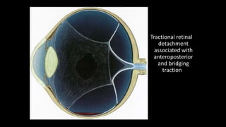
TRACTIONAL RETINAL
DETACHMENT
Dr Nikhil R P
Vitreous adhesions
1 Normal. The peripheral cortical vitreous is loosely attached to the
internal limiting membrane (ILM) of the sensory retina. Stronger
adhesions occur at the following sites:
• Vitreous base, where they are very strong.
• Around the optic nerve head, where they are fairly strong.
• Around the fovea, where they are fairly weak, except in eyes
with vitreomacular traction and macular hole formation.
• Along peripheral blood vessels, where they are usually weak.
2 Abnormal adhesions at the following sites may be associated with
retinal tear formation as a result of dynamic vitreoretinal traction
associated with acute PVD.
• Posterior border of islands of lattice degeneration.
• Retinal pigment clumps.
• Peripheral paravascular condensations.
• Vitreous base anomalies such as tongue-like extensions and
posterior islands.
Vitreoretinal traction
• Vitreoretinal traction is a force exerted on the retina by structures
originating in the vitreous, and may be dynamic or static. The difference
between the two is crucial in understanding the pathogenesis of the
various types of RD.
1 Dynamic traction is induced by eye movements and exerts a centripetal
force towards the vitreous cavity. It plays an important role in the
pathogenesis of retinal tears and rhegmatogenous RD.
2 Static traction is independent of ocular movements. It plays a key role in
the pathogenesis of tractional RD and proliferative vitreoretinopathy.
TRACTIONAL RETINAL
DETACHMENT
• The main causes of tractional RD are
(a) proliferative retinopathy due to diabetes
(b) penetrating posterior segment trauma
Other causes – Retinopathy of prematurity, vascular lesions,
familial exudative vitreoretinopathy, idiopathic vasculitis
Pathogenesis of diabetic tractional
retinal detachment
Tractional RD is caused by progressive contraction of fibrovascular membranes
over large areas of vitreoretinal adhesion.
- In contrast to acute PVD in eyes with rhegmatogenous RD, PVD in diabetic eyes
is gradual and frequently incomplete.
- It is thought to be caused by leakage of plasma constituents into the vitreous
gel from a fibrovascular network adherent to the posterior vitreous surface.
• 2 Static vitreoretinal traction of the following three types is recognized.
a Tangential traction is caused by the contraction of epiretinal
fibrovascular membranes with puckering of the retina and distortion of
retinal blood vessels.
b Anteroposterior traction is caused by the contraction of fibrovascular
membranes extending from the posterior retina, usually in association with
the major arcades, to the vitreous base anteriorly.
c Bridging (trampoline) traction is the result of contraction of fibrovascular
membranes which stretch from one part of the posterior retina to another or
between the vascular arcades, tending to pull the two involved points
together.
Tractional retinal
detachment
associated with
anteroposterior
and bridging
traction
Clinical findings characteristic of TRD
• Detached retina has a concave configuration and smooth appearance
and is relatively immobile
• Anterior limit to detachment can be traced
• The detachment is usually, rarely extends to ora serrate
• Retinal breaks are not seen unless there is combined RRD with TRD
• No demarcation lines or intraretinal cysts even in long standing cases
• Often a fibrovascular tissue is seen pulling the retina
Diagnosis
1 Symptoms.
Photopsia and floaters are usually absent because vitreoretinal traction
develops insidiously and is not associated with acute PVD.
The visual field defect usually progresses slowly and may become
stationary for months or even years.
2 Signs.
• The RD has a concave configuration and breaks are absent.
• Retinal mobility is severely reduced and shifting fluid is absent.
• The SRF is shallower than in a rhegmatogenous RD and seldom
extends to the ora serrata.
• The highest elevation of the retina occurs at sites of
vitreoretinal traction.
• If a tractional RD develops a break it assumes the
characteristics of a rhegmatogenous RD and progresses
more quickly (combined tractional-rhegmatogenous RD).
(A) Tractional
retinal
detachment in
severe
proliferative
diabetic
retinopathy;
(B) B-scan image
of another
patient shows a
shallow
tractional
retinal
detachment
Proliferative vitreoretinopathy
• Proliferative vitreoretinopathy (PVR) is caused by epiretinal and subretinal
membrane formation.
• Cell-mediated contraction of these membranes causes tangential retinal
traction and fixed retinal folds.
• Usually, PVR occurs following surgery for rhegmatogenous RD or
penetrating injury.
• However, it may also occur in eyes with rhegmatogenous RD that have not had
previous vitreoretinal surgery.
• The main features are retinal folds and rigidity so that retinal mobility induced by
eye movements or scleral indentation is decreased.
• Localized contracture in the periphery is referred to as a star fold
• A similar process in the posterior pole is referred to as a macular pucker
Retina society PVR classification
Symptoms
• The classic premonitory symptoms reported in about 60% of patients
with spontaneous rhegmatogenous RD are flashing lights and vitreous
floaters caused by acute PVD with collapse.
• After a variable period of time the patient notices a relative
peripheral visual field defect which may progress to involve central
vision.
• 1 Photopsia is the subjective sensation of a flash of light.
- In eyes with acute PVD it is probably caused by traction at sites of vitreoretinal
adhesion.
-
- The cessation of photopsia is the result of either separation of the adhesion or
complete tearing away of a piece of retina (operculum).
- In PVD the photopsia is often described as an arc of golden or white light
induced by eye movements and is more noticeable in dim illumination. It tends
to be projected into the patient's temporal peripheral visual field.
- Occasionally photopsia precedes PVD by 24–48 hours.
2 Floaters are moving vitreous opacities which are perceived when they cast
shadows on the retina.
• Vitreous opacities in eyes with acute PVD are of the following three types:
a Weiss ring is a solitary floater consisting of the detached annular
attachment of vitreous to the margin of the optic disc. Its presence does
not necessarily indicate total PVD, nor does its absence confirm absence of
PVD since it may be destroyed during the process of separation.
b Cobwebs are caused by condensation of collagen fibres
within the collapsed vitreous cortex.
c A sudden shower of minute red-coloured or dark spots
usually indicates vitreous hemorrhage secondary to tearing of
a peripheral retinal blood vessel. Vitreous haemorrhage
associated with acute PVD is usually sparse due to the small
calibre of peripheral retinal vessels.
(A) Weiss ring;
(B) B-scan
shows a
Weiss ring
associated
with
posterior
vitreous
detachment
3 A visual field defect is perceived as a ‘black curtain’.
- In some patients it may not be present on waking in the
morning, due to spontaneous absorption of SRF while lying
inactive overnight, only to reappear later in the day.
- A lower field defect is usually appreciated more quickly by
the patient than an upper field defect.
- The quadrant of the visual field in which the field defect
first appears is useful in predicting the location of the
primary retinal break, which will be in the opposite
quadrant.
- Loss of central vision may be due either to involvement
of the fovea by SRF or, less frequently, obstruction of the
visual axis by a large upper bullous RD.
Signs
General
1 Marcus Gunn pupil (relative afferent pupillary defect) is
present in an eye with an extensive RD irrespective of the type.
2 Intraocular pressure is usually lower by about 5 mmHg
compared with the normal eye. If the intraocular pressure is
extremely low, an associated choroidal detachment may be
present.
3 Iritis is very common but usually mild. Occasionally it may be
severe enough to cause posterior synechiae. In these cases the
underlying RD may be overlooked and the poor visual acuity
incorrectly ascribed to some other cause.
4 ‘Tobacco dust’ consisting of pigment cells is seen in the
anterior vitreous.
5 Retinal breaks appear as discontinuities in the retinal
surface. They are usually red because of the colour contrast
between the sensory retina and underlying choroid. However,
in eyes with hypopigmented choroid (as in high myopia), the
colour contrast is decreased and small breaks may be
overlooked unless careful slit-lamp and indirect
ophthalmoscopic examination is performed.
6 Retinal signs depend on the duration of RD and the
presence or absence of proliferative vitreoretinopathy (PVR) as
described below.
‘Tobacco
dust’ in
the
anterior
vitreous
Ultrasonography
• B-scan ultrasonography (US) is very useful in the diagnosis of
RD in eyes with opaque media, particularly severe vitreous
haemorrhage
• Utilises high frequency sound waves ranging from 8-10 MHz.
• Gain adjusts the amplification of the echo signal,
similar to volume control of a radio.
• Higher gain increases the sensitivity of the instrument
in displaying weak echoes such as vitreous opacities.
• Lower gain only allows display of strong echoes such as
the retina and sclera, though improves resolution
because it narrows the beam.
POSTERIOR VITREOUS DETACHMENT
membranous lesion
with no/some
attachments to the
optic disc
POSTERIOR VITREOUS DETACHMENT
Mobility of PVD is
more than RD.
The spike of RD is
more than PVD.
PVD becomes more
prominent in higher
gain settings
RETINAL DETACHMENT
The detachment produces a
bright continuous, folded
appearance with insertion into
the disc and ora serrata.
It is to determine the
configuration of the
detachment as shallow, flat or
bullous
RHEGMATOGENOUS RD
CLOSED FUNNEL RD
WITH RETINAL CYST
Retinal Tear
PROLIFERATIVE VITERO RETINOPATHY
Retinal Reattachment Surgery
• Scleral Buckling Surgery
• Pars Plana Vitrectomy
• Pneumatic retinopexy
• Retinal breaks – cryotherapy / laser photocoagulation
INDICATIONS FOR BED REST
• To prevent macular involvement by subretinal fluid.
• To promote resorption of SRF.
• To unroll mobile flap of giant retinal tear.
Indications of scleral buckling
• Radial buckling
• Large U shaped tears
• Posterior breaks because sutures are easier to insert.
• Segmental circumferential buckling
• Multiple breaks
• Anterior breaks
• Wide breaks such as dialysis and giant tears.
Indications of pneumatic retinopexy
• Short, minimally invasive, OPD procedure
• Indications
• Retinal break smaller than one clock hour
• Multiple breaks within one clock hour
• All breaks in superior 8 clock hours
• Hypotony following drainage of SRF.
• Fishmouthing of large retinal tear
• Radial retinal folds
• Macular hole giving rise to RD.
Primary Vitrectomy in RD
Pars Plana Vitrectomy
• Indicated in
• Media opacities- VH & advanced PVR
• Posteriorly located breaks
• RD with giant retinal tear or macular hole
• Tractional RD
• Scarring from penetrating trauma
• Relative contraindications
• Relatively simple phakic RD
PRINCIPLES OF VITRECTOMY
• The principles of vitrectomy to treat RRD are release of
tractional forces that precipitated the retinal break,
and the closure and reattachment of breaks to the
underlying RPE .
• The surgical procedure requires: (1) removal of the vitreous
gel and preretinal tractional membrane; (2) intraoperative
flattening of the detached retina; (3) application of
retinopexy; and (4) placement of a tamponade in vitreous
cavity.
PPV
Compared to SB, PPV offers several advantages.
• The view of the retinal periphery is enhanced,
• Identification of retinal breaks is rendered easier,
• Achievement of complete intraoperative retinal attachment is
possible, the risks of hemorrhage or retinal incarceration
inherent to external drainage procedure applied during SB is
eliminated, and the technique is less likely to cause a
refractive change.
Procedure
• video
Vitreous substitutes
• Intra ocular gases : Air, Sulphur hexafluoride (SF6),
• Silicon oil – Polydimethyl siloxane, trifluoromethyl siloxane
• Perfluorocarbon liquids ( PFCL )
Sutureless Microincision Vitrectomy
• Transconjunctival sutureless MIVS using 23G/ 25G
instrumentation
• Advantages
• Shorter surgical time
• Less surgically induced astigmatism
• Reduced risk of post-operative corneal astigmatism
• Greater rigidity, better illumination, improved fluidics with 23 G
• IOP compensation via direct control of infusion pressure
• Wide angle viewing systems
Management of Tractional Retinal
Detachment
• TRD progresses very slowly, may reattach spontaneously
• Localized TRD away from macula- observation
• Indications for surgery
• Macular threatened or detached
• Vitreous haemorrhage
• Retinal holes
• Surgical Principles
• To relax the vitreoretinal traction
• Closure of retinal holes
• Drainage of SRF
• PPV- to clear media, release of AP & tangential traction
• ERM- peeling/ segmentation/ delamination
• Enblock excision of traction membranes
• Retinotomy with internal drainage of SRF, internal tamponade with
gas/silicone oil injection
• Endodiathermy & endophotocoagulation- new vessels & retinopexy
Comparison of various surgical techniques
Method Reattachment Rate Limitations/Complications Benefits
Scleral Buckling 94% Morbidity, infection, buckle
extrusion, ocular motility
disturbances
Excellent long term
anatomic success, good
visual outcome
Pars Plana Vitrectomy 71-92% (1˚ success
rate)
94% (2˚ success rate)
Iatrogenic retinal breaks, PVR,
lens trauma, cataract
progression
Visualization of all
breaks, removal of
opacities/synechiae,
anatomic success in
complicated
detachments
Pneumatic Retinopexy 64% (1˚ success rate)
91% (2˚ success rate)
Limited use only in
uncomplicated RRD with
superior breaks
Post-op positioning,
iatrogenic breaks
In-office procedure,
minimally invasive,
↓ Recovery time, better
post-op VA
SB / PR / PPV
• Complicated detachments are usually managed with PPV ,
whereas localized, relatively simple cases are usually
managed with a “walling-off” (demarcating) procedure
employing laser or cryotherapy, with PR, or with a small
and localized scleral buckling procedure.
Management of PVR
• Upto grade C1 and C2 -------- high encircling scleral buckling
• C3 or greater ----- pars plana vitrectomy
Tractional RD

Tractional RD

  • 1.
  • 2.
    Vitreous adhesions 1 Normal.The peripheral cortical vitreous is loosely attached to the internal limiting membrane (ILM) of the sensory retina. Stronger adhesions occur at the following sites: • Vitreous base, where they are very strong. • Around the optic nerve head, where they are fairly strong. • Around the fovea, where they are fairly weak, except in eyes with vitreomacular traction and macular hole formation. • Along peripheral blood vessels, where they are usually weak.
  • 3.
    2 Abnormal adhesionsat the following sites may be associated with retinal tear formation as a result of dynamic vitreoretinal traction associated with acute PVD. • Posterior border of islands of lattice degeneration. • Retinal pigment clumps. • Peripheral paravascular condensations. • Vitreous base anomalies such as tongue-like extensions and posterior islands.
  • 4.
    Vitreoretinal traction • Vitreoretinaltraction is a force exerted on the retina by structures originating in the vitreous, and may be dynamic or static. The difference between the two is crucial in understanding the pathogenesis of the various types of RD. 1 Dynamic traction is induced by eye movements and exerts a centripetal force towards the vitreous cavity. It plays an important role in the pathogenesis of retinal tears and rhegmatogenous RD. 2 Static traction is independent of ocular movements. It plays a key role in the pathogenesis of tractional RD and proliferative vitreoretinopathy.
  • 5.
  • 6.
    • The maincauses of tractional RD are (a) proliferative retinopathy due to diabetes (b) penetrating posterior segment trauma Other causes – Retinopathy of prematurity, vascular lesions, familial exudative vitreoretinopathy, idiopathic vasculitis
  • 7.
    Pathogenesis of diabetictractional retinal detachment Tractional RD is caused by progressive contraction of fibrovascular membranes over large areas of vitreoretinal adhesion. - In contrast to acute PVD in eyes with rhegmatogenous RD, PVD in diabetic eyes is gradual and frequently incomplete. - It is thought to be caused by leakage of plasma constituents into the vitreous gel from a fibrovascular network adherent to the posterior vitreous surface.
  • 8.
    • 2 Staticvitreoretinal traction of the following three types is recognized. a Tangential traction is caused by the contraction of epiretinal fibrovascular membranes with puckering of the retina and distortion of retinal blood vessels. b Anteroposterior traction is caused by the contraction of fibrovascular membranes extending from the posterior retina, usually in association with the major arcades, to the vitreous base anteriorly. c Bridging (trampoline) traction is the result of contraction of fibrovascular membranes which stretch from one part of the posterior retina to another or between the vascular arcades, tending to pull the two involved points together.
  • 9.
  • 10.
    Clinical findings characteristicof TRD • Detached retina has a concave configuration and smooth appearance and is relatively immobile • Anterior limit to detachment can be traced • The detachment is usually, rarely extends to ora serrate • Retinal breaks are not seen unless there is combined RRD with TRD • No demarcation lines or intraretinal cysts even in long standing cases • Often a fibrovascular tissue is seen pulling the retina
  • 11.
    Diagnosis 1 Symptoms. Photopsia andfloaters are usually absent because vitreoretinal traction develops insidiously and is not associated with acute PVD. The visual field defect usually progresses slowly and may become stationary for months or even years. 2 Signs. • The RD has a concave configuration and breaks are absent. • Retinal mobility is severely reduced and shifting fluid is absent. • The SRF is shallower than in a rhegmatogenous RD and seldom extends to the ora serrata.
  • 12.
    • The highestelevation of the retina occurs at sites of vitreoretinal traction. • If a tractional RD develops a break it assumes the characteristics of a rhegmatogenous RD and progresses more quickly (combined tractional-rhegmatogenous RD).
  • 13.
    (A) Tractional retinal detachment in severe proliferative diabetic retinopathy; (B)B-scan image of another patient shows a shallow tractional retinal detachment
  • 15.
    Proliferative vitreoretinopathy • Proliferativevitreoretinopathy (PVR) is caused by epiretinal and subretinal membrane formation. • Cell-mediated contraction of these membranes causes tangential retinal traction and fixed retinal folds. • Usually, PVR occurs following surgery for rhegmatogenous RD or penetrating injury.
  • 16.
    • However, itmay also occur in eyes with rhegmatogenous RD that have not had previous vitreoretinal surgery. • The main features are retinal folds and rigidity so that retinal mobility induced by eye movements or scleral indentation is decreased. • Localized contracture in the periphery is referred to as a star fold • A similar process in the posterior pole is referred to as a macular pucker
  • 19.
    Retina society PVRclassification
  • 29.
    Symptoms • The classicpremonitory symptoms reported in about 60% of patients with spontaneous rhegmatogenous RD are flashing lights and vitreous floaters caused by acute PVD with collapse. • After a variable period of time the patient notices a relative peripheral visual field defect which may progress to involve central vision.
  • 30.
    • 1 Photopsiais the subjective sensation of a flash of light. - In eyes with acute PVD it is probably caused by traction at sites of vitreoretinal adhesion. - - The cessation of photopsia is the result of either separation of the adhesion or complete tearing away of a piece of retina (operculum). - In PVD the photopsia is often described as an arc of golden or white light induced by eye movements and is more noticeable in dim illumination. It tends to be projected into the patient's temporal peripheral visual field. - Occasionally photopsia precedes PVD by 24–48 hours.
  • 31.
    2 Floaters aremoving vitreous opacities which are perceived when they cast shadows on the retina. • Vitreous opacities in eyes with acute PVD are of the following three types: a Weiss ring is a solitary floater consisting of the detached annular attachment of vitreous to the margin of the optic disc. Its presence does not necessarily indicate total PVD, nor does its absence confirm absence of PVD since it may be destroyed during the process of separation.
  • 32.
    b Cobwebs arecaused by condensation of collagen fibres within the collapsed vitreous cortex. c A sudden shower of minute red-coloured or dark spots usually indicates vitreous hemorrhage secondary to tearing of a peripheral retinal blood vessel. Vitreous haemorrhage associated with acute PVD is usually sparse due to the small calibre of peripheral retinal vessels.
  • 33.
    (A) Weiss ring; (B)B-scan shows a Weiss ring associated with posterior vitreous detachment
  • 34.
    3 A visualfield defect is perceived as a ‘black curtain’. - In some patients it may not be present on waking in the morning, due to spontaneous absorption of SRF while lying inactive overnight, only to reappear later in the day. - A lower field defect is usually appreciated more quickly by the patient than an upper field defect.
  • 35.
    - The quadrantof the visual field in which the field defect first appears is useful in predicting the location of the primary retinal break, which will be in the opposite quadrant. - Loss of central vision may be due either to involvement of the fovea by SRF or, less frequently, obstruction of the visual axis by a large upper bullous RD.
  • 36.
    Signs General 1 Marcus Gunnpupil (relative afferent pupillary defect) is present in an eye with an extensive RD irrespective of the type. 2 Intraocular pressure is usually lower by about 5 mmHg compared with the normal eye. If the intraocular pressure is extremely low, an associated choroidal detachment may be present. 3 Iritis is very common but usually mild. Occasionally it may be severe enough to cause posterior synechiae. In these cases the underlying RD may be overlooked and the poor visual acuity incorrectly ascribed to some other cause.
  • 37.
    4 ‘Tobacco dust’consisting of pigment cells is seen in the anterior vitreous. 5 Retinal breaks appear as discontinuities in the retinal surface. They are usually red because of the colour contrast between the sensory retina and underlying choroid. However, in eyes with hypopigmented choroid (as in high myopia), the colour contrast is decreased and small breaks may be overlooked unless careful slit-lamp and indirect ophthalmoscopic examination is performed. 6 Retinal signs depend on the duration of RD and the presence or absence of proliferative vitreoretinopathy (PVR) as described below.
  • 38.
  • 39.
    Ultrasonography • B-scan ultrasonography(US) is very useful in the diagnosis of RD in eyes with opaque media, particularly severe vitreous haemorrhage • Utilises high frequency sound waves ranging from 8-10 MHz.
  • 40.
    • Gain adjuststhe amplification of the echo signal, similar to volume control of a radio. • Higher gain increases the sensitivity of the instrument in displaying weak echoes such as vitreous opacities. • Lower gain only allows display of strong echoes such as the retina and sclera, though improves resolution because it narrows the beam.
  • 41.
    POSTERIOR VITREOUS DETACHMENT membranouslesion with no/some attachments to the optic disc
  • 42.
    POSTERIOR VITREOUS DETACHMENT Mobilityof PVD is more than RD. The spike of RD is more than PVD. PVD becomes more prominent in higher gain settings
  • 43.
    RETINAL DETACHMENT The detachmentproduces a bright continuous, folded appearance with insertion into the disc and ora serrata. It is to determine the configuration of the detachment as shallow, flat or bullous
  • 45.
  • 46.
    CLOSED FUNNEL RD WITHRETINAL CYST Retinal Tear
  • 47.
  • 49.
    Retinal Reattachment Surgery •Scleral Buckling Surgery • Pars Plana Vitrectomy • Pneumatic retinopexy • Retinal breaks – cryotherapy / laser photocoagulation
  • 50.
    INDICATIONS FOR BEDREST • To prevent macular involvement by subretinal fluid. • To promote resorption of SRF. • To unroll mobile flap of giant retinal tear.
  • 51.
    Indications of scleralbuckling • Radial buckling • Large U shaped tears • Posterior breaks because sutures are easier to insert. • Segmental circumferential buckling • Multiple breaks • Anterior breaks • Wide breaks such as dialysis and giant tears.
  • 52.
    Indications of pneumaticretinopexy • Short, minimally invasive, OPD procedure • Indications • Retinal break smaller than one clock hour • Multiple breaks within one clock hour • All breaks in superior 8 clock hours • Hypotony following drainage of SRF. • Fishmouthing of large retinal tear • Radial retinal folds • Macular hole giving rise to RD.
  • 53.
  • 54.
    Pars Plana Vitrectomy •Indicated in • Media opacities- VH & advanced PVR • Posteriorly located breaks • RD with giant retinal tear or macular hole • Tractional RD • Scarring from penetrating trauma • Relative contraindications • Relatively simple phakic RD
  • 55.
    PRINCIPLES OF VITRECTOMY •The principles of vitrectomy to treat RRD are release of tractional forces that precipitated the retinal break, and the closure and reattachment of breaks to the underlying RPE . • The surgical procedure requires: (1) removal of the vitreous gel and preretinal tractional membrane; (2) intraoperative flattening of the detached retina; (3) application of retinopexy; and (4) placement of a tamponade in vitreous cavity.
  • 56.
    PPV Compared to SB,PPV offers several advantages. • The view of the retinal periphery is enhanced, • Identification of retinal breaks is rendered easier, • Achievement of complete intraoperative retinal attachment is possible, the risks of hemorrhage or retinal incarceration inherent to external drainage procedure applied during SB is eliminated, and the technique is less likely to cause a refractive change.
  • 57.
  • 60.
    Vitreous substitutes • Intraocular gases : Air, Sulphur hexafluoride (SF6), • Silicon oil – Polydimethyl siloxane, trifluoromethyl siloxane • Perfluorocarbon liquids ( PFCL )
  • 62.
    Sutureless Microincision Vitrectomy •Transconjunctival sutureless MIVS using 23G/ 25G instrumentation • Advantages • Shorter surgical time • Less surgically induced astigmatism • Reduced risk of post-operative corneal astigmatism • Greater rigidity, better illumination, improved fluidics with 23 G • IOP compensation via direct control of infusion pressure • Wide angle viewing systems
  • 63.
    Management of TractionalRetinal Detachment • TRD progresses very slowly, may reattach spontaneously • Localized TRD away from macula- observation • Indications for surgery • Macular threatened or detached • Vitreous haemorrhage • Retinal holes • Surgical Principles • To relax the vitreoretinal traction • Closure of retinal holes • Drainage of SRF
  • 64.
    • PPV- toclear media, release of AP & tangential traction • ERM- peeling/ segmentation/ delamination • Enblock excision of traction membranes • Retinotomy with internal drainage of SRF, internal tamponade with gas/silicone oil injection • Endodiathermy & endophotocoagulation- new vessels & retinopexy
  • 65.
    Comparison of varioussurgical techniques Method Reattachment Rate Limitations/Complications Benefits Scleral Buckling 94% Morbidity, infection, buckle extrusion, ocular motility disturbances Excellent long term anatomic success, good visual outcome Pars Plana Vitrectomy 71-92% (1˚ success rate) 94% (2˚ success rate) Iatrogenic retinal breaks, PVR, lens trauma, cataract progression Visualization of all breaks, removal of opacities/synechiae, anatomic success in complicated detachments Pneumatic Retinopexy 64% (1˚ success rate) 91% (2˚ success rate) Limited use only in uncomplicated RRD with superior breaks Post-op positioning, iatrogenic breaks In-office procedure, minimally invasive, ↓ Recovery time, better post-op VA
  • 66.
    SB / PR/ PPV • Complicated detachments are usually managed with PPV , whereas localized, relatively simple cases are usually managed with a “walling-off” (demarcating) procedure employing laser or cryotherapy, with PR, or with a small and localized scleral buckling procedure.
  • 67.
    Management of PVR •Upto grade C1 and C2 -------- high encircling scleral buckling • C3 or greater ----- pars plana vitrectomy