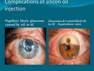
Presenting author-Dr Priyanka (DNB RESIDENT)
DEPARTMENT OF OPHTHALMOLOGY
Definition
Retinal detachment (RD) refers to separation of
the neurosensory layer of retina from the pigment
epithelial layer, to which it is normally loosely
attached.
Layers of retina-
inside out
1) Internal limiting membrane
2) Nerve fibre layer
3) Ganglion cell layer
4) Inner plexiform layer
5) Inner nuclear layer
6) Outer plexiform layer
7) Outer nuclear layer
8) External limiting membrane
9) Photoreceptors
10) Retinal Pigment epithelium
Types of RD
Four types-
1) Rhegmatogenous Retinal detachment (RRD)
2) Tractional retinal detachment
3) Exudative / serous retinal detachment
4) Combined tractional-rhegmatogenous retinal
detachment
RHEGMATOGENOUS RD
 Most common type, affects 1 in 10,000 of population ,
with BE affected in about 10%. M>F, 40-70 yrs
 Greek: rhegma , meaning a rent or a fissure or a break
 Characterized by the presence of a retinal break in
concert with vitreoretinal traction that allows
accumulation of liquified vitreous under the
neurosensory retina , separating it from the RPE.
 Predisposing factors in pathogenesis of RD- Over
40% of RRD occur in myopic eyes(>-6.0D/ AL
26mm). Presence of PVD, H/O trauma ,
predisposing peripheral retinal lesions, Vitreous loss
during cataract surgery and laser capsulotomy carry
a higher risk
 SYMPTOMS- flashing lights and floaters reported
in about 60% of pts., Curtain like relative peripheral
visual field defect
 SIGNS- RAPD (Marcus Gunn pupil) in extensive RD,
low IOP, mild iritis, tobacco dust like pigments cells
seen in anterior chamber
Fresh rhegmatogenous RD
Macular hole
Surrounded by
sub-retinal
fluid
Fresh RRD signs- convex
configuration
U tear with ST detachment
Typical corrugated appearance
of detached retina
Tobacco dust in anterior vitreous(
shaffer’s sign)- pathognomic of a
retinal tear
Long standing RRD signs-
Retinal cysts multiple cysts B-scan
Demarcation line
Demarcation line surrounding
localized fluid with a small hole
Proliferative vitreoretinopathy-
sign of RRD
 Usually, PVR occurs following surgery for
rhegmatogenous RD or penetrating injury , though
may occur in eyes with RRD without previous
retinal surgery
 Main features- retinal folds and rigidity, causing
decreased retinal mobility following eye movements
Staging of PVR-
GRADE A- early retinal
wrinkling
GRADE B- Rolled retinal
break edges
Staging of PVR
GRADE C with prominent
star fold
Advanced disease with characteristic
funnel shaped detachment
Tractional RD
Neurosensory retina is pulled away from the RPE by
contracting vitreoretinal membranes in the absence of
a retinal break
MAIN CAUSES: Proliferative retinopathy like DM,
ROP and penetrating posterior segment trauma
SYMPTOMS- slowly progressive visual field defect,
Photopsia and floaters usually absent.
SIGNS- retinal mobility severely reduced, shifting
fluid absent,
If Tractional RD develops a break, it assumes
characteristics of rhegmatogenous RD and progresses
rapidly, known as combined tractional-
rhegmatogenous RD
Diagnosis of TRD –
Tractional RD – concave
configuration
Secondary to Proliferative
Diabetic retinopathy
 Severe TRD
OCT showing concave
configuration of Tractional RD
Tractional RD USG B-scan:
incomplete posterior vitreous detachment
and relatively immobile retina
Exudative retinal detachment
• characterized by accumulation of SRF in
the absence of retinal breaks or traction
• May occur in a variety of vascular,
inflammatory, and neoplastic diseases
involving the retina, RPE and the choroid in
which fluid leaks outside the vessel and
accumulate under the retina
CAUSES OF Exudative RD-
 Choroidal tm like melanomas, haemangiomas ,
metastases.
 Inflammation such as harada ds , posterior scleritis.
 Bullous central serous chorioretinopathy – rare cause
 Iatrogenic causes- RD surgery, PRP
 Choroidal neovascularization – may leak to give rise to
ERD
 Hypertensive choroidopathy as in toxemia of pregnancy
 Idiopathic ,such as uveal effusion syndrome
Exudative RD – convex configuration,
choroidal melanoma
Exudative RD- Diagnosis
 SYMPTOMS : Floaters may be present if a/s vitritis,
visual field defects may develop suddenly and
progress rapidly.
No vitreoretinal traction , so photopsia absent
SIGNS : convex configuration like RRD but its surface
are smooth and not corrugated, detached retina is
very mobile and exhibits the phenomenon of
‘shifting fluid’ .
ERD WITH SHIFTING FLUID
Inferior collection of SRF
with pt. sitting
Shifting of SRF upwards
when the pt. is supine
Resolution of exudative RD- ‘leopard
spot’ pigmentation
Differential diagnosis of RD-
 Degenerative retinoschisis- present in about 5% of
population over age 20 years, prevalent in
hypermetropia. Elevation is convex, smooth, thin and
relatively immobile
 Choroidal detachment- include low IOP, elevations
are brown, smooth , relatively immobile and do not
extend to the posterior pole
 Uveal effusion syndrome- MC in middle aged
hypermetropes
 Vascularized vitreous membranes( d/t PDR, Vos etc)
 Old organized vitreous hemorrhage
Management of RD-
 Identification of the causative retinal break
 Selection of surgical procedure with least
morbidity
 Evaluation of the fellow eye to plan for any
prophylaxis (laser, cryotherapy)
 Medical evaluation for presurgical fitness
Lincoff’s for finding the primary
break in RD
Selection of surgical procedure
 Scleral buckling (minimal invasive/ classical)- gold
standard for most cases of uncomplicated RRD
 Vitrectomy (classical/ sutureless; using gas/ silicone oil
and if needed, an encircling silicone band)- PPV is
required in cases which are complicated by significant
media opacities like cataract, VH, or advanced PVR
 Pneumoretinopexy- out patient procedure in which an
intravitreal gas bubble together with cryotherapy or
laser are used to seal a retinal break
Scleral buckling
 Reattachment rate- 94%
 Limitations/ complications- morbidity, infection,
buckle extrusion, ocular motility distubances
 Benefits- excellent long term anatomic success
Scleral buckling
Circumferential explant
Buckle induced
circumferential explant
Pars plana vitrectomy
 Reattachment rate- 71-92%
 Limitations/ complications- iatrogenic retinal
breaks, PVR, lens trauma, cataract progression
 Benefits- visualization of all breaks, removal of
opacities/ synechiae, anatomic success in
complicated detachment
Giant retinal tear large posterior tear severe PVR
• Reattachment rate- 64%
•Limitations/ complications- limited to uncomplicated RRD
with superior breaks, need for post operative positioning,
creation of iatrogenic breaks.
•Benefits- in-office procedure, minimally invasive, reduced
recovery time, better post operative VA
Pneumatic retinopexy
Gas bubble in vitreous
cavity
‘Fish eggs’ due to gas
bubble break up
Exudative RD t/t
 Some cases resolve
spontaneously
 Others treated with
systemic corticosteroids(
harada ds, posterior
scleritis)
 Laser photocoagulation- in
bullous central serous
chorioretinopathy
Gases tried in vitreoretinal surgery
Nonexpansile gases( after
PPV) Expansile gases in
pneumoretinopexy
 Air - avg duration 3 days
 Nitrogen
 Helium
 Oxygen
 Argon
 Xenon
 krypton
 SF6 - avg duration 12 days
 C3F8- Longer acting
perfluoropropane, avg
duration 38 days
 C4F10
 CF4
 C2F6
 C4F10
 C5F12
Tamponading agents/ vitreous
subtitute used in vitreoretinal
surgery
 Intraocular gases
 Silicone oil
 Perfluorocarbons (PFCL)
Post operative complications-
 Raised IOP - due to overexpansion of intraocular
gas, silicone oil associated glaucoma
 Cataract- gas induced, silicone induced, following
vitrectomy
 Band keratopathy- due to extended silicone oil
tamponade
Complications of scleral buckling
Buckle extrusion
Buckle intrusion
subretinally
Complications of silicone oil
injection
Cataract with an inverted
pseudo-hypopyon
(hyperoleon)
Band keratopathy
Complications of silicon oil
injection
Pupillary block glaucoma
caused by oil in AC
Glaucoma d/t emulsified oil
in AC , hyperoleon seen
Thank You

Retinal detachment

  • 1.
    Presenting author-Dr Priyanka(DNB RESIDENT) DEPARTMENT OF OPHTHALMOLOGY
  • 2.
    Definition Retinal detachment (RD)refers to separation of the neurosensory layer of retina from the pigment epithelial layer, to which it is normally loosely attached.
  • 3.
    Layers of retina- insideout 1) Internal limiting membrane 2) Nerve fibre layer 3) Ganglion cell layer 4) Inner plexiform layer 5) Inner nuclear layer 6) Outer plexiform layer 7) Outer nuclear layer 8) External limiting membrane 9) Photoreceptors 10) Retinal Pigment epithelium
  • 4.
    Types of RD Fourtypes- 1) Rhegmatogenous Retinal detachment (RRD) 2) Tractional retinal detachment 3) Exudative / serous retinal detachment 4) Combined tractional-rhegmatogenous retinal detachment
  • 5.
    RHEGMATOGENOUS RD  Mostcommon type, affects 1 in 10,000 of population , with BE affected in about 10%. M>F, 40-70 yrs  Greek: rhegma , meaning a rent or a fissure or a break  Characterized by the presence of a retinal break in concert with vitreoretinal traction that allows accumulation of liquified vitreous under the neurosensory retina , separating it from the RPE.
  • 6.
     Predisposing factorsin pathogenesis of RD- Over 40% of RRD occur in myopic eyes(>-6.0D/ AL 26mm). Presence of PVD, H/O trauma , predisposing peripheral retinal lesions, Vitreous loss during cataract surgery and laser capsulotomy carry a higher risk  SYMPTOMS- flashing lights and floaters reported in about 60% of pts., Curtain like relative peripheral visual field defect  SIGNS- RAPD (Marcus Gunn pupil) in extensive RD, low IOP, mild iritis, tobacco dust like pigments cells seen in anterior chamber
  • 7.
    Fresh rhegmatogenous RD Macularhole Surrounded by sub-retinal fluid
  • 8.
    Fresh RRD signs-convex configuration U tear with ST detachment Typical corrugated appearance of detached retina
  • 9.
    Tobacco dust inanterior vitreous( shaffer’s sign)- pathognomic of a retinal tear
  • 10.
    Long standing RRDsigns- Retinal cysts multiple cysts B-scan
  • 11.
    Demarcation line Demarcation linesurrounding localized fluid with a small hole
  • 12.
    Proliferative vitreoretinopathy- sign ofRRD  Usually, PVR occurs following surgery for rhegmatogenous RD or penetrating injury , though may occur in eyes with RRD without previous retinal surgery  Main features- retinal folds and rigidity, causing decreased retinal mobility following eye movements
  • 13.
    Staging of PVR- GRADEA- early retinal wrinkling GRADE B- Rolled retinal break edges
  • 14.
    Staging of PVR GRADEC with prominent star fold Advanced disease with characteristic funnel shaped detachment
  • 15.
    Tractional RD Neurosensory retinais pulled away from the RPE by contracting vitreoretinal membranes in the absence of a retinal break MAIN CAUSES: Proliferative retinopathy like DM, ROP and penetrating posterior segment trauma
  • 16.
    SYMPTOMS- slowly progressivevisual field defect, Photopsia and floaters usually absent. SIGNS- retinal mobility severely reduced, shifting fluid absent, If Tractional RD develops a break, it assumes characteristics of rhegmatogenous RD and progresses rapidly, known as combined tractional- rhegmatogenous RD Diagnosis of TRD –
  • 17.
    Tractional RD –concave configuration Secondary to Proliferative Diabetic retinopathy  Severe TRD
  • 18.
  • 19.
    Tractional RD USGB-scan: incomplete posterior vitreous detachment and relatively immobile retina
  • 20.
    Exudative retinal detachment •characterized by accumulation of SRF in the absence of retinal breaks or traction • May occur in a variety of vascular, inflammatory, and neoplastic diseases involving the retina, RPE and the choroid in which fluid leaks outside the vessel and accumulate under the retina
  • 21.
    CAUSES OF ExudativeRD-  Choroidal tm like melanomas, haemangiomas , metastases.  Inflammation such as harada ds , posterior scleritis.  Bullous central serous chorioretinopathy – rare cause  Iatrogenic causes- RD surgery, PRP  Choroidal neovascularization – may leak to give rise to ERD  Hypertensive choroidopathy as in toxemia of pregnancy  Idiopathic ,such as uveal effusion syndrome
  • 22.
    Exudative RD –convex configuration, choroidal melanoma
  • 23.
    Exudative RD- Diagnosis SYMPTOMS : Floaters may be present if a/s vitritis, visual field defects may develop suddenly and progress rapidly. No vitreoretinal traction , so photopsia absent SIGNS : convex configuration like RRD but its surface are smooth and not corrugated, detached retina is very mobile and exhibits the phenomenon of ‘shifting fluid’ .
  • 24.
    ERD WITH SHIFTINGFLUID Inferior collection of SRF with pt. sitting Shifting of SRF upwards when the pt. is supine
  • 25.
    Resolution of exudativeRD- ‘leopard spot’ pigmentation
  • 26.
    Differential diagnosis ofRD-  Degenerative retinoschisis- present in about 5% of population over age 20 years, prevalent in hypermetropia. Elevation is convex, smooth, thin and relatively immobile  Choroidal detachment- include low IOP, elevations are brown, smooth , relatively immobile and do not extend to the posterior pole  Uveal effusion syndrome- MC in middle aged hypermetropes  Vascularized vitreous membranes( d/t PDR, Vos etc)  Old organized vitreous hemorrhage
  • 27.
    Management of RD- Identification of the causative retinal break  Selection of surgical procedure with least morbidity  Evaluation of the fellow eye to plan for any prophylaxis (laser, cryotherapy)  Medical evaluation for presurgical fitness
  • 28.
    Lincoff’s for findingthe primary break in RD
  • 29.
    Selection of surgicalprocedure  Scleral buckling (minimal invasive/ classical)- gold standard for most cases of uncomplicated RRD  Vitrectomy (classical/ sutureless; using gas/ silicone oil and if needed, an encircling silicone band)- PPV is required in cases which are complicated by significant media opacities like cataract, VH, or advanced PVR  Pneumoretinopexy- out patient procedure in which an intravitreal gas bubble together with cryotherapy or laser are used to seal a retinal break
  • 30.
    Scleral buckling  Reattachmentrate- 94%  Limitations/ complications- morbidity, infection, buckle extrusion, ocular motility distubances  Benefits- excellent long term anatomic success
  • 31.
    Scleral buckling Circumferential explant Buckleinduced circumferential explant
  • 32.
    Pars plana vitrectomy Reattachment rate- 71-92%  Limitations/ complications- iatrogenic retinal breaks, PVR, lens trauma, cataract progression  Benefits- visualization of all breaks, removal of opacities/ synechiae, anatomic success in complicated detachment
  • 33.
    Giant retinal tearlarge posterior tear severe PVR
  • 34.
    • Reattachment rate-64% •Limitations/ complications- limited to uncomplicated RRD with superior breaks, need for post operative positioning, creation of iatrogenic breaks. •Benefits- in-office procedure, minimally invasive, reduced recovery time, better post operative VA
  • 35.
    Pneumatic retinopexy Gas bubblein vitreous cavity ‘Fish eggs’ due to gas bubble break up
  • 36.
    Exudative RD t/t Some cases resolve spontaneously  Others treated with systemic corticosteroids( harada ds, posterior scleritis)  Laser photocoagulation- in bullous central serous chorioretinopathy
  • 37.
    Gases tried invitreoretinal surgery Nonexpansile gases( after PPV) Expansile gases in pneumoretinopexy  Air - avg duration 3 days  Nitrogen  Helium  Oxygen  Argon  Xenon  krypton  SF6 - avg duration 12 days  C3F8- Longer acting perfluoropropane, avg duration 38 days  C4F10  CF4  C2F6  C4F10  C5F12
  • 38.
    Tamponading agents/ vitreous subtituteused in vitreoretinal surgery  Intraocular gases  Silicone oil  Perfluorocarbons (PFCL)
  • 39.
    Post operative complications- Raised IOP - due to overexpansion of intraocular gas, silicone oil associated glaucoma  Cataract- gas induced, silicone induced, following vitrectomy  Band keratopathy- due to extended silicone oil tamponade
  • 40.
    Complications of scleralbuckling Buckle extrusion Buckle intrusion subretinally
  • 41.
    Complications of siliconeoil injection Cataract with an inverted pseudo-hypopyon (hyperoleon) Band keratopathy
  • 42.
    Complications of siliconoil injection Pupillary block glaucoma caused by oil in AC Glaucoma d/t emulsified oil in AC , hyperoleon seen
  • 43.