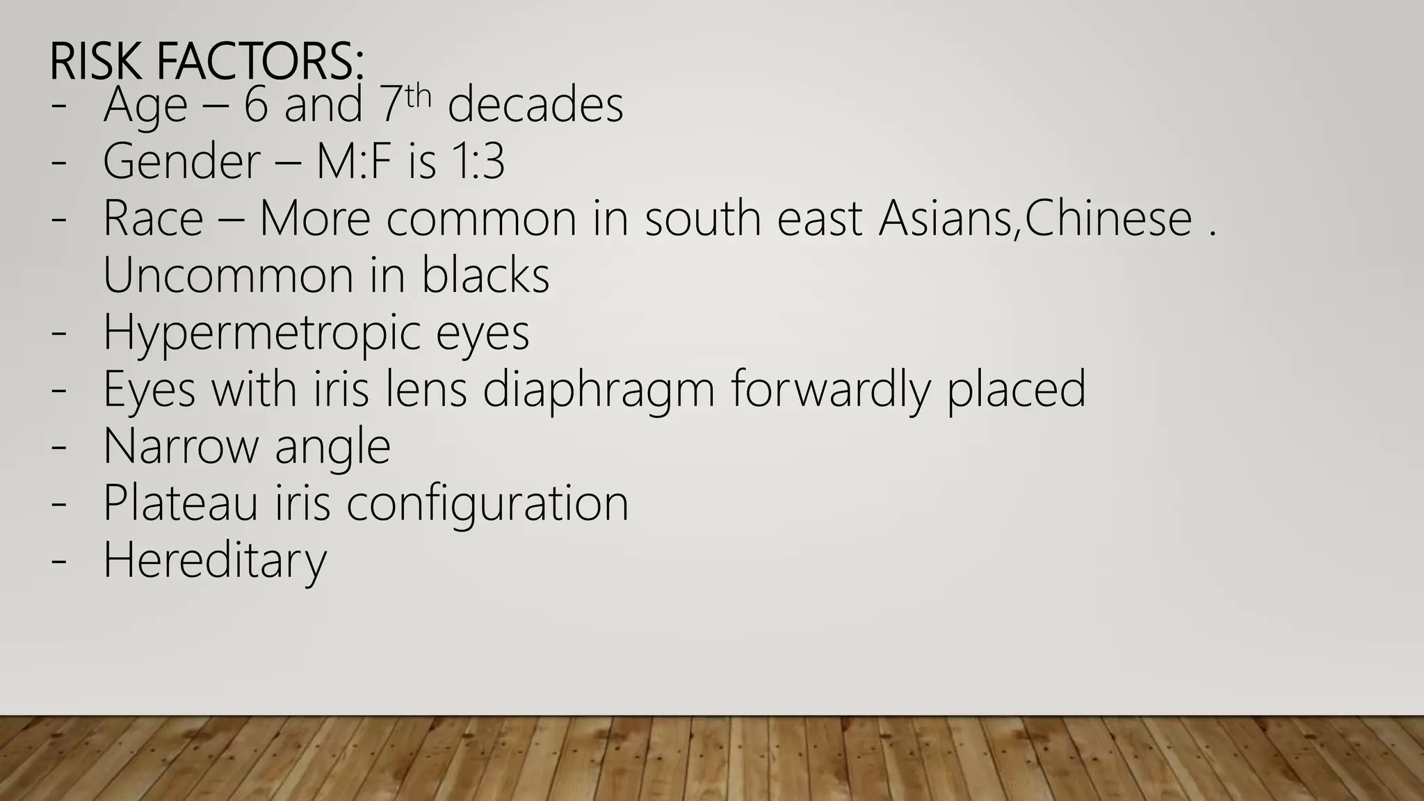
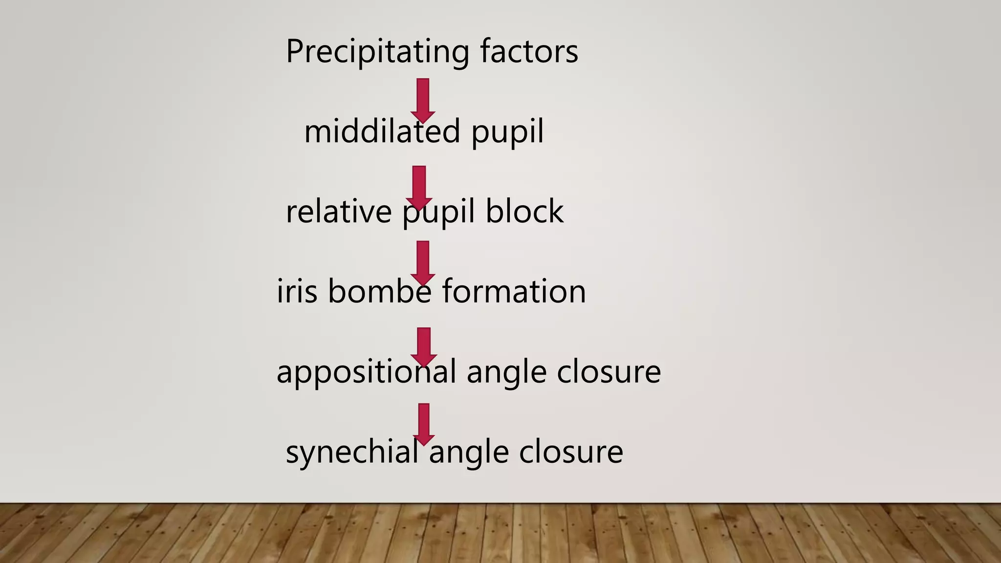
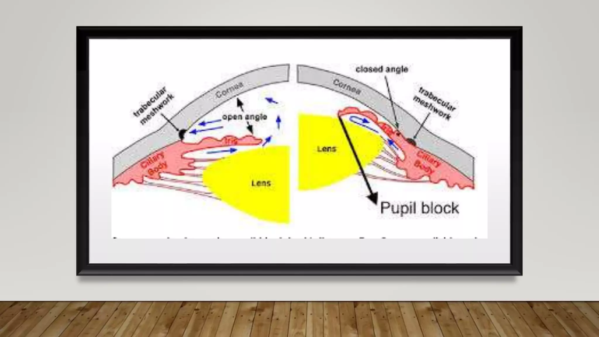
PRIMARY ANGLE
CLOSURE GLAUCOMA
DR. PRAKRITI YAGNAM. K
Primary angle closure disease – apposition of peripheral
iris against trabecular meshwork resulting in aqueous flow
obstruction
-If optic disc changes and visual field defects present called
primary angle closure glaucoma
- Major cause of world glaucoma blindness
- For every 10 PACG suspects one case occurs
- Chronic PACG more common than acute
- India POAG:PACG is 1:1
RISK FACTORS:
- Age – 6 and 7th decades
- Gender – M:F is 1:3
- Race – More common in south east Asians,Chinese .
Uncommon in blacks
- Hypermetropic eyes
- Eyes with iris lens diaphragm forwardly placed
- Narrow angle
- Plateau iris configuration
- Hereditary
Pathogenesis :
1. Pupillary block mechanism
2.Plateau iris syndrome
3.Phacomorphic
Pupillary block :
i. Precipitating factors :
Physiological mydriasis
Pharmacological mydriasis
Pharmacological miosis
Valsalva manoevure
Precipitating factors
middilated pupil
relative pupil block
iris bombe formation
appositional angle closure
synechial angle closure
2. Plateau iris syndrome :
- Due to anteriorly placed ciliary process pushing
peripheral iris anteriorly
- Due to pushing mechanism anterior chamber is closed
- Angle closure glaucoma without pupillary block
- Treated with miotics and laser peripheral iridotomy
3. Phacomorphic :
- Abnormal lens may cause pupillary block or pushes iris
anteriorly causing closure angle glaucoma
- Treatment - lens extraction
Classification :ISGEO
Primary angle closure suspect
Primary angle closure
Primary angle closure glaucoma
Primary angle closure suspect :
- Latent primary angle closure glaucoma
- Symptoms absent
- Fellow eye may already had an attack
- Signs – Eclipse sign – pen torch method
- Slit lamp – decreased axial anterior chamber depth
- Convex shaped iris lens diaphragm
- Peripheral proximity of iris and cornea
Von Herick grading :
Grade 4 wide angle – ¾ to 1 CT
Grade 3 Mild narrow – ¼ to ½ CT
Grade 2 Moderate narrow – ¼ CT
Grade 1 extremely narrow - <1/4 CT
Grade 0 closed angle
Diagnosis : IOP
Gonioscopy
Ultrasonic biomicroscopy
Ant segment OCT
Optic disc evaluation
Visual field analysis
Diagnostic criteria :
- IOP normal
- No PAS
- Iridotrabecular contact present
- Disc and fields normal
Provocative tests : Prone darkroom or mydriatic tests
Treatment :
- Prophylactic laser iridotomy
- Periodic follow up
Primary angle closure : Subacute , acute , chronic
- Iridocorneal contact present with PAS
- IOP raised
- No optic disc or field changes
Subacute :
- Intermittent attacks present lasting for few minutes to 1-2
hours
- IOP upto 50mm Hg
- Precipitating factors present
- Symptoms : Unilateral transient blurring of vision
- Colored halos around light not broken by finchams test
- Self termination by physiological miosis
- Recurrent attacks common
Treatment : Peripheral laser iridotomy
Acute primary angle closure :
- Sudden closure of angle
Symptoms : Pain with nausea and vomitings
- Rapid deterioration of vision with redness and photophobia
- Past history of subacute attacks present
Signs : Lid edematous
- Conjunctiva chemosed and congestion present
- Cornea is edematous
- AC shallow with cells and flare
- Angle occluded completely
- Iris discolored
- Pupil midilated fixed
- IOP upto 70mm Hg
- Optic disc edema and hyperemia
- Fellow eye may also have occludable angle
DD:
- Acute red eye
- Acute secondary glaucomas
Management :
To lower IOP - IV Mannitol(1gm/kg bodywt.)or Oral Glycerol
IV acetazolamide 500mg.stat f/by 250 mg PO
TID
Topical antiglaucoma drugs
Pilocarpine QID after IOP lowered
Analgesics and antiemetics
Compressive gonioscopy
Topical steroids
Definitive :
- Laser peripheral iridotomy
- Filtration surgery – Trabeculectomy
- Clear lens extraction
- Prophylactic treatment of normal fellow eye
- Follow up
Sequelae :
- Post surgical – Normalised with PI or trabeculectomy
- Spontaneous angle reopening
- Ciliary body shutdown
- Due to ischemia of ciliary epithelium
- Recovery causes rise in IOP with glaucomatic changes
- Treatment : - Topical steroids
- Laser PI
- Trabeculectomy
Vogts triad :
1.Glaucomoflecken
2.Iris atrophic patches
3.Slightly dilated non reacting pupil
3. Primary angle closure glaucoma :
- Gradual synechial closure of angle
- Untreated PAC may convert to PACG
- Divided into subacute,acute,chronic
- Acute and subacute similar to their counterparts in angle
closure disease along with disc and field changes
Chronic PACG – similar to POAG with closed angles
- Symptoms - eyeball white and painless
- IOP raised
- Gonioscopy reveals closed angles
- DISC and field changes present
Diagnostic criteria :
- Iridocorneal contact with PAS
- IOP elevated
- Disc and field changes
Treatment : Laser. PI along with medical therapy
Trabeculectomy
Prophylactic laser iridotomy
Absolute PACG :
- Untreated cases
- Painful blind eye – no PL
- Perilimbal reddish blue zone
- Caput medusae
- Cornea hazy goes into bullous keratopathy or filamentary
keratitis
- AC shallow
- Iris atrophic
- Pupil fixed and dilated
- IOP high and eye stony hard
Complications :
- Corneal ulceration
- Staphyloma formation
- Atrophic bulbi
Treatment :
- Retrobulbar alcohol injection
- Destruction of ciliary epithelium(secretory)-
cyclocryotherapy
- Enucleation
THANKYOU!!!

Primary Angle Closure Glaucoma

  • 1.
  • 2.
    Primary angle closuredisease – apposition of peripheral iris against trabecular meshwork resulting in aqueous flow obstruction -If optic disc changes and visual field defects present called primary angle closure glaucoma - Major cause of world glaucoma blindness - For every 10 PACG suspects one case occurs - Chronic PACG more common than acute - India POAG:PACG is 1:1
  • 3.
    RISK FACTORS: - Age– 6 and 7th decades - Gender – M:F is 1:3 - Race – More common in south east Asians,Chinese . Uncommon in blacks - Hypermetropic eyes - Eyes with iris lens diaphragm forwardly placed - Narrow angle - Plateau iris configuration - Hereditary
  • 4.
    Pathogenesis : 1. Pupillaryblock mechanism 2.Plateau iris syndrome 3.Phacomorphic Pupillary block : i. Precipitating factors : Physiological mydriasis Pharmacological mydriasis Pharmacological miosis Valsalva manoevure
  • 5.
    Precipitating factors middilated pupil relativepupil block iris bombe formation appositional angle closure synechial angle closure
  • 10.
    2. Plateau irissyndrome : - Due to anteriorly placed ciliary process pushing peripheral iris anteriorly - Due to pushing mechanism anterior chamber is closed - Angle closure glaucoma without pupillary block - Treated with miotics and laser peripheral iridotomy
  • 12.
    3. Phacomorphic : -Abnormal lens may cause pupillary block or pushes iris anteriorly causing closure angle glaucoma - Treatment - lens extraction
  • 13.
    Classification :ISGEO Primary angleclosure suspect Primary angle closure Primary angle closure glaucoma
  • 14.
    Primary angle closuresuspect : - Latent primary angle closure glaucoma - Symptoms absent - Fellow eye may already had an attack - Signs – Eclipse sign – pen torch method - Slit lamp – decreased axial anterior chamber depth - Convex shaped iris lens diaphragm - Peripheral proximity of iris and cornea
  • 16.
    Von Herick grading: Grade 4 wide angle – ¾ to 1 CT Grade 3 Mild narrow – ¼ to ½ CT Grade 2 Moderate narrow – ¼ CT Grade 1 extremely narrow - <1/4 CT Grade 0 closed angle Diagnosis : IOP Gonioscopy Ultrasonic biomicroscopy Ant segment OCT
  • 20.
    Optic disc evaluation Visualfield analysis Diagnostic criteria : - IOP normal - No PAS - Iridotrabecular contact present - Disc and fields normal Provocative tests : Prone darkroom or mydriatic tests
  • 21.
    Treatment : - Prophylacticlaser iridotomy - Periodic follow up Primary angle closure : Subacute , acute , chronic - Iridocorneal contact present with PAS - IOP raised - No optic disc or field changes
  • 22.
    Subacute : - Intermittentattacks present lasting for few minutes to 1-2 hours - IOP upto 50mm Hg - Precipitating factors present - Symptoms : Unilateral transient blurring of vision - Colored halos around light not broken by finchams test - Self termination by physiological miosis - Recurrent attacks common Treatment : Peripheral laser iridotomy
  • 23.
    Acute primary angleclosure : - Sudden closure of angle Symptoms : Pain with nausea and vomitings - Rapid deterioration of vision with redness and photophobia - Past history of subacute attacks present Signs : Lid edematous - Conjunctiva chemosed and congestion present - Cornea is edematous - AC shallow with cells and flare - Angle occluded completely
  • 24.
    - Iris discolored -Pupil midilated fixed - IOP upto 70mm Hg - Optic disc edema and hyperemia - Fellow eye may also have occludable angle DD: - Acute red eye - Acute secondary glaucomas
  • 26.
    Management : To lowerIOP - IV Mannitol(1gm/kg bodywt.)or Oral Glycerol IV acetazolamide 500mg.stat f/by 250 mg PO TID Topical antiglaucoma drugs Pilocarpine QID after IOP lowered Analgesics and antiemetics Compressive gonioscopy Topical steroids
  • 27.
    Definitive : - Laserperipheral iridotomy - Filtration surgery – Trabeculectomy - Clear lens extraction - Prophylactic treatment of normal fellow eye - Follow up Sequelae : - Post surgical – Normalised with PI or trabeculectomy - Spontaneous angle reopening - Ciliary body shutdown
  • 30.
    - Due toischemia of ciliary epithelium - Recovery causes rise in IOP with glaucomatic changes - Treatment : - Topical steroids - Laser PI - Trabeculectomy Vogts triad : 1.Glaucomoflecken 2.Iris atrophic patches 3.Slightly dilated non reacting pupil
  • 32.
    3. Primary angleclosure glaucoma : - Gradual synechial closure of angle - Untreated PAC may convert to PACG - Divided into subacute,acute,chronic - Acute and subacute similar to their counterparts in angle closure disease along with disc and field changes Chronic PACG – similar to POAG with closed angles - Symptoms - eyeball white and painless - IOP raised - Gonioscopy reveals closed angles - DISC and field changes present
  • 33.
    Diagnostic criteria : -Iridocorneal contact with PAS - IOP elevated - Disc and field changes Treatment : Laser. PI along with medical therapy Trabeculectomy Prophylactic laser iridotomy
  • 34.
    Absolute PACG : -Untreated cases - Painful blind eye – no PL - Perilimbal reddish blue zone - Caput medusae - Cornea hazy goes into bullous keratopathy or filamentary keratitis - AC shallow - Iris atrophic - Pupil fixed and dilated - IOP high and eye stony hard
  • 36.
    Complications : - Cornealulceration - Staphyloma formation - Atrophic bulbi Treatment : - Retrobulbar alcohol injection - Destruction of ciliary epithelium(secretory)- cyclocryotherapy - Enucleation
  • 38.