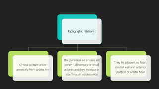
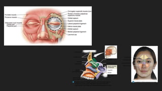
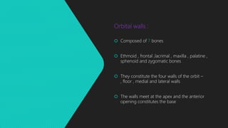
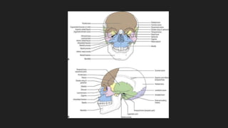
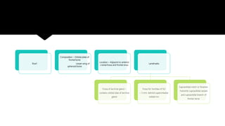
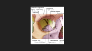
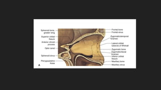
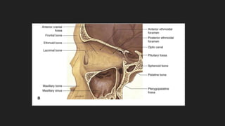
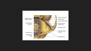
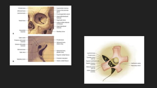
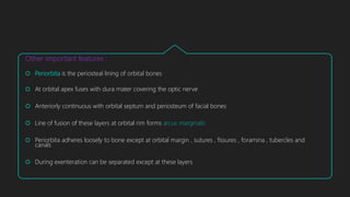
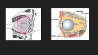
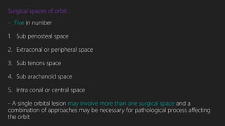
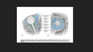
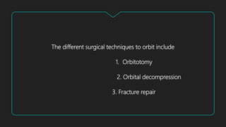
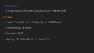
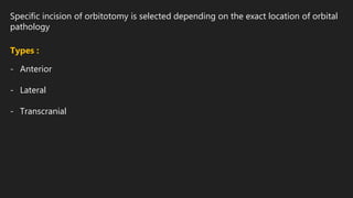
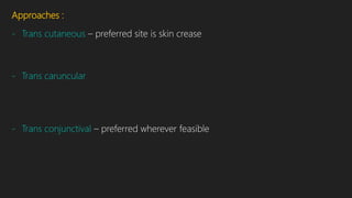
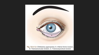
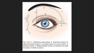
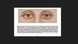
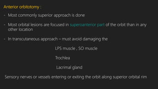
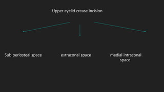
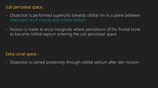
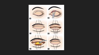
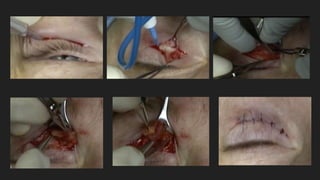
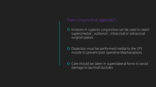
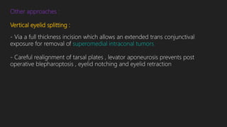
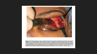
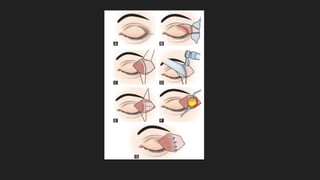
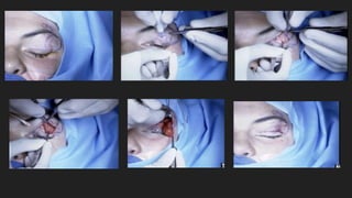
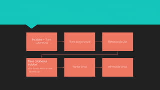
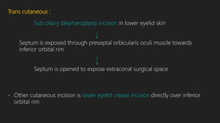
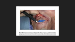
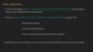
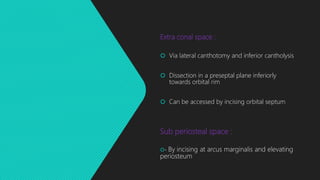
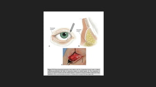
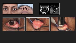
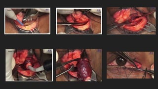
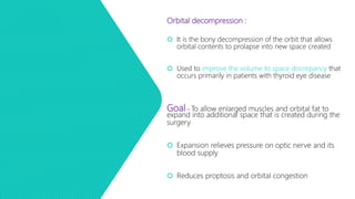
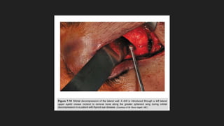
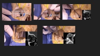
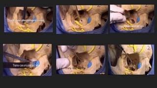
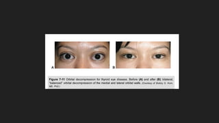
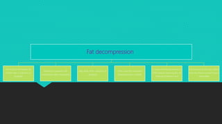
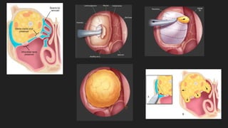
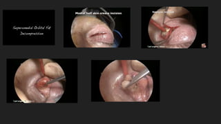
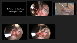
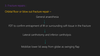
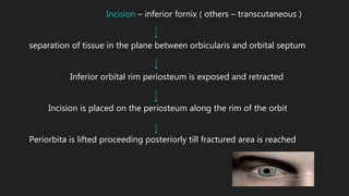
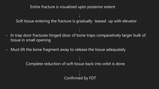
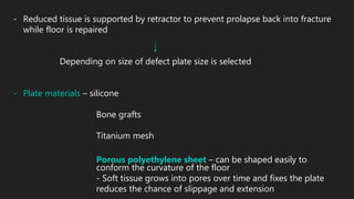
ORBITAL SURGERIES
DR.PRAKRITI YAGNAM .K
MODERATOR : DR. CH. MOHANRAO
Orbit :
 Bony cavities that contain globes , EOM ,
, fat and blood vessels
 Each orbit is pear shaped ( quadrilateral
pyramid ) tapered posteriorly to the apex and
the optic canal
 Medial orbital walls are parallel and separated
by 25 mm. in an average adult
 Widest dimension of the orbit is approximately
1 cm. behind the anterior orbital rim
Average dimensions of adult orbit :
 Volume – 30 cc.
 Entrance height – 35 mm.
 Entrance width – 40 to 45 mm.
 Medial wall length – 40 to 45 mm.
 Distance from posterior globe to optic foramen – 18 mm.
 Length of orbital segment of optic nerve – 25 to 30 mm.
Topographic relations :
Orbital septum arises
anteriorly from orbital rim
The paranasal air sinuses are
either rudimentary or small
at birth and they increase in
size through adolescence
They lie adjacent to floor ,
medial wall and anterior
portion of orbital floor
Orbital walls :
 Composed of 7 bones
 Ethmoid , frontal ,lacrimal , maxilla , palatine ,
sphenoid and zygomatic bones
 They constitute the four walls of the orbit –
, floor , medial and lateral walls
 The walls meet at the apex and the anterior
opening constitutes the base
Roof :
Composition – Orbital plate of
frontal bone
Lesser wing of
sphenoid bone
Location – Adjacent to anterior
cranial fossa and frontal sinus
Landmarks
Fossa of lacrimal gland –
contains orbital lobe of lacrimal
gland
Fossa for trochlea of SO
– 5 mm. behind superomedial
orbital rim
Supraorbital notch or foramen
Transmits supraorbital vessels
and supraorbital branch of
frontal nerve
Lateral wall :
•Thickest and strongest
Composed of – Zygomatic bone
Greater wing of sphenoid
•Separated from the roof by superior orbital fissure
Location – Adjacent to middle cranial fossa and temporal fossa
Extends anteriorly to the equator of the globe helping to protect
posterior half of the eye while still allowing wide peripheral vision
Landmarks
- Lateral orbital tubercle of Whitnall - Attachment to lateral canthal tendon
Lateral horn of levator aponeurosis
Check ligament of lateral rectus
Lockwood ligament
- Whitnall ligament inserts onto lateral orbital wall mm. above lateral orbital
tubercle in attachment to lacrimal gland fascia
- Frontozygomatic suture is located 1 cm. above the tubercle
Medial wall :
Composition – Orbital plate of ethmoid
Lacrimal bone
Frontal process of maxillary bone
Lesser wing of sphenoid bone
- Only the lacrimal bone is wholly within the orbital confines
Location – Adjacent to ethmoid and sphenoid sinuses and nasal cavity
Landmarks
- Frontoethmoid sutures – Approximate level of cribriform plate
Roof of ethmoids
Floor of anterior cranial fossa
Exit of anterior and posterior ethmoidal vessels
Lamina papyracea - Thinnest walls of the orbit
 Between orbits and ethmoidal sinuses along medial wall and maxillary bone ( posterior
medial portion )
 Frequently fractured as a result of indirect or blow out fractures
 Acute bacterial infections of ethmoidal sinus may extend through lamina papyracea or
neurovasular perforation to form subperiosteal abscess and extend into. Orbital soft tissues
Floor :
Composition – Maxillary bone
Palatine bone
Orbital plate of zygomatic bone
- Forms the roof of maxillary sinus
- Does not extend to orbital apex but ends at pterygopalatine fossa – shortest
of all walls
Landmark
- Infraorbital groove and canal which transmits infraorbital artery and maxillary
division of trigeminal nerve
Apertures :
Ethmoidal foramina
- Anterior and posterior ethmoidal foramina present in the medial wall along
frontoethmoidal suture
- Anterior and posterior ethmoidal arteries pass through corresponding
foramina
Importance
- Surgical landmark for superior extent of medial wall surgery
- Damage to medial wall above this level may disrupt a plane superior to
cribriform plate
 Potential route of entry into orbit for pathogens
and neoplasms from the sinus
Others :
 Superior orbital fissure
 Inferior orbital fissure
 Zygomatico facial and Zygomatico temporal
canals
 Nasolacrimal canal
 Optic canal
Other important features :
 Periorbita is the periosteal lining of orbital bones
 At orbital apex fuses with dura mater covering the optic nerve
 Anteriorly continuous with orbital septum and periosteum of facial bones
 Line of fusion of these layers at orbital rim forms arcus marginalis
 Periorbita adheres loosely to bone except at orbital margin , sutures , fissures , foramina , tubercles and
canals
 During exenteration can be separated except at these layers
Surgical spaces of orbit :
- Five in number
1. Sub periosteal space
2. Extraconal or peripheral space
3. Sub tenons space
4. Sub arachanoid space
5. Intra conal or central space
- A single orbital lesion may involve more than one surgical space and a
combination of approaches may be necessary for pathological process affecting
the orbit
The different surgical techniques to orbit include
1. Orbitotomy
2. Orbital decompression
3. Fracture repair
1. Orbitotomy :
- It is procedure of making a surgical incision into the orbit
Indications :
- Complete removal ( excision biopsy ) of orbital tumor
- Incision biopsy of tumor
- Removal of IOFB
- Drainage of orbital abscess or hematoma
Specific incision of orbitotomy is selected depending on the exact location of orbital
pathology
Types :
- Anterior
- Lateral
- Transcranial
Approaches :
- Trans cutaneous – preferred site is skin crease
- Trans caruncular
- Trans conjunctival – preferred wherever feasible
Trans cutaneous incisions :
If lesion in anterior two thirds of the orbit , placed superiorly
- Subbrow Benedicts incision
- Upper eyelid crease incision
Supero medial orbit – Lid split incision
Medial orbit – Lynch incision
Sea gull incision
Inferior orbit – Lower lid subciliary incision
Superotemporal orbit – Stallard – wright incision
Extension of upperlid crease incision
Berkes incision
Anterior orbitotomy :
- Most commonly superior approach is done
- Most orbital lesions are focused in superoanterior part of the orbit than in any
other location
- In transcutaneous approach – must avoid damaging the
LPS muscle , SO muscle
Trochlea
Lacrimal gland
Sensory nerves or vessels entering or exiting the orbit along superior orbital rim
Upper eyelid crease incision
Sub periosteal space extraconal space medial intraconal
space
Sub periosteal space :
- Dissection is performed superiorly towards orbital rim in a plane between
orbicularis oculi muscle and orbital septum
- Incision is made at arcus marginalis where periosteum of the frontal bone
to become orbital septum entering the sub periosteal space
Extra conal space :
- Dissection is carried posteriorly through orbital septum after skin incision
Medial intraconal space :
 Dissection is kept medial to medial edge of levator muscle after opening the orbital septum
 Proceed between medial and central pad of fat pads through inter muscular septum extending from
superior rectus to medial rectus
 It is used for - biopsy of optic nerve
for optic nerve sheath fenestration in cases of IIH
for accessing intraconal lesions medial to optic nerve
Technique :
- Via upper lid crease incision
- It provides better cosmosis than incision over superior orbital rim
- Sub periosteal and extraconal spaces are approached
General anaesthesia
Skin incision in upper lid crease
Dissection is carried in plane between orbicularis anteriorly and
orbital septum posteriorly
Periorbita of the roof is lifted off with Freer elevator and is pushed inferiorly
If inadequate exposure
Small amount of superior orbital rim is nibbled away
T shaped incision is placed on periorbita with sharp Westcott scissors
Corners retracted to expose superior orbit
Lesion is visualized and incision biopsy is taken
If circumscribed lesion – Blunt dissection done all around and lesion removed
Traction may be placed on tumor with cryoprobe also
Orbital rim periosteum is repaired
Lid crease is reformed
Skin incision is sutured
Trans conjunctival approach :
 Incisions in superior conjunctiva can be used to reach
superomedial , subtenon , intraconal or extraconal
surgical spaces
 Dissection must be performed medial to the LPS
muscle to prevent post operative blepharoptosis
 Care should be taken in superolateral fornix to avoid
damage to lacrimal ductules
Other approaches :
Vertical eyelid splitting :
- Via a full thickness incision which allows an extended trans conjunctival
exposure for removal of superomedial intraconal tumors
- Careful realignment of tarsal plates , levator aponeurosis prevents post
operative blepharoptosis , eyelid notching and eyelid retraction
Lateral orbitotomy :
- To access
Lateral intraconal Fossa of lacrimal gland
space behind equator of the
globe
- In children as orbit is relatively shallower extensive exposure may be achieved
without the need for bone removal
Technique :
- Via lateral extension of upper lid crease incision
General anaesthesia
Incision – Upper lid crease in lateral one third and extended laterally
to lateral orbital rim
Soft tissue is dissected to expose lateral orbital rim with
reflection of temporalis muscle and periosteum of orbit
Periosteum is incised and retracted
Orbital rim bone , zygomatic process of frontal bone and frontal process
of zygomatic bone are exposed
Periorbita is lifted and pushed medially pushing orbital contents
away from the rim
Metal guard is placed between periorbita or orbital contents and
bony rim before cutting the bone
Bone in incised just below the frontozygomatic suture and inferiorly
at lower end of lateral orbital rim
Cut segment is held with bone holding rongeur and fractured away
temporally to give exposure to orbit – MARGINOTOMY
T shaped incision is placed on periorbita to expose orbital contents
LR is needed to be identified and retracted for an intraconal mass
Incision or excision biopsy is done
Fragment of orbital rim may be replaced by wiring
Periosteum is closed with 6 – 0 vicryl sutures
Skin is repaired in layers
- Complete hemostasis is achieved before closure
- To help prevent post operative intra orbital hemorrhage an external drain may
be placed in the deep orbit
Medial orbitotomy :
Indications – repair of
medial wall trauma
orbital decompression
access to lacrimal sac or
sino – nasal tumors with
orbital involvement
Avoid damage to medial
canthal tendon , lacrimal
canaliculi and sac
SO tendon and muscle ,
trochlea , IO muscle
sensory nerves and
vessels along medial
aspect of
superior orbital rim
Incisions – Trans
cutaneous Trans conjunctival Retrocanalicular
Trans cutaneous
incision :
• For tumors within or near
lacrimal sac
frontal sinus ethmoidal sinus
Technique :
- Approached through frontoethmoidal skin incision placed vertically just medial
to insertion of medial canthal tendon
- Enter the sub periosteal space by reflecting medial canthal tendon in
with periosteum preserving lacrimal drainage apparatus
Trans conjunctival :
 Bulbar conjunctiva incision
 To approach – Extra conal space
Subtenons space
Intra conal space – If MR is detached
In the region of anterior optic nerve
for exploration or biopsy
Retro caruncular incision :
Indications – Medial wall fractures
Medial orbital bone decompression
Medial sub periosteal abscess
Technique :
- Incision posterior to caruncle – excellent exposure of medial orbit
Blunt dissection carried medially followed by incision and elevation of
periosteum sub periosteal space
 Retro caruncular + Inferior trans conjunctival incision gives panoramic exposure of
inferior and medial orbit
 IO muscle may be divided at its origin along infero medial orbital rim and reattached at
the end of the surgery
 Has better cosmesis than transcutaneous incision
 Care must be taken to remain posterior to lacrimal drainage apparatus
 Combined lateral and medial orbitotomies can be done for posterior optic nerve
approach or muscle cone assessment
 Lateral orbitotomy with removal of lateral orbital wall allows globe to be displaced
temporally maximizing medial exposure to deeper orbit
Inferior orbitotomy :
Indications – Masses that are visible or palpable in the inferior conjunctival
fornix of lower eyelid
Deep inferior extra conal or intra conal orbital masses
To approach orbital floor for fracture repair or decompression
Tran cutaneous
Approaches
Trans conjunctival
Trans cutaneous :
Sub ciliary blepharoplasty incision in lower eyelid skin
Septum is exposed through preseptal orbicularis oculi muscle towards
inferior orbital rim
Septum is opened to expose extraconal surgical space
- Other cutaneous incision is lower eyelid crease incision directly over inferior
orbital rim
Trans conjunctival :
- Incision through inferior conjunctiva and lower eyelid retractors where extra
space and orbital floor are exposed
- Incision below inferior tarsal border in conjunctival fornix is given for
extraconal space
sub periosteal space
intra conal space and sub tenons spaces
- Avoid injury to IO muscle , IR muscle and infra orbital neuro vascular bundle
Extra conal space :
 Via lateral canthotomy and inferior cantholysis
 Dissection in a preseptal plane inferiorly
towards orbital rim
 Can be accessed by incising orbital septum
Sub periosteal space :
- By incising at arcus marginalis and elevating
periosteum
Intra conal space :
By dissection between
rectus and lateral rectus
Sub tenon space :
Incision of bulbar conjunctiva
and tenon capsule
Also used to gain access to
intraconal space by
or reflecting inferior rectus
muscle from the globe
Orbital decompression :
 It is the bony decompression of the orbit that allows
orbital contents to prolapse into new space created
 Used to improve the volume to space discrepancy that
occurs primarily in patients with thyroid eye disease
Goal – To allow enlarged muscles and orbital fat to
expand into additional space that is created during the
surgery
 Expansion relieves pressure on optic nerve and its
blood supply
 Reduces proptosis and orbital congestion
Methods :
Historical – Removal of medial
orbital wall and much of
orbital floor extending to apex
including maxilla ethmoidal
strut
It allows orbital tissues to
expand into ethmoid and
maxillary sinuses
Approach is by maxillary
vestibular or transcutaneous
incisions
Post operative complaints :
 Globe ptosis and upper eyelid retraction could
be exacerbated in patients with large restricted
inferior rectus muscle
 Could also disrupt globe excursion due to
prolapse of muscles into sinus space and
displacement of orbital content
Current approach :
- Combination of one or more discrete incisions that allow access to lateral , inferior
and / or medial walls
- Lateral and inferior orbit – Upper eyelid crease incision
Extended lateral canthotomy incision
Inferior trans conjunctival incision combined
with a lateral canthotomy or inferior cantholysis
- Decompression of lateral wall includes removal of bone along the sphenoid wing
by using rongever and / or a drill
- Medial wall – Retrocaruncular + trans conjunctival incision
Trans nasal endoscopic approach via ethmoid sinus
Balanced decompression :
- Removing bones from opposite walls
- Will reduce the risk of worsening or new onset diplopia
For further decompression – removal of lateral orbital rim and / or reposition it anteriorly at time of closure is done
Anterior displacement of lateral canthus aid in reduction of eyelid retraction
Each additional wall provides 2 mm. reduction in proptosis
The generalized technique –
After reaching orbital rim periosteum is incised and lifted off and orbital wall is removed
Periosteum is incised at each area
Fat decompression :
Removal of retrobulbar fat
further aids in reduction of
proptosis
Beneficial in patients with
compressive optic neuropathy
May allow 3mm. reduction of
proptosis
Intra conal fat is removed
preserving extra conal fat
It causes minimal disturbance in
EOM balance reducing the risk
inducing strabismus and
Decompression through orbital
roof into anterior cranial fossa is
inadvisable
3. Fracture repairs :
Orbital floor or blow out fracture repair –
General anaesthesia
FDT to confirm entrapment of IR or surrounding soft tissue in the fracture
Lateral canthotomy and inferior cantholysis
Mobilize lower lid away from globe as swinging flap
Incision – inferior fornix ( others – transcutaneous )
separation of tissue in the plane between orbicularis and orbital septum
Inferior orbital rim periosteum is exposed and retracted
Incision is placed on the periosteum along the rim of the orbit
Periorbita is lifted proceeding posteriorly till fractured area is reached
Entire fracture is visualized upto posterior extent
Soft tissue entering the fracture is gradually teased up with elevator
- In trap door fractures hinged door of bone traps comparatively larger bulk of
tissue in small opening
- Must lift the bone fragment away to release the tissue adequately
Complete reduction of soft tissue back into orbit is done
Confirmed by FDT
- Reduced tissue is supported by retractor to prevent prolapse back into fracture
while floor is repaired
Depending on size of defect plate size is selected
- Plate materials – silicone
Bone grafts
Titanium mesh
Porous polyethylene sheet – can be shaped easily to
conform the curvature of the floor
- Soft tissue grows into pores over time and fixes the plate
reduces the chance of slippage and extension
- Plate must be large enough to extend into intact bone beyond the defect
- If there is large defect in periorbita – barrier plate is used
Plate is placed and retractor is removed and tissue is replaced
- If anterior rim is projecting or FDT is positive
Plate may be too large or placed too superficially
Trimmed or position is corrected
Finally fixed with metal screws to orbital rim
- If silicone plate is used tissue adhesives can be used
Periosteum is repaired with interrupted 6 – 0 vicryl sutures
Lower lid retractors and inferior fornix is also repaired with interrupted
6 - 0 vicryl suturing
Lateral canthus is repaired
- Some surgeons use lower lid skin crease which is a more direct route to floor
but leaves visible scar
Porous polyethylene Silicone
Special surgical techniques in the orbit :
1. Fine Needle Aspiration and Biopsy – Lymphoid lesions
Secondary tumors invading orbit from sinuses
Suspected metastatic tumors
Blind eyes with optic N. tumors
- Not effective for obtaining tissue from fibrous inflammatory lesions because of
difficulty in successfully aspirating cells
- Lymphoproliferative disorders – FNAB + flow cytometry + monoclonal antibody
or PCR analysis
2. Endoscopic trans nasal surgery :
 Utilizing the anatomic relationship of ethmoid and maxillary
sinus to medial and inferior respectively
 Permits biopsy or resection of intraconal pathology
 May also be combined with open orbitotomy to allow
improved access to apical processes
 Used in TED or non traumatic compressive optic neuropathy
for decompression of orbit or optic canal
 May also be used for drainage of medial sub periosteal orbital
abscess in patients with sinusitis or for debridement of
tissue in patients with fungal sinusitis
3. Tumors or traumatic injuries of skull base involving superior and posterior orbit
- Access via frontal cranitotomy or fronto temporal orbito zygomatic approach
- Requires a multidisciplinary team
- Neuro surgeon gives access to deep superior and lateral orbit by removing
frontal bone and orbital roof
- Indications – Decompression of optic canal
Resection of meningioma , fibrous dysplasia , schwannomas
gliomas , cavernous venous malformations and solitary fibrous
tumors
Post operative care for orbital surgeries :
To reduce postop edema – Elevation of head
Ice compressors over eyelids
Administration of systemic corticosteroids
optimal placement of drain
- Ice packs minimize swelling and allow for observation of operative site and
monitoring of visual acuity
- Prophylactic systemic antibiotics may also be given
Preoperative and intraoperative requisites :
 Complete preoperative evaluation
 Orbital imaging
 Choosing appropriate surgical approach
 Obtaining adequate exposure
 Manipulating the tissues carefully
 Employing proper illumination and instrumentation
 Maintaining excellent hemostasis and team approach
Complications :
Decreased or complete loss of vision
- Most dreaded complication
- May be due to - Excessive traction on the globe or optic nerve
- Contusion of optic nerve
- Post op hemorrhage
Raised orbital pressure Ischemic injury to ON
- Post op infection
- Patient with severe orbital pain post op must be evaluated immediately for
possible orbital hemorrhage
- Pain + decreased vision + proptosis + ecchymosis + increased IOP + afferent
pupillary defect
Surgeon should open the wound to minimize the effect of orbital
compression syndrome
Any hematoma is evacuated or any active bleeding is controlled
Other complications :
 Blepharoptosis
 CSF leak
 Ciliary ganglion dysfunction with loss of accommodation
 Cranial neuropathy resulting in EOM weakness or palsy
 Hypoaesthesia in the distribution of trigeminal nerve ( V1 and
V2 )- seen in floor decompression due to damage to
infraorbital nerve
 Infraplacement of globe after decompression – preservation
medial strut of bone between medial wall and floor may help
preventing it
 Keratitis sicca
 Motility disturbance – diplopia
 Neuroparalytic keratopathy
 Orbital cellulitis
 Pupillary dysfunction
 Retinal detachment
 Vitreous hemorrhage
Instruments used in orbital surgery :
- Quality of surgery depends on availability of appropriate instruments
- Operating light – External surgical lamp – ceiling or stand mounted
- Magnification – Microscope or surgical loupe is used
2.5 X or 3.0 X
- Suction apparatus
- Cautery – standard bipolar cautery for hemostasis
- Surgical unit – Monopolar radiofrequency or electrosurgical hand piece
Others :
- Cryoprobe
- Microdrill
- Oscillating saw
Others :
THANK YOU !!!

Orbital surgeries

  • 1.
    ORBITAL SURGERIES DR.PRAKRITI YAGNAM.K MODERATOR : DR. CH. MOHANRAO
  • 2.
    Orbit :  Bonycavities that contain globes , EOM , , fat and blood vessels  Each orbit is pear shaped ( quadrilateral pyramid ) tapered posteriorly to the apex and the optic canal  Medial orbital walls are parallel and separated by 25 mm. in an average adult  Widest dimension of the orbit is approximately 1 cm. behind the anterior orbital rim
  • 4.
    Average dimensions ofadult orbit :  Volume – 30 cc.  Entrance height – 35 mm.  Entrance width – 40 to 45 mm.  Medial wall length – 40 to 45 mm.  Distance from posterior globe to optic foramen – 18 mm.  Length of orbital segment of optic nerve – 25 to 30 mm.
  • 6.
    Topographic relations : Orbitalseptum arises anteriorly from orbital rim The paranasal air sinuses are either rudimentary or small at birth and they increase in size through adolescence They lie adjacent to floor , medial wall and anterior portion of orbital floor
  • 8.
    Orbital walls : Composed of 7 bones  Ethmoid , frontal ,lacrimal , maxilla , palatine , sphenoid and zygomatic bones  They constitute the four walls of the orbit – , floor , medial and lateral walls  The walls meet at the apex and the anterior opening constitutes the base
  • 10.
    Roof : Composition –Orbital plate of frontal bone Lesser wing of sphenoid bone Location – Adjacent to anterior cranial fossa and frontal sinus Landmarks Fossa of lacrimal gland – contains orbital lobe of lacrimal gland Fossa for trochlea of SO – 5 mm. behind superomedial orbital rim Supraorbital notch or foramen Transmits supraorbital vessels and supraorbital branch of frontal nerve
  • 12.
    Lateral wall : •Thickestand strongest Composed of – Zygomatic bone Greater wing of sphenoid •Separated from the roof by superior orbital fissure Location – Adjacent to middle cranial fossa and temporal fossa Extends anteriorly to the equator of the globe helping to protect posterior half of the eye while still allowing wide peripheral vision
  • 13.
    Landmarks - Lateral orbitaltubercle of Whitnall - Attachment to lateral canthal tendon Lateral horn of levator aponeurosis Check ligament of lateral rectus Lockwood ligament - Whitnall ligament inserts onto lateral orbital wall mm. above lateral orbital tubercle in attachment to lacrimal gland fascia - Frontozygomatic suture is located 1 cm. above the tubercle
  • 15.
    Medial wall : Composition– Orbital plate of ethmoid Lacrimal bone Frontal process of maxillary bone Lesser wing of sphenoid bone - Only the lacrimal bone is wholly within the orbital confines Location – Adjacent to ethmoid and sphenoid sinuses and nasal cavity
  • 16.
    Landmarks - Frontoethmoid sutures– Approximate level of cribriform plate Roof of ethmoids Floor of anterior cranial fossa Exit of anterior and posterior ethmoidal vessels
  • 17.
    Lamina papyracea -Thinnest walls of the orbit  Between orbits and ethmoidal sinuses along medial wall and maxillary bone ( posterior medial portion )  Frequently fractured as a result of indirect or blow out fractures  Acute bacterial infections of ethmoidal sinus may extend through lamina papyracea or neurovasular perforation to form subperiosteal abscess and extend into. Orbital soft tissues
  • 19.
    Floor : Composition –Maxillary bone Palatine bone Orbital plate of zygomatic bone - Forms the roof of maxillary sinus - Does not extend to orbital apex but ends at pterygopalatine fossa – shortest of all walls Landmark - Infraorbital groove and canal which transmits infraorbital artery and maxillary division of trigeminal nerve
  • 21.
    Apertures : Ethmoidal foramina -Anterior and posterior ethmoidal foramina present in the medial wall along frontoethmoidal suture - Anterior and posterior ethmoidal arteries pass through corresponding foramina Importance - Surgical landmark for superior extent of medial wall surgery - Damage to medial wall above this level may disrupt a plane superior to cribriform plate
  • 22.
     Potential routeof entry into orbit for pathogens and neoplasms from the sinus Others :  Superior orbital fissure  Inferior orbital fissure  Zygomatico facial and Zygomatico temporal canals  Nasolacrimal canal  Optic canal
  • 24.
    Other important features:  Periorbita is the periosteal lining of orbital bones  At orbital apex fuses with dura mater covering the optic nerve  Anteriorly continuous with orbital septum and periosteum of facial bones  Line of fusion of these layers at orbital rim forms arcus marginalis  Periorbita adheres loosely to bone except at orbital margin , sutures , fissures , foramina , tubercles and canals  During exenteration can be separated except at these layers
  • 26.
    Surgical spaces oforbit : - Five in number 1. Sub periosteal space 2. Extraconal or peripheral space 3. Sub tenons space 4. Sub arachanoid space 5. Intra conal or central space - A single orbital lesion may involve more than one surgical space and a combination of approaches may be necessary for pathological process affecting the orbit
  • 28.
    The different surgicaltechniques to orbit include 1. Orbitotomy 2. Orbital decompression 3. Fracture repair
  • 29.
    1. Orbitotomy : -It is procedure of making a surgical incision into the orbit Indications : - Complete removal ( excision biopsy ) of orbital tumor - Incision biopsy of tumor - Removal of IOFB - Drainage of orbital abscess or hematoma
  • 30.
    Specific incision oforbitotomy is selected depending on the exact location of orbital pathology Types : - Anterior - Lateral - Transcranial
  • 31.
    Approaches : - Transcutaneous – preferred site is skin crease - Trans caruncular - Trans conjunctival – preferred wherever feasible
  • 33.
    Trans cutaneous incisions: If lesion in anterior two thirds of the orbit , placed superiorly - Subbrow Benedicts incision - Upper eyelid crease incision Supero medial orbit – Lid split incision Medial orbit – Lynch incision Sea gull incision
  • 34.
    Inferior orbit –Lower lid subciliary incision Superotemporal orbit – Stallard – wright incision Extension of upperlid crease incision Berkes incision
  • 37.
    Anterior orbitotomy : -Most commonly superior approach is done - Most orbital lesions are focused in superoanterior part of the orbit than in any other location - In transcutaneous approach – must avoid damaging the LPS muscle , SO muscle Trochlea Lacrimal gland Sensory nerves or vessels entering or exiting the orbit along superior orbital rim
  • 38.
    Upper eyelid creaseincision Sub periosteal space extraconal space medial intraconal space
  • 39.
    Sub periosteal space: - Dissection is performed superiorly towards orbital rim in a plane between orbicularis oculi muscle and orbital septum - Incision is made at arcus marginalis where periosteum of the frontal bone to become orbital septum entering the sub periosteal space Extra conal space : - Dissection is carried posteriorly through orbital septum after skin incision
  • 40.
    Medial intraconal space:  Dissection is kept medial to medial edge of levator muscle after opening the orbital septum  Proceed between medial and central pad of fat pads through inter muscular septum extending from superior rectus to medial rectus  It is used for - biopsy of optic nerve for optic nerve sheath fenestration in cases of IIH for accessing intraconal lesions medial to optic nerve
  • 41.
    Technique : - Viaupper lid crease incision - It provides better cosmosis than incision over superior orbital rim - Sub periosteal and extraconal spaces are approached General anaesthesia Skin incision in upper lid crease Dissection is carried in plane between orbicularis anteriorly and orbital septum posteriorly
  • 42.
    Periorbita of theroof is lifted off with Freer elevator and is pushed inferiorly If inadequate exposure Small amount of superior orbital rim is nibbled away T shaped incision is placed on periorbita with sharp Westcott scissors Corners retracted to expose superior orbit Lesion is visualized and incision biopsy is taken
  • 43.
    If circumscribed lesion– Blunt dissection done all around and lesion removed Traction may be placed on tumor with cryoprobe also Orbital rim periosteum is repaired Lid crease is reformed Skin incision is sutured
  • 46.
    Trans conjunctival approach:  Incisions in superior conjunctiva can be used to reach superomedial , subtenon , intraconal or extraconal surgical spaces  Dissection must be performed medial to the LPS muscle to prevent post operative blepharoptosis  Care should be taken in superolateral fornix to avoid damage to lacrimal ductules
  • 47.
    Other approaches : Verticaleyelid splitting : - Via a full thickness incision which allows an extended trans conjunctival exposure for removal of superomedial intraconal tumors - Careful realignment of tarsal plates , levator aponeurosis prevents post operative blepharoptosis , eyelid notching and eyelid retraction
  • 49.
    Lateral orbitotomy : -To access Lateral intraconal Fossa of lacrimal gland space behind equator of the globe - In children as orbit is relatively shallower extensive exposure may be achieved without the need for bone removal
  • 50.
    Technique : - Vialateral extension of upper lid crease incision General anaesthesia Incision – Upper lid crease in lateral one third and extended laterally to lateral orbital rim Soft tissue is dissected to expose lateral orbital rim with reflection of temporalis muscle and periosteum of orbit Periosteum is incised and retracted
  • 51.
    Orbital rim bone, zygomatic process of frontal bone and frontal process of zygomatic bone are exposed Periorbita is lifted and pushed medially pushing orbital contents away from the rim Metal guard is placed between periorbita or orbital contents and bony rim before cutting the bone Bone in incised just below the frontozygomatic suture and inferiorly at lower end of lateral orbital rim
  • 52.
    Cut segment isheld with bone holding rongeur and fractured away temporally to give exposure to orbit – MARGINOTOMY T shaped incision is placed on periorbita to expose orbital contents LR is needed to be identified and retracted for an intraconal mass Incision or excision biopsy is done
  • 53.
    Fragment of orbitalrim may be replaced by wiring Periosteum is closed with 6 – 0 vicryl sutures Skin is repaired in layers - Complete hemostasis is achieved before closure - To help prevent post operative intra orbital hemorrhage an external drain may be placed in the deep orbit
  • 58.
    Medial orbitotomy : Indications– repair of medial wall trauma orbital decompression access to lacrimal sac or sino – nasal tumors with orbital involvement Avoid damage to medial canthal tendon , lacrimal canaliculi and sac SO tendon and muscle , trochlea , IO muscle sensory nerves and vessels along medial aspect of superior orbital rim
  • 59.
    Incisions – Trans cutaneousTrans conjunctival Retrocanalicular Trans cutaneous incision : • For tumors within or near lacrimal sac frontal sinus ethmoidal sinus
  • 60.
    Technique : - Approachedthrough frontoethmoidal skin incision placed vertically just medial to insertion of medial canthal tendon - Enter the sub periosteal space by reflecting medial canthal tendon in with periosteum preserving lacrimal drainage apparatus
  • 61.
    Trans conjunctival : Bulbar conjunctiva incision  To approach – Extra conal space Subtenons space Intra conal space – If MR is detached In the region of anterior optic nerve for exploration or biopsy
  • 63.
    Retro caruncular incision: Indications – Medial wall fractures Medial orbital bone decompression Medial sub periosteal abscess Technique : - Incision posterior to caruncle – excellent exposure of medial orbit Blunt dissection carried medially followed by incision and elevation of periosteum sub periosteal space
  • 66.
     Retro caruncular+ Inferior trans conjunctival incision gives panoramic exposure of inferior and medial orbit  IO muscle may be divided at its origin along infero medial orbital rim and reattached at the end of the surgery  Has better cosmesis than transcutaneous incision  Care must be taken to remain posterior to lacrimal drainage apparatus
  • 68.
     Combined lateraland medial orbitotomies can be done for posterior optic nerve approach or muscle cone assessment  Lateral orbitotomy with removal of lateral orbital wall allows globe to be displaced temporally maximizing medial exposure to deeper orbit
  • 69.
    Inferior orbitotomy : Indications– Masses that are visible or palpable in the inferior conjunctival fornix of lower eyelid Deep inferior extra conal or intra conal orbital masses To approach orbital floor for fracture repair or decompression Tran cutaneous Approaches Trans conjunctival
  • 70.
    Trans cutaneous : Subciliary blepharoplasty incision in lower eyelid skin Septum is exposed through preseptal orbicularis oculi muscle towards inferior orbital rim Septum is opened to expose extraconal surgical space - Other cutaneous incision is lower eyelid crease incision directly over inferior orbital rim
  • 72.
    Trans conjunctival : -Incision through inferior conjunctiva and lower eyelid retractors where extra space and orbital floor are exposed - Incision below inferior tarsal border in conjunctival fornix is given for extraconal space sub periosteal space intra conal space and sub tenons spaces - Avoid injury to IO muscle , IR muscle and infra orbital neuro vascular bundle
  • 73.
    Extra conal space:  Via lateral canthotomy and inferior cantholysis  Dissection in a preseptal plane inferiorly towards orbital rim  Can be accessed by incising orbital septum Sub periosteal space : - By incising at arcus marginalis and elevating periosteum
  • 74.
    Intra conal space: By dissection between rectus and lateral rectus Sub tenon space : Incision of bulbar conjunctiva and tenon capsule Also used to gain access to intraconal space by or reflecting inferior rectus muscle from the globe
  • 78.
    Orbital decompression : It is the bony decompression of the orbit that allows orbital contents to prolapse into new space created  Used to improve the volume to space discrepancy that occurs primarily in patients with thyroid eye disease Goal – To allow enlarged muscles and orbital fat to expand into additional space that is created during the surgery  Expansion relieves pressure on optic nerve and its blood supply  Reduces proptosis and orbital congestion
  • 79.
    Methods : Historical –Removal of medial orbital wall and much of orbital floor extending to apex including maxilla ethmoidal strut It allows orbital tissues to expand into ethmoid and maxillary sinuses Approach is by maxillary vestibular or transcutaneous incisions
  • 80.
    Post operative complaints:  Globe ptosis and upper eyelid retraction could be exacerbated in patients with large restricted inferior rectus muscle  Could also disrupt globe excursion due to prolapse of muscles into sinus space and displacement of orbital content
  • 81.
    Current approach : -Combination of one or more discrete incisions that allow access to lateral , inferior and / or medial walls - Lateral and inferior orbit – Upper eyelid crease incision Extended lateral canthotomy incision Inferior trans conjunctival incision combined with a lateral canthotomy or inferior cantholysis
  • 82.
    - Decompression oflateral wall includes removal of bone along the sphenoid wing by using rongever and / or a drill - Medial wall – Retrocaruncular + trans conjunctival incision Trans nasal endoscopic approach via ethmoid sinus Balanced decompression : - Removing bones from opposite walls - Will reduce the risk of worsening or new onset diplopia
  • 84.
    For further decompression– removal of lateral orbital rim and / or reposition it anteriorly at time of closure is done Anterior displacement of lateral canthus aid in reduction of eyelid retraction Each additional wall provides 2 mm. reduction in proptosis The generalized technique – After reaching orbital rim periosteum is incised and lifted off and orbital wall is removed Periosteum is incised at each area
  • 88.
    Fat decompression : Removalof retrobulbar fat further aids in reduction of proptosis Beneficial in patients with compressive optic neuropathy May allow 3mm. reduction of proptosis Intra conal fat is removed preserving extra conal fat It causes minimal disturbance in EOM balance reducing the risk inducing strabismus and Decompression through orbital roof into anterior cranial fossa is inadvisable
  • 92.
    3. Fracture repairs: Orbital floor or blow out fracture repair – General anaesthesia FDT to confirm entrapment of IR or surrounding soft tissue in the fracture Lateral canthotomy and inferior cantholysis Mobilize lower lid away from globe as swinging flap
  • 93.
    Incision – inferiorfornix ( others – transcutaneous ) separation of tissue in the plane between orbicularis and orbital septum Inferior orbital rim periosteum is exposed and retracted Incision is placed on the periosteum along the rim of the orbit Periorbita is lifted proceeding posteriorly till fractured area is reached
  • 94.
    Entire fracture isvisualized upto posterior extent Soft tissue entering the fracture is gradually teased up with elevator - In trap door fractures hinged door of bone traps comparatively larger bulk of tissue in small opening - Must lift the bone fragment away to release the tissue adequately Complete reduction of soft tissue back into orbit is done Confirmed by FDT
  • 95.
    - Reduced tissueis supported by retractor to prevent prolapse back into fracture while floor is repaired Depending on size of defect plate size is selected - Plate materials – silicone Bone grafts Titanium mesh Porous polyethylene sheet – can be shaped easily to conform the curvature of the floor - Soft tissue grows into pores over time and fixes the plate reduces the chance of slippage and extension
  • 96.
    - Plate mustbe large enough to extend into intact bone beyond the defect - If there is large defect in periorbita – barrier plate is used Plate is placed and retractor is removed and tissue is replaced - If anterior rim is projecting or FDT is positive Plate may be too large or placed too superficially Trimmed or position is corrected
  • 97.
    Finally fixed withmetal screws to orbital rim - If silicone plate is used tissue adhesives can be used Periosteum is repaired with interrupted 6 – 0 vicryl sutures Lower lid retractors and inferior fornix is also repaired with interrupted 6 - 0 vicryl suturing Lateral canthus is repaired - Some surgeons use lower lid skin crease which is a more direct route to floor but leaves visible scar
  • 99.
  • 103.
    Special surgical techniquesin the orbit : 1. Fine Needle Aspiration and Biopsy – Lymphoid lesions Secondary tumors invading orbit from sinuses Suspected metastatic tumors Blind eyes with optic N. tumors - Not effective for obtaining tissue from fibrous inflammatory lesions because of difficulty in successfully aspirating cells - Lymphoproliferative disorders – FNAB + flow cytometry + monoclonal antibody or PCR analysis
  • 105.
    2. Endoscopic transnasal surgery :  Utilizing the anatomic relationship of ethmoid and maxillary sinus to medial and inferior respectively  Permits biopsy or resection of intraconal pathology  May also be combined with open orbitotomy to allow improved access to apical processes  Used in TED or non traumatic compressive optic neuropathy for decompression of orbit or optic canal  May also be used for drainage of medial sub periosteal orbital abscess in patients with sinusitis or for debridement of tissue in patients with fungal sinusitis
  • 106.
    3. Tumors ortraumatic injuries of skull base involving superior and posterior orbit - Access via frontal cranitotomy or fronto temporal orbito zygomatic approach - Requires a multidisciplinary team - Neuro surgeon gives access to deep superior and lateral orbit by removing frontal bone and orbital roof - Indications – Decompression of optic canal Resection of meningioma , fibrous dysplasia , schwannomas gliomas , cavernous venous malformations and solitary fibrous tumors
  • 107.
    Post operative carefor orbital surgeries : To reduce postop edema – Elevation of head Ice compressors over eyelids Administration of systemic corticosteroids optimal placement of drain - Ice packs minimize swelling and allow for observation of operative site and monitoring of visual acuity - Prophylactic systemic antibiotics may also be given
  • 108.
    Preoperative and intraoperativerequisites :  Complete preoperative evaluation  Orbital imaging  Choosing appropriate surgical approach  Obtaining adequate exposure  Manipulating the tissues carefully  Employing proper illumination and instrumentation  Maintaining excellent hemostasis and team approach
  • 109.
    Complications : Decreased orcomplete loss of vision - Most dreaded complication - May be due to - Excessive traction on the globe or optic nerve - Contusion of optic nerve - Post op hemorrhage Raised orbital pressure Ischemic injury to ON - Post op infection
  • 110.
    - Patient withsevere orbital pain post op must be evaluated immediately for possible orbital hemorrhage - Pain + decreased vision + proptosis + ecchymosis + increased IOP + afferent pupillary defect Surgeon should open the wound to minimize the effect of orbital compression syndrome Any hematoma is evacuated or any active bleeding is controlled
  • 111.
    Other complications : Blepharoptosis  CSF leak  Ciliary ganglion dysfunction with loss of accommodation  Cranial neuropathy resulting in EOM weakness or palsy  Hypoaesthesia in the distribution of trigeminal nerve ( V1 and V2 )- seen in floor decompression due to damage to infraorbital nerve  Infraplacement of globe after decompression – preservation medial strut of bone between medial wall and floor may help preventing it
  • 112.
     Keratitis sicca Motility disturbance – diplopia  Neuroparalytic keratopathy  Orbital cellulitis  Pupillary dysfunction  Retinal detachment  Vitreous hemorrhage
  • 113.
    Instruments used inorbital surgery : - Quality of surgery depends on availability of appropriate instruments - Operating light – External surgical lamp – ceiling or stand mounted - Magnification – Microscope or surgical loupe is used 2.5 X or 3.0 X - Suction apparatus - Cautery – standard bipolar cautery for hemostasis - Surgical unit – Monopolar radiofrequency or electrosurgical hand piece
  • 114.
    Others : - Cryoprobe -Microdrill - Oscillating saw
  • 115.
  • 116.