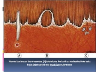
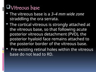
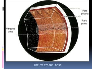
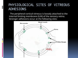
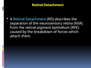
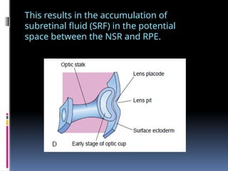
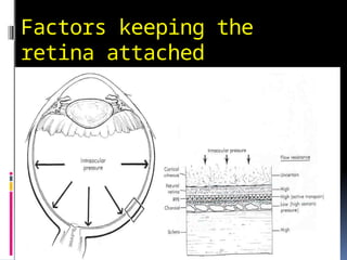
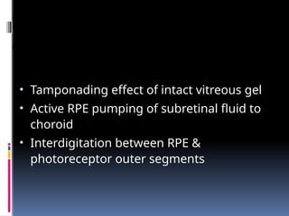
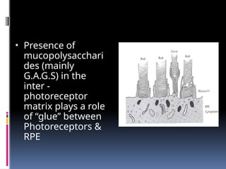
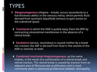
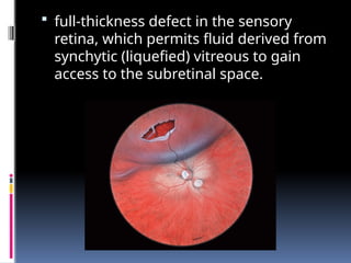
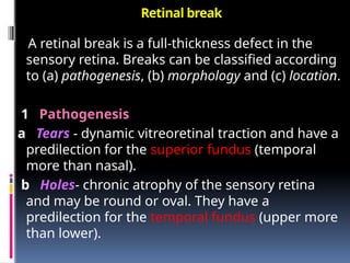
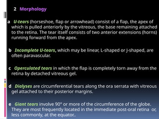
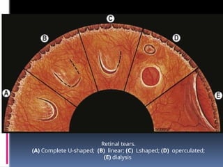
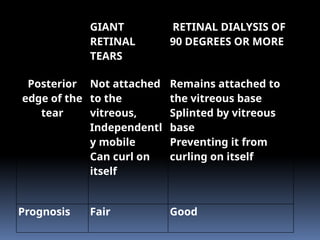
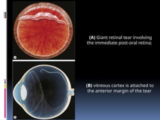
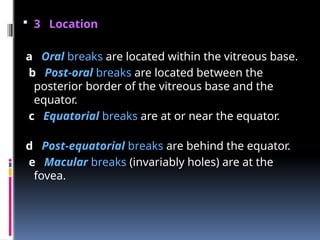
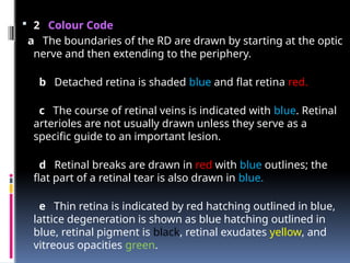
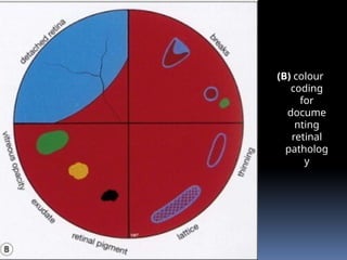
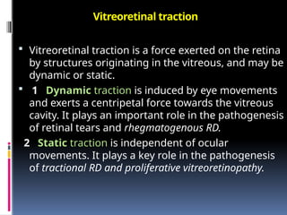
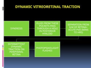
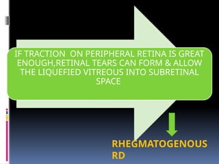
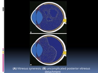
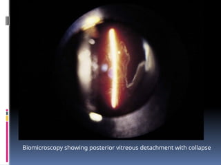
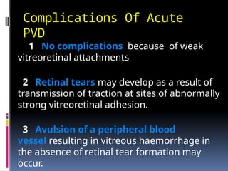
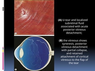
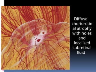
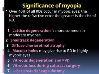
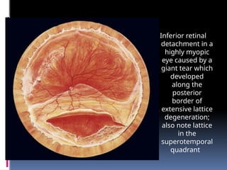
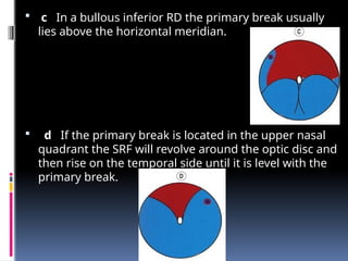
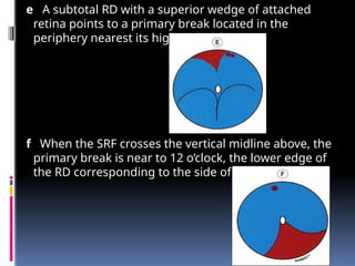
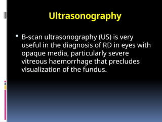
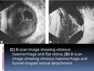
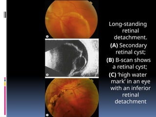
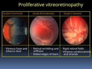
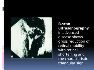
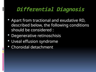
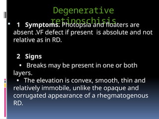
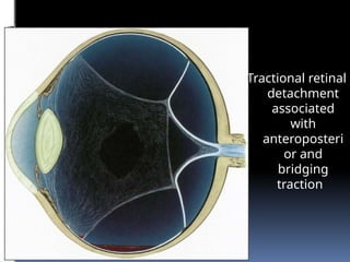
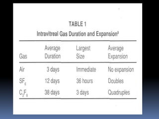
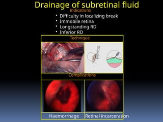
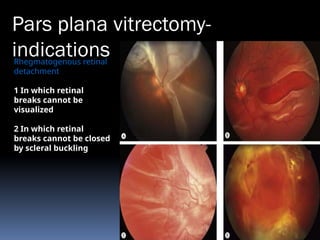
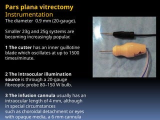
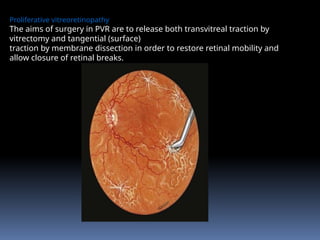
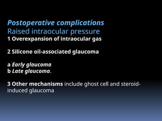
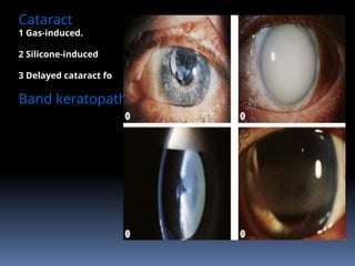
RETINAL
DETACHMENT
TITLES
 Relevant Anatomy
 Introduction
 Types
 Mechanism
 Clinical evaluation
 Ultrasonography
 Each type in detail
 Management
ANATOMY OF THE PERIPHERAL RETINA
 Pars Plana
 The ciliary body starts 1 mm from the limbus and
extends posteriorly for about 6 mm.
 The first 2 mm consist of the pars plicata and the
remaining 4 mm comprises the flattened pars plana.
 In order not to endanger the lens or retina, the optimal
location for a pars plana surgical incision is 4 mm from
the limbus in phakic eyes and 3.5 mm from the limbus in
pseudophakic eyes.
Ora Serrata
 The ora serrata forms the junction between the
retina and ciliary body.
Vitreous base
 The vitreous base is a 3–4 mm wide zone
straddling the ora serrata.
 The cortical vitreous is strongly attached at
the vitreous base, so that following acute
posterior vitreous detachment (PVD), the
posterior hyaloid face remains attached to
the posterior border of the vitreous base.
 Pre-existing retinal holes within the vitreous
base do not lead to RD.
The vitreous base
PHYSIOLOGICAL SITES OF VITREOUS
ADHESIONS
The peripheral cortical vitreous is loosely attached to the
internal limiting membrane (ILM) of the sensory retina.
Stronger adhesions occur at the following sites:
MICROSCOPIC LAYERS OF
RETINA
1. Retinal pigment epithelium
2. Rods and Cones layer
3. External limiting membrane
4. Outer nuclear layer
5. Outer plexiform layer
6. Inner nuclear layer- 1st
order neuron(bipolar cells)
7. Inner plexiform
8. Ganglion cell layer- 2nd
order neuron
9. Nerve fibre layer
10. Internal limiting memb
RETINAL DETACHMENT
Retinal Detachment
 A Retinal Detachment (RD) describes the
separation of the neurosensory retina (NSR)
from the retinal pigment epithelium (RPE)
caused by the breakdown of forces which
attach them.
This results in the accumulation of
subretinal fluid (SRF) in the potential
space between the NSR and RPE.
Factors keeping the
retina attached
Factors keeping the retina
attached
• Tamponading effect of intact vitreous gel
• Active RPE pumping of subretinal fluid to
choroid
• Interdigitation between RPE &
photoreceptor outer segments
Inter Photoreceptor Matrix
• Presence of
mucopolysacchari
des (mainly
G.A.G.S) in the
inter -
photoreceptor
matrix plays a role
of “glue” between
Photoreceptors &
RPE
TYPES
1 Rhegmatogenous (rhegma – break), occurs secondarily to a
full-thickness defect in the sensory retina, which permits fluid
derived from synchytic (liquefied) vitreous to gain access to
the subretinal space.
2 Tractional in which the NSR is pulled away from the RPE by
contracting vitreoretinal membranes in the absence of a
retinal break.
3 Exudative (serous, secondary) is caused neither by a break
nor traction; the SRF is derived from fluid in the vessels of the
NSR or choroid, or both.
4 Combined tractional-rhegmatogenous, as the name
implies, is the result of a combination of a retinal break and
retinal traction. The retinal break is caused by traction from an
adjacent area of fibrovascular proliferation and is most
commonly seen in advanced proliferative diabetic retinopathy.
Posterior vitreous detachment
 A posterior vitreous detachment (PVD) is a separation of the
cortical vitreous from the internal limiting membrane (ILM) of
the NSR posterior to the vitreous base. PVD can be classified
according to the following characteristics:
1 Onset.
 Acute PVD - most common. It develops suddenly and usually
becomes complete soon after onset.
 Chronic PVD occurs gradually and may take weeks or months to
become complete.
2 Extent
a Complete PVD in which the entire vitreous cortex detaches up
to the posterior margin of the vitreous base.
b Incomplete PVD in which residual vitreoretinal attachments
remain posterior to the vitreous base.
 Rhegmatogenous RD is usually
associated with acute PVD
 Tractional RD is associated with
chronic, incomplete PVD
 Exudative RD is unrelated to the
presence of PVD.
RHEGMATOGENOUS
RETINAL DETACHMENT
 full-thickness defect in the sensory
retina, which permits fluid derived from
synchytic (liquefied) vitreous to gain
access to the subretinal space.
 Pathogenesis
 Rhegmatogenous RD affects about 1 in 10 000 of
the population each year and both eyes may
eventually be involved in about 10% of patients.
Three pre-requisites for the development of RRD
 RETINAL BREAKS
 DYNAMIC VITREORETINAL TRACTION
 LIQUEFIED VITREOUS
Retinal break
A retinal break is a full-thickness defect in the
sensory retina. Breaks can be classified according
to (a) pathogenesis, (b) morphology and (c) location.
1 Pathogenesis
a Tears - dynamic vitreoretinal traction and have a
predilection for the superior fundus (temporal
more than nasal).
b Holes- chronic atrophy of the sensory retina
and may be round or oval. They have a
predilection for the temporal fundus (upper more
than lower).
2 Morphology
a U-tears (horseshoe, flap or arrowhead) consist of a flap, the apex of
which is pulled anteriorly by the vitreous, the base remaining attached
to the retina. The tear itself consists of two anterior extensions (horns)
running forward from the apex.
b Incomplete U-tears, which may be linear, L-shaped or J-shaped, are
often paravascular.
c Operculated tears in which the flap is completely torn away from the
retina by detached vitreous gel.
d Dialyses are circumferential tears along the ora serrata with vitreous
gel attached to their posterior margins.
e Giant tears involve 90° or more of the circumference of the globe.
They are most frequently located in the immediate post-oral retina or,
less commonly, at the equator..
Retinal tears.
(A) Complete U-shaped; (B) linear; (C) Lshaped; (D) operculated;
(E) dialysis
GIANT
RETINAL
TEARS
RETINAL DIALYSIS OF
90 DEGREES OR MORE
Posterior
edge of the
tear
Not attached
to the
vitreous,
Independentl
y mobile
Can curl on
itself
Remains attached to
the vitreous base
Splinted by vitreous
base
Preventing it from
curling on itself
Prognosis Fair Good
(A) Giant retinal tear involving
the immediate post-oral retina;
(B) vitreous cortex is attached to
the anterior margin of the tear
 3 Location
a Oral breaks are located within the vitreous base.
b Post-oral breaks are located between the
posterior border of the vitreous base and the
equator.
c Equatorial breaks are at or near the equator.
d Post-equatorial breaks are behind the equator.
e Macular breaks (invariably holes) are at the
fovea.
CLINICAL EXAMINATION
Appearance of retinal breaks in detached
retina. (A) Without scleral indentation; (B) with
indentation
Technique of scleral
indentation.
(A) Insertion of
indenter;
(B) indentation;
(C) mound created by
indentation
Fundus Drawing
 1 Technique.
 The image seen with indirect ophthalmoscopy is
vertically inverted and laterally reversed.
 This phenomenon can be compensated for when
viewing the fundus if the top of the chart is
placed towards the patient's feet (i.e. upside
down).
Technique of drawing retinal lesions. (A) Position of the
chart in relation to the eye;
 2 Colour Code
a The boundaries of the RD are drawn by starting at the optic
nerve and then extending to the periphery.
b Detached retina is shaded blue and flat retina red.
c The course of retinal veins is indicated with blue. Retinal
arterioles are not usually drawn unless they serve as a
specific guide to an important lesion.
d Retinal breaks are drawn in red with blue outlines; the
flat part of a retinal tear is also drawn in blue.
e Thin retina is indicated by red hatching outlined in blue,
lattice degeneration is shown as blue hatching outlined in
blue, retinal pigment is black, retinal exudates yellow, and
vitreous opacities green.
(B) colour
coding
for
docume
nting
retinal
patholog
y
 Even if a retinal break is present, a
RD will not occur if---
vitreous is not at least partially
liquefied and
if the necessary traction is not
present.
Vitreous adhesions
Abnormal adhesions at the following sites may
be associated with retinal tear formation as a
result of dynamic vitreoretinal traction associated
with acute PVD.
• Posterior border of islands of lattice
degeneration.
• Retinal pigment clumps.
• Peripheral paravascular condensations.
• Vitreous base anomalies such as tongue-like
extensions and posterior islands.
• ‘White with pressure’ and ‘white without
pressure’.
Vitreoretinal traction
 Vitreoretinal traction is a force exerted on the retina
by structures originating in the vitreous, and may be
dynamic or static.
 1 Dynamic traction is induced by eye movements
and exerts a centripetal force towards the vitreous
cavity. It plays an important role in the pathogenesis
of retinal tears and rhegmatogenous RD.
2 Static traction is independent of ocular
movements. It plays a key role in the pathogenesis
of tractional RD and proliferative vitreoretinopathy.
SYNERESIS
FLUID FROM THESE
POCKETS PASS
THROUGH A HOLE
IN POSTERIOR
HYALOID
SEPARATION FROM
ILM OF RETINA=
ACUTE PVD (MINS
TO HRS)
INTERMITTENT
DYNAMIC
TRACTION ON
PERIPHERAL
RETINA
PHOTOPSIASLIGHT
FLASHES
DYNAMIC VITREORETINAL TRACTION
IF TRACTION ON PERIPHERAL RETINA IS GREAT
ENOUGH,RETINAL TEARS CAN FORM & ALLOW
THE LIQUEFIED VITREOUS INTO SUBRETINAL
SPACE
RHEGMATOGENOUS
RD
(A) Vitreous syneresis; (B) uncomplicated posterior vitreous
detachment
Biomicroscopy showing posterior vitreous detachment with collapse
Complications Of Acute
PVD
1 No complications because of weak
vitreoretinal attachments
2 Retinal tears may develop as a result of
transmission of traction at sites of abnormally
strong vitreoretinal adhesion.
3 Avulsion of a peripheral blood
vessel resulting in vitreous haemorrhage in
the absence of retinal tear formation may
occur.
PRESENTATION
• Age at onset is typically 45–65 years in the
general population but may occur earlier in
myopic or otherwise predisposed individuals
(e.g. trauma, uveitis).
• The fellow eye frequently becomes affected
within 6 months to 2 years
(A) U-tear and localized
subretinal fluid
associated with acute
posterior vitreous
detachment;
(B) the vitreous shows
syneresis, posterior
vitreous detachment
with partial collapse,
and retained
attachment of cortical
vitreous to the flap of
the tear
 About 60% of all breaks develop in areas of the
peripheral retina that show specific changes.
 Retinal holes are round or oval, usually smaller
than tears and carry a lower risk of RD.
 Retinal detachment without PVD is usually
associated with either retinal dialysis, or round
holes predominantly in young female myopes.
PREDISPOSING FACTORS
• Presence of posterior vitreous
detachment
• High myopia
• History of ocular surgery
• History of trauma
• Predisposing peripheral retinal lesions
Factors predisposing to development RD
LATTICE
DEGENERATI
ON
SNAIL-TRACK
DEGENERATI
ON
DIFFUSE
CHORIORETI
NAL
ATROPHY
DEGENERATI
VE
RETINOSCHI
SIS
WHITE WITH
PRESSURE
AND WHITE
WITHOUT
PRESSURE
Lattice degeneration
1 Prevalence-8% of the population.
 peak incidence during the second and third decades.
 moderate
myopes ,bilateral,temporal>nasal,Superior>inferior
 MOST IMPORTANT DEGENERATION DIRECTLY RELATED
TO RD
 Lattice is present in about 40% of eyes with RD.
2 Pathology-discontinuity of the internal limiting
membrane with variable atrophy of the underlying NSR.
 The vitreous overlying an area of lattice is synchytic but
the vitreous attachments around the margins are
exaggerated.
Vitreous changes associated with lattice
3 Signs
• Spindle-shaped areas of retinal thinning, commonly
located between the equator and the posterior border of
the vitreous base.
• A characteristic feature is an arborizing network of
white lines within the islands.
• Some lattice lesions may be associated with
‘snowflakes’ (remnants of degenerate Müller cells.)
• Associated hyperplasia of the RPE is common.
• Small holes within lattice lesions are common and
usually innocuous.
Clinical features of
lattice
degeneration.
(A) Small island of
lattice with an
arborizing
network of
white lines;
(B) lattice
associated with
‘snowflakes’;
(C) lattice
associated with
RPE changes;
(D) small holes
within lattice
seen on scleral
indentation
 4 Complications
a No complications are encountered in most
patients.
b Tears may occasionally develop in eyes with acute
PVD at the posterior edge of lattice
c Atrophic holes may rarely lead to RD, particularly
in young myopes.
Complications of
lattice
degeneration.
(A) Atypical radial
lattice without
breaks;
(B) two U-tears, the
larger one of
which shows a
small patch of
lattice on its flap
and is surrounded
by a small puddle
of subretinal fluid;
(C) linear tear along
the posterior
margin of lattice;
(D) multiple small
holes within
islands of lattice
Snailtrack degeneration
 Snailtrack degeneration is characterized by
sharply demarcated bands of tightly packed
‘snowflakes’ which give the peripheral retina a
white frost-like appearance.
 The islands are usually longer than in lattice
degeneration and associated with overlying
vitreous liquefaction.
 Tractional U-tears rarely occur, although round
holes within the snailtracks may be present.
Islands of
snailtrack
degeneration,
some of which
contain holes
Degenerative retinoschisis
 1 Prevalence. 5% of the population over the age of 20
years and is particularly prevalent in Hypermetropes
(70% of patients are hypermetropic).
 Bilateral
2 Pathology. Coalescence of cystic lesions as a result
of degeneration of neuroretinal and glial supporting
elements within areas of peripheral cystoid
degeneration.
 results in separation or splitting of the NSR into an inner
(vitreous) layer and an outer (choroidal) layer with
severing of neurones and complete loss of visual
function in the affected area.
 1. Typical retinoschisis- the split is in the outer plexiform
layer, 2.Reticular retinoschisis-less common, split at the
Microcystoid
degeneration.
(A) Histology shows
spaces in the nerve
fibre layer
delineated by
delicate vertical
columns of Müller
cells;
(B) circumferential
microcystoid
degeneration and
mild retinoschisis in
the inferotemporal
and superotemporal
quadrants
Retinoschisis
with breaks
in both
layers. The
inner layer
shows
snowflakes
and ‘silver-
wiring’ of
blood
vessels, and
the cavity is
bridged by
torn grey-
white tissue
 4 Complications
a No complications occur in most cases and the
condition is asymptomatic and innocuous.
b Breaks. Inner layer breaks are small and round,
whilst the less common outer layer breaks are
usually larger, with rolled edges and located behind
the equator.
c RD may occasionally develop in eyes with breaks
in both layers, especially in the presence of PVD
d Vitreous haemorrhage is uncommon.
Retinoschisis
with
multiple
outer layer
breaks
White-without-pressure
Indications for prophylaxis - giant tear in other eye
Translucent grey appearance of
retina
Occasional giant tear formation along
posterior margin of lesion
Diffuse chorioretinal atrophy
 Diffuse chorioretinal atrophy is
characterized by choroidal depigmentation
and thinning of the overlying retina in the
equatorial area of highly myopic eyes.
 Retinal holes developing in the atrophic
retina may lead to RD.
Diffuse
chorioretin
al atrophy
with holes
and
localized
subretinal
fluid
Significance of myopia
 Over 40% of all RDs occur in myopic eyes; the
higher the refractive error the greater is the risk of
RD.
1 Lattice degeneration is more common in
moderate myopes
2 Snailtrack degeneration
3 Diffuse chorioretinal atrophy
4 Macular holes may give rise to RD in highly
myopic eyes
5 Vitreous degeneration and PVD
6 Vitreous loss during cataract surgery
7 Laser posterior capsulotomy
Inferior retinal
detachment in a
highly myopic
eye caused by a
giant tear which
developed
along the
posterior
border of
extensive lattice
degeneration;
also note lattice
in the
superotemporal
quadrant
Symptoms
 1 Photopsia is the subjective sensation of a flash of light.
- caused by traction at sites of vitreoretinal adhesion.
- The cessation of photopsia is the result of either separation
of the adhesion or complete tearing away of a piece of
retina (operculum).
- In PVD the photopsia is often described as an arc of golden
or white light induced by eye movements and is more
noticeable in dim illumination. It tends to be projected into
the patient's temporal peripheral visual field.
- Occasionally photopsia precedes PVD by 24–48 hours.
2 Floaters are moving vitreous opacities which are
perceived when they cast shadows on the retina.
 Three types:
a Weiss ring is a solitary floater consisting of the detached
annular attachment of vitreous to the margin of the optic
disc
b Cobwebs are caused by condensation of collagen fibres
within the collapsed vitreous cortex.
c A sudden shower of minute red-coloured or dark spots
usually indicates vitreous haemorrhage secondary to
tearing of a peripheral retinal blood vessel.
(A) Weiss ring;
(B) B-scan
shows a
Weiss ring
associated
with
posterior
vitreous
detachment
3 A visual field defect is perceived as a ‘black
curtain’.
- A lower field defect is usually appreciated more
quickly by the patient than an upper field defect.
- The quadrant of the visual field in which the field
defect first appears is useful in predicting the
location of the primary retinal break, which will be
in the opposite quadrant.
- Loss of central vision may be due either to
involvement of the fovea by SRF or, less
frequently, obstruction of the visual axis by a
large upper bullous RD.
Signs
General
1 Marcus Gunn pupil
2 Intraocular pressure is usually lower by about
5 mmHg
3 Iritis
4 ‘Tobacco dust’ ,Schaffers sign-consisting of pigment
cells is seen in the anterior vitreous.
5 Retinal breaks appear as discontinuities in the retinal
surface. They are usually red because of the colour
contrast between the sensory retina and underlying
choroid.
‘Tobacco
dust’ in
the
anterior
vitreous
Finding the primary break
 The primary break is the one responsible for
the RD.
 A secondary break is not responsible for the
RD because it was either present before the
development of the RD or formed after the
retina is detached.
• About 50% of eyes with RD have more than
one break, and in most eyes these are located
within 90° of each other.
DISTRIBUTION OF BREAKS
15
60
10
15
15
 2 Configuration of SRF - SRF spreads in a
gravitational fashion, and its shape is governed by
anatomical limits (ora serrata and optic nerve) and by
the location of the primary retinal break.
• If the primary break is located superiorly, the SRF first
spreads inferiorly on the same side as the break
and then spreads superiorly on the opposite side of the
fundus.
• The likely location of the primary retinal break can
therefore be predicted by studying the shape of the RD.
LINCOFF’S RULES
Lincoff showed
how the location of
retinal break
determines the
distribution of
subretinal fluid.
 a A shallow inferior RD in which the SRF
is slightly higher on the temporal side
points to a primary break located
inferiorly on that side.
 b A primary break located at 6 o’clock
will cause an inferior RD with equal
fluid levels.
 c In a bullous inferior RD the primary break usually
lies above the horizontal meridian.
 d If the primary break is located in the upper nasal
quadrant the SRF will revolve around the optic disc and
then rise on the temporal side until it is level with the
primary break.
e A subtotal RD with a superior wedge of attached
retina points to a primary break located in the
periphery nearest its highest border.
f When the SRF crosses the vertical midline above, the
primary break is near to 12 o’clock, the lower edge of
the RD corresponding to the side of the break .
Ultrasonography
 B-scan ultrasonography (US) is very
useful in the diagnosis of RD in eyes with
opaque media, particularly severe
vitreous haemorrhage that precludes
visualization of the fundus.
(C) B-scan image showing vitreous
haemorrhage and flat retina; (D) B-scan
image showing vitreous haemorrhage and
funnel-shaped retinal detachment
Rhegmatogenous RD
• Fresh
• Longstanding
• Proliferative vitreoretinopathy (PVR)
Fresh retinal detachment
 Grayish white,opaque,raised detached retina(convex
configuration) and corrugated appppearance(due to
intraretinal edema) with loss of underlying choroidal
pattern
 Dark retinal vessels with no central light reflex.
 No shifting of fliud
 Oscillation of multiple retinal folds with movement of eye
 One or more retinal breaks or holes in upper temporal
region
 Vitreous show pigment in anterior vitreous(tobacco
dusting or shafer sign)with posterior detachmnet.
 Associated degeneration,pigmentation and haemorrhage
in retina.
 B-scan ultrasonography shows good mobility of the retina
Fresh retinal detachment.
(A) U-tear in detached
retina;
(B) superior bullous
retinal detachment;
(C) shallow temporal
retinal detachment;
(D) B-scan shows a totally
detached retina with
linear echogenic
structures inserting
onto the optic nerve
head to form an open
funnel
Long-standing retinal
detachment
 The following are the main features of a long-standing
rhegmatogenous RD:
1 Retinal thinning secondary to atrophy is a
characteristic finding which must not be mistaken for
retinoschisis.
2 Secondary intraretinal cysts may develop if the RD
has been present for about 1 year; these tend to disappear
after retinal reattachment.
3 Subretinal demarcation lines (‘high water marks’)
caused by proliferation of RPE cells at the junction of flat
and detached retina are common and take about 3 months
to develop. Demarcation lines are convex with respect to
the ora serrata.
4 Multiple opaque strands of subretinal
Long-standing
retinal
detachment.
(A) Secondary
retinal cyst;
(B) B-scan shows
a retinal cyst;
(C) ‘high water
mark’ in an eye
with an inferior
retinal
detachment
Proliferative
vitreoretinopathy
 Proliferative vitreoretinopathy (PVR) is caused
by epiretinal and subretinal membrane
formation.
 Cell-mediated contraction of these membranes
causes tangential retinal traction and fixed
retinal folds.
 PVR occurs following surgery for
rhegmatogenous RD or penetrating injury,
rhegmatogenous RD that have not had
previous vitreoretinal surgery.
Development of proliferative
vitreoretinopathy (PVR).
(A) Extensive vitreous
syneresis;
(B) total retinal detachment
without PVR; shrunken
vitreous is condensed and
attached to the equator of
the retina;
(C) early PVR with anteriorly
retracted vitreous gel and
equatorial circumferential
retinal folds;
(D) advanced PVR with a
funnel-like retinal
detachment bridged by
dense vitreous
membranes
 The term "proliferative vitreoretinopathy" was coined in
1983 by the Retina Society Terminology Committee.
 Silicone Study Group classification (1991).
 Grade A is limited to the presence of vitreous cells or haze.
 Grade B is defined by the presence of rolled or irregular
edges of a tear or inner retinal surface wrinkling, denoting
subclinical contraction.
 Grade C is recognized by the presence of preretinal or
subretinal membranes.
 Grade C is further delineated as being anterior to the equator
(grade Ca) or posterior to the equator (grade Cp) and by the
number of clock hours involved (1 to 12).
Proliferative vitreoretinopathy
• Vitreous haze and
tobacco dust
Grade A (minimal)
• Rigid retinal folds
• Vitreous condensations
and strands
Grade C (severe)
• Retinal wrinkling and
stiffness
• Rolled edges of tears
Grade B (moderate)
B-scan
ultrasonography
in advanced
disease shows
gross reduction of
retinal mobility
with retinal
shortening and
the characteristic
triangular sign .
Differential Diagnosis
 Apart from tractional and exudative RD,
described below, the following conditions
should be considered :
 Degenerative retinoschisis
 Uveal effusion syndrome
 Choroidal detachment
Degenerative
retinoschisis
 1 Symptoms. Photopsia and floaters are
absent .VF defect if present is absolute and not
relative as in RD.
2 Signs
• Breaks may be present in one or both
layers.
• The elevation is convex, smooth, thin and
relatively immobile, unlike the opaque and
corrugated appearance of a rhegmatogenous
RD.
Uveal effusion syndrome
 The uveal effusion syndrome is a rare, idiopathic
condition which most frequently affects middle-aged
hypermetropic men.
1 Signs
• Ciliochoroidal detachment followed by exudative
RD which may be bilateral.
• Following resolution, the RPE frequently shows a
characteristic residual ‘leopard spot’ mottling caused by
degenerative changes in the RPE associated with high
concentration of protein in the SRF.
2 Differential diagnosis includes RD complicated by
choroidal detachment and ring melanoma of the
anterior choroid.
uveal effusion
characterized by
choroidal
detachment and
exudative
retinal
detachment
Choroidal detachment
1 Symptoms. Photopsia and floaters are absent because there is
no vitreoretinal traction. A visual field defect may be noticed if the
choroidal detachment is extensive.
2 Signs
• Low intraocular pressure is common as a result of
concomitant detachment of the ciliary body.
• The anterior chamber may be shallow in eyes with extensive
choroidal detachments.
• The elevations are brown, convex, smooth and relatively
immobile. Temporal and nasal bullae tend to be most prominent.
• Large ‘kissing’ choroidal detachments may obscure the view
of the fundus.
• The elevations do not extend to the posterior pole because
they are limited by the firm adhesion between the suprachoroidal
lamellae where the vortex veins enter their scleral canals.
(A) Choroidal
detachment.
(B) B-scan
image of
extensive
choroidal
detachment
almost
touching in
the middle of
the vitreous
cavity
TRACTIONAL RETINAL
DETACHMENT
DEFINITION
 The NSR is pulled away from RPE by
contracting vitreoretinal membranes in
the absence of a retinal break.
 CAUSES
(a) proliferative retinopathy such as diabetic
and retinopathy of prematurity
(b) penetrating posterior segment trauma
Pathogenesis of diabetic
tractional retinal
detachment
 CHRONIC PVD ,gradual and frequently incomplete
- Leakage of plasma constituents into the vitreous gel
from a fibrovascular network adherent to the posterior
vitreous surface.
- strong adhesions of the cortical vitreous to areas of
fibrovascular proliferation, PVD is usually incomplete.
- Tractional RD is caused by progressive contraction of
fibrovascular membranes over large areas of vitreoretinal
adhesion.
- In the very rare event of a subsequent complete PVD, the
new blood vessels are avulsed and RD does not develop.
 2 Static vitreoretinal traction of the following three
types is recognized.
a Tangential traction is caused by the contraction of
epiretinal fibrovascular membranes with puckering of the
retina and distortion of retinal blood vessels.
b Anteroposterior traction is caused by the contraction
of fibrovascular membranes extending from the posterior
retina, usually in association with the major arcades, to the
vitreous base anteriorly.
c Bridging (trampoline) traction is the result of
contraction of fibrovascular membranes between the
vascular arcades.
Tractional retinal
detachment
associated
with
anteroposteri
or and
bridging
traction
Diagnosis
1 Symptoms.
 Photopsia and floaters are usually absent because vitreoretinal
traction develops insidiously and is not associated with acute PVD.
 Visual field defect
2 Signs.
• The RD has a concave configuration and breaks are absent.
• Retinal mobility is severely reduced and shifting fluid is
absent.
• The SRF is shallower than in a rhegmatogenous RD and
seldom extends to the ora serrata.
• The highest elevation of the retina occurs at sites of
vitreoretinal traction.
• If a tractional RD develops a break it assumes the
characteristics of a rhegmatogenous RD and progresses more
quickly (combined tractional-rhegmatogenous RD).
(A) Tractional retinal
detachment in
severe
proliferative
diabetic
retinopathy;
(B) B-scan image of
another patient
shows incomplete
posterior vitreous
detachment and a
shallow
tractionalretinal
detachment,relativ
ely immobile
retina
TREATMENT
 Pars plana vitrectomy.
EXUDATIVE RETINAL
DETACHMENT
DEFINITION
 Serous/secondary RD
 SRF is derived from the vessels of the NSR
and/or choroid.
 Exudative RD is characterized by the
accumulation of SRF in the absence of
retinal breaks or traction
Pathogenesis
 Vascular, inflammatory and neoplastic diseases involving
the NSR, RPE and choroid in which fluid leaks outside the
vessels and accumulates under the retina.
 Compensated normally by RPE pump.
 When the normal RPE pump is overwhelmed, or if the
RPE activity is decreased, then fluid starts to accumulate
in the subretinal space
CAUSES:
1 Choroidal tumours - melanomas, haemangiomas and
metastases;
2 Inflammation such as Harada disease and posterior scleritis.
3 Bullous central serous chorioretinopathy - rare cause.
4 Iatrogenic causes include retinal detachment surgery and
panretinal photocoagulation.
5 Choroidal neovascularization -SRF at posterior pole.
6 Hypertensive choroidopathy- toxaemia of pregnancy
7 Idiopathic such as the uveal effusion syndrome.
Diagnosis
1 Symptoms
Photopsia is absent because there is no
vitreoretinal traction
 floaters -vitritis.
 visual field defect may develop suddenly and
progress rapidly.
 bilateral involvement (e.g. Harada disease).
2 Signs
Exudative retinal
detachment
with shifting
fluid.
(A) Inferior
collection of
subretinal fluid
with the
patient sitting;
(B) the subretinal
fluid shifts
upwards when
the patient
assumes the
supine
position
Exudative
retinal
detachmen
t caused by
a choroidal
melanoma
‘Leopard spot’
pigmentatio
n following
resolution of
exudative
retinal
detachment
TREATMENT
 Depends on the cause
 Systemic corticosteroids(Harada
disease and posterior scleritis
 Bullous CSR-Laser photocoagualtion to
seal the leak in RPE.
MANAGEMENT
 Scleral Buckling
 Pneumatic retinopexy
 Primary pars plana vitrectomy
(with or without sclera buckling)
THANK YOU…
Asymptomatic retinal
detachment
 range from conservative observation to
prophylactic surgery.
 demarcation laser photocoagulation of
shallow, macula-sparing, RRD without
associated PVR to be a reasonable
alternative to surgical repair.
CHOICE
 individual clinical situation combined
with each surgeon's experience bias and
comfort level with a particular procedure.
 Without a randomized clinical trial
comparing the 3 modalities, and in
various clinical scenarios, definitive
answer as to which procedure is superior
will be impossible.
Basic Rationale of repair of RRD ---
 The regardless of the procedure
chosen, the surgery aims to identify
and close all the retinal breaks with
minimum iatrogenic damage.
Retinal Reattachment Surgeries
GONIN’S
PRINCIPLES-
-SEARCH FOR
THE BREAK
-SEE A BREAK
-SEAL THE
BREAK
Break closure
1)to bring the edges of the retinal break
into contact with the underlying RPE –
 by bringing the eye wall closer to the
detached retina (a scleral buckle)
 pushing the detached retina toward the
RPE (intraocular tamponade with a gas/
PFCL expand bubble).
2)to create a strong chorio-retinal
adhesion around the breaks;
 cryotherapy,
 laser photocoagulation
 diathermy.
THE USE OF SCLERAL BUCKLES IN CONJUNCTION
WITH CHORIORETINAL ADHESIONS FORMS THE
BASIS OF THERAPY FOR MANY UNCOMPLICATED
RHEGMATOGENOUS RETINAL DETACHMENTS…….
• Pneumatic retinopexy is a
procedure in which an intravitreal
expanding gas bubble is used to
• seal a retinal break and reattach the
retina without scleral buckling.
• Advantage- quick, minimally
invasive, ‘office-based’ procedure.
• uncomplicated RD with a small
retinal break or a cluster of
• breaks extending over an area of
less than two clock hours situation
in the upper two-thirds of the
peripheral
• retina.
RETINOPEXY
Drainage of subretinal fluid
Indications
Haemorrhage
• Difficulty in localizing break
• Immobile retina
• Longstanding RD
• Inferior RD
Retinal incarceration
Complications
Technique
Rhegmatogenous retinal
detachment
1 In which retinal
breaks cannot be
visualized
2 In which retinal
breaks cannot be closed
by scleral buckling
Pars plana vitrectomy-
indications
Pars plana vitrectomy
Instrumentation
The diameter 0.9 mm (20-gauge).
Smaller 23g and 25g systems are
becoming increasingly popular.
1 The cutter has an inner guillotine
blade which oscillates at up to 1500
times/minute.
2 The intraocular illumination
source is through a 20-gauge
fibreoptic probe 80–150 W bulb.
3 The infusion cannula usually has an
intraocular length of 4 mm, although
in special circumstances
such as choroidal detachment or eyes
with opaque media, a 6 mm cannula
4 Accessory instruments
include scissors, forceps, flute
needle, and endodiathermy
and endolaser
delivery systems.
5 Wide-angle viewing
system consists of an indirect
lens beneath the operating
microscope and an
incorporated series of prisms
to reinvert the image. The
field of view extends
almost to the ora serrata;
higher magnification lenses
are also available for macular
Tamponading agents
1 Purpose is to achieve intraoperative retinal flattening by fluid–
gas exchange combined with internal
drainage of SRF, and to produce internal tamponade of retinal
breaks during the postoperative period.
2 Expanding gases
Most tamponading agents are used at an isovolumetric
concentration (e.g. 20–30% for SF6 and 12–16% for
C3F8).
3 Heavy liquids (perfluorocarbons) have a high specific gravity –
they are heavier than water – and
thus remain in a dependent position when injected into the
vitreous cavity.
4 Silicone oils
Technique
Basic vitrectomy
a Following limbal peritomy an infusion
cannula is secured to the sclera (3.5 mm
behind the limbus in
pseudophakic or aphakic eyes and 4 mm in
phakic eyes) at the level of the inferior
border of the lateral
rectus muscle.
b Further sclerotomies are made at the 10
and 2 o’clock positions. These can be
standard stab incisions
made with an MVR blade, or self-sealing
sclerotomies.
c The cutter and fibreoptic light pipe enter
through the upper two sclerotomies.
d The central vitreous gel and posterior
hyaloid face are excised.
Closure of giant tears
a Fluid-air exchange is performed to
flatten the retina (hydraulic retinal
reattachment).
b The flap of a giant tear is unrolled by
injecting a heavy liquid over the optic
disc .
c Retinopexy of the now flat retinal
breaks is performed with either trans-
scleral cryotherapy or
endolaser using minimal energy.
d Prolonged internal tamponade is
achieved by replacing air with a non-
expansile concentration of
sulphur hexafluoride (SF6) or
perfluoropropane (C3F8) gases, or with
silicone oil. The non-expansile
mixture of gas and air is prepared in a
large (50 mL) syringe and the air-filled
Proliferative vitreoretinopathy
The aims of surgery in PVR are to release both transvitreal traction by
vitrectomy and tangential (surface)
traction by membrane dissection in order to restore retinal mobility and
allow closure of retinal breaks.
Postoperative complications
Raised intraocular pressure
1 Overexpansion of intraocular gas
2 Silicone oil-associated glaucoma
a Early glaucoma
b Late glaucoma.
3 Other mechanisms include ghost cell and steroid-
induced glaucoma
Cataract
1 Gas-induced.
2 Silicone-induced
3 Delayed cataract formation.
Band keratopathy

retinal detachment evaluation and management

  • 1.
  • 2.
    TITLES  Relevant Anatomy Introduction  Types  Mechanism  Clinical evaluation  Ultrasonography  Each type in detail  Management
  • 3.
    ANATOMY OF THEPERIPHERAL RETINA  Pars Plana  The ciliary body starts 1 mm from the limbus and extends posteriorly for about 6 mm.  The first 2 mm consist of the pars plicata and the remaining 4 mm comprises the flattened pars plana.  In order not to endanger the lens or retina, the optimal location for a pars plana surgical incision is 4 mm from the limbus in phakic eyes and 3.5 mm from the limbus in pseudophakic eyes.
  • 4.
    Ora Serrata  Theora serrata forms the junction between the retina and ciliary body.
  • 6.
    Vitreous base  Thevitreous base is a 3–4 mm wide zone straddling the ora serrata.  The cortical vitreous is strongly attached at the vitreous base, so that following acute posterior vitreous detachment (PVD), the posterior hyaloid face remains attached to the posterior border of the vitreous base.  Pre-existing retinal holes within the vitreous base do not lead to RD.
  • 7.
  • 8.
    PHYSIOLOGICAL SITES OFVITREOUS ADHESIONS The peripheral cortical vitreous is loosely attached to the internal limiting membrane (ILM) of the sensory retina. Stronger adhesions occur at the following sites:
  • 9.
    MICROSCOPIC LAYERS OF RETINA 1.Retinal pigment epithelium 2. Rods and Cones layer 3. External limiting membrane 4. Outer nuclear layer 5. Outer plexiform layer 6. Inner nuclear layer- 1st order neuron(bipolar cells) 7. Inner plexiform 8. Ganglion cell layer- 2nd order neuron 9. Nerve fibre layer 10. Internal limiting memb
  • 10.
  • 11.
    Retinal Detachment  ARetinal Detachment (RD) describes the separation of the neurosensory retina (NSR) from the retinal pigment epithelium (RPE) caused by the breakdown of forces which attach them.
  • 12.
    This results inthe accumulation of subretinal fluid (SRF) in the potential space between the NSR and RPE.
  • 13.
  • 14.
    Factors keeping theretina attached • Tamponading effect of intact vitreous gel • Active RPE pumping of subretinal fluid to choroid • Interdigitation between RPE & photoreceptor outer segments
  • 15.
    Inter Photoreceptor Matrix •Presence of mucopolysacchari des (mainly G.A.G.S) in the inter - photoreceptor matrix plays a role of “glue” between Photoreceptors & RPE
  • 16.
    TYPES 1 Rhegmatogenous (rhegma– break), occurs secondarily to a full-thickness defect in the sensory retina, which permits fluid derived from synchytic (liquefied) vitreous to gain access to the subretinal space. 2 Tractional in which the NSR is pulled away from the RPE by contracting vitreoretinal membranes in the absence of a retinal break. 3 Exudative (serous, secondary) is caused neither by a break nor traction; the SRF is derived from fluid in the vessels of the NSR or choroid, or both. 4 Combined tractional-rhegmatogenous, as the name implies, is the result of a combination of a retinal break and retinal traction. The retinal break is caused by traction from an adjacent area of fibrovascular proliferation and is most commonly seen in advanced proliferative diabetic retinopathy.
  • 17.
    Posterior vitreous detachment A posterior vitreous detachment (PVD) is a separation of the cortical vitreous from the internal limiting membrane (ILM) of the NSR posterior to the vitreous base. PVD can be classified according to the following characteristics: 1 Onset.  Acute PVD - most common. It develops suddenly and usually becomes complete soon after onset.  Chronic PVD occurs gradually and may take weeks or months to become complete. 2 Extent a Complete PVD in which the entire vitreous cortex detaches up to the posterior margin of the vitreous base. b Incomplete PVD in which residual vitreoretinal attachments remain posterior to the vitreous base.
  • 18.
     Rhegmatogenous RDis usually associated with acute PVD  Tractional RD is associated with chronic, incomplete PVD  Exudative RD is unrelated to the presence of PVD.
  • 19.
  • 20.
     full-thickness defectin the sensory retina, which permits fluid derived from synchytic (liquefied) vitreous to gain access to the subretinal space.
  • 21.
     Pathogenesis  RhegmatogenousRD affects about 1 in 10 000 of the population each year and both eyes may eventually be involved in about 10% of patients. Three pre-requisites for the development of RRD  RETINAL BREAKS  DYNAMIC VITREORETINAL TRACTION  LIQUEFIED VITREOUS
  • 22.
    Retinal break A retinalbreak is a full-thickness defect in the sensory retina. Breaks can be classified according to (a) pathogenesis, (b) morphology and (c) location. 1 Pathogenesis a Tears - dynamic vitreoretinal traction and have a predilection for the superior fundus (temporal more than nasal). b Holes- chronic atrophy of the sensory retina and may be round or oval. They have a predilection for the temporal fundus (upper more than lower).
  • 23.
    2 Morphology a U-tears(horseshoe, flap or arrowhead) consist of a flap, the apex of which is pulled anteriorly by the vitreous, the base remaining attached to the retina. The tear itself consists of two anterior extensions (horns) running forward from the apex. b Incomplete U-tears, which may be linear, L-shaped or J-shaped, are often paravascular. c Operculated tears in which the flap is completely torn away from the retina by detached vitreous gel. d Dialyses are circumferential tears along the ora serrata with vitreous gel attached to their posterior margins. e Giant tears involve 90° or more of the circumference of the globe. They are most frequently located in the immediate post-oral retina or, less commonly, at the equator..
  • 24.
    Retinal tears. (A) CompleteU-shaped; (B) linear; (C) Lshaped; (D) operculated; (E) dialysis
  • 25.
    GIANT RETINAL TEARS RETINAL DIALYSIS OF 90DEGREES OR MORE Posterior edge of the tear Not attached to the vitreous, Independentl y mobile Can curl on itself Remains attached to the vitreous base Splinted by vitreous base Preventing it from curling on itself Prognosis Fair Good
  • 26.
    (A) Giant retinaltear involving the immediate post-oral retina; (B) vitreous cortex is attached to the anterior margin of the tear
  • 27.
     3 Location aOral breaks are located within the vitreous base. b Post-oral breaks are located between the posterior border of the vitreous base and the equator. c Equatorial breaks are at or near the equator. d Post-equatorial breaks are behind the equator. e Macular breaks (invariably holes) are at the fovea.
  • 28.
  • 29.
    Appearance of retinalbreaks in detached retina. (A) Without scleral indentation; (B) with indentation
  • 30.
    Technique of scleral indentation. (A)Insertion of indenter; (B) indentation; (C) mound created by indentation
  • 31.
    Fundus Drawing  1Technique.  The image seen with indirect ophthalmoscopy is vertically inverted and laterally reversed.  This phenomenon can be compensated for when viewing the fundus if the top of the chart is placed towards the patient's feet (i.e. upside down).
  • 32.
    Technique of drawingretinal lesions. (A) Position of the chart in relation to the eye;
  • 33.
     2 ColourCode a The boundaries of the RD are drawn by starting at the optic nerve and then extending to the periphery. b Detached retina is shaded blue and flat retina red. c The course of retinal veins is indicated with blue. Retinal arterioles are not usually drawn unless they serve as a specific guide to an important lesion. d Retinal breaks are drawn in red with blue outlines; the flat part of a retinal tear is also drawn in blue. e Thin retina is indicated by red hatching outlined in blue, lattice degeneration is shown as blue hatching outlined in blue, retinal pigment is black, retinal exudates yellow, and vitreous opacities green.
  • 34.
  • 35.
     Even ifa retinal break is present, a RD will not occur if--- vitreous is not at least partially liquefied and if the necessary traction is not present.
  • 36.
    Vitreous adhesions Abnormal adhesionsat the following sites may be associated with retinal tear formation as a result of dynamic vitreoretinal traction associated with acute PVD. • Posterior border of islands of lattice degeneration. • Retinal pigment clumps. • Peripheral paravascular condensations. • Vitreous base anomalies such as tongue-like extensions and posterior islands. • ‘White with pressure’ and ‘white without pressure’.
  • 37.
    Vitreoretinal traction  Vitreoretinaltraction is a force exerted on the retina by structures originating in the vitreous, and may be dynamic or static.  1 Dynamic traction is induced by eye movements and exerts a centripetal force towards the vitreous cavity. It plays an important role in the pathogenesis of retinal tears and rhegmatogenous RD. 2 Static traction is independent of ocular movements. It plays a key role in the pathogenesis of tractional RD and proliferative vitreoretinopathy.
  • 38.
    SYNERESIS FLUID FROM THESE POCKETSPASS THROUGH A HOLE IN POSTERIOR HYALOID SEPARATION FROM ILM OF RETINA= ACUTE PVD (MINS TO HRS) INTERMITTENT DYNAMIC TRACTION ON PERIPHERAL RETINA PHOTOPSIASLIGHT FLASHES DYNAMIC VITREORETINAL TRACTION
  • 39.
    IF TRACTION ONPERIPHERAL RETINA IS GREAT ENOUGH,RETINAL TEARS CAN FORM & ALLOW THE LIQUEFIED VITREOUS INTO SUBRETINAL SPACE RHEGMATOGENOUS RD
  • 40.
    (A) Vitreous syneresis;(B) uncomplicated posterior vitreous detachment
  • 41.
    Biomicroscopy showing posteriorvitreous detachment with collapse
  • 42.
    Complications Of Acute PVD 1No complications because of weak vitreoretinal attachments 2 Retinal tears may develop as a result of transmission of traction at sites of abnormally strong vitreoretinal adhesion. 3 Avulsion of a peripheral blood vessel resulting in vitreous haemorrhage in the absence of retinal tear formation may occur.
  • 43.
    PRESENTATION • Age atonset is typically 45–65 years in the general population but may occur earlier in myopic or otherwise predisposed individuals (e.g. trauma, uveitis). • The fellow eye frequently becomes affected within 6 months to 2 years
  • 44.
    (A) U-tear andlocalized subretinal fluid associated with acute posterior vitreous detachment; (B) the vitreous shows syneresis, posterior vitreous detachment with partial collapse, and retained attachment of cortical vitreous to the flap of the tear
  • 45.
     About 60%of all breaks develop in areas of the peripheral retina that show specific changes.  Retinal holes are round or oval, usually smaller than tears and carry a lower risk of RD.  Retinal detachment without PVD is usually associated with either retinal dialysis, or round holes predominantly in young female myopes.
  • 46.
    PREDISPOSING FACTORS • Presenceof posterior vitreous detachment • High myopia • History of ocular surgery • History of trauma • Predisposing peripheral retinal lesions
  • 47.
    Factors predisposing todevelopment RD LATTICE DEGENERATI ON SNAIL-TRACK DEGENERATI ON DIFFUSE CHORIORETI NAL ATROPHY DEGENERATI VE RETINOSCHI SIS WHITE WITH PRESSURE AND WHITE WITHOUT PRESSURE
  • 48.
    Lattice degeneration 1 Prevalence-8%of the population.  peak incidence during the second and third decades.  moderate myopes ,bilateral,temporal>nasal,Superior>inferior  MOST IMPORTANT DEGENERATION DIRECTLY RELATED TO RD  Lattice is present in about 40% of eyes with RD. 2 Pathology-discontinuity of the internal limiting membrane with variable atrophy of the underlying NSR.  The vitreous overlying an area of lattice is synchytic but the vitreous attachments around the margins are exaggerated.
  • 49.
  • 50.
    3 Signs • Spindle-shapedareas of retinal thinning, commonly located between the equator and the posterior border of the vitreous base. • A characteristic feature is an arborizing network of white lines within the islands. • Some lattice lesions may be associated with ‘snowflakes’ (remnants of degenerate Müller cells.) • Associated hyperplasia of the RPE is common. • Small holes within lattice lesions are common and usually innocuous.
  • 51.
    Clinical features of lattice degeneration. (A)Small island of lattice with an arborizing network of white lines; (B) lattice associated with ‘snowflakes’; (C) lattice associated with RPE changes; (D) small holes within lattice seen on scleral indentation
  • 52.
     4 Complications aNo complications are encountered in most patients. b Tears may occasionally develop in eyes with acute PVD at the posterior edge of lattice c Atrophic holes may rarely lead to RD, particularly in young myopes.
  • 53.
    Complications of lattice degeneration. (A) Atypicalradial lattice without breaks; (B) two U-tears, the larger one of which shows a small patch of lattice on its flap and is surrounded by a small puddle of subretinal fluid; (C) linear tear along the posterior margin of lattice; (D) multiple small holes within islands of lattice
  • 54.
    Snailtrack degeneration  Snailtrackdegeneration is characterized by sharply demarcated bands of tightly packed ‘snowflakes’ which give the peripheral retina a white frost-like appearance.  The islands are usually longer than in lattice degeneration and associated with overlying vitreous liquefaction.  Tractional U-tears rarely occur, although round holes within the snailtracks may be present.
  • 55.
  • 56.
    Degenerative retinoschisis  1Prevalence. 5% of the population over the age of 20 years and is particularly prevalent in Hypermetropes (70% of patients are hypermetropic).  Bilateral 2 Pathology. Coalescence of cystic lesions as a result of degeneration of neuroretinal and glial supporting elements within areas of peripheral cystoid degeneration.  results in separation or splitting of the NSR into an inner (vitreous) layer and an outer (choroidal) layer with severing of neurones and complete loss of visual function in the affected area.  1. Typical retinoschisis- the split is in the outer plexiform layer, 2.Reticular retinoschisis-less common, split at the
  • 57.
    Microcystoid degeneration. (A) Histology shows spacesin the nerve fibre layer delineated by delicate vertical columns of Müller cells; (B) circumferential microcystoid degeneration and mild retinoschisis in the inferotemporal and superotemporal quadrants
  • 58.
    Retinoschisis with breaks in both layers.The inner layer shows snowflakes and ‘silver- wiring’ of blood vessels, and the cavity is bridged by torn grey- white tissue
  • 59.
     4 Complications aNo complications occur in most cases and the condition is asymptomatic and innocuous. b Breaks. Inner layer breaks are small and round, whilst the less common outer layer breaks are usually larger, with rolled edges and located behind the equator. c RD may occasionally develop in eyes with breaks in both layers, especially in the presence of PVD d Vitreous haemorrhage is uncommon.
  • 60.
  • 61.
    White-without-pressure Indications for prophylaxis- giant tear in other eye Translucent grey appearance of retina Occasional giant tear formation along posterior margin of lesion
  • 62.
    Diffuse chorioretinal atrophy Diffuse chorioretinal atrophy is characterized by choroidal depigmentation and thinning of the overlying retina in the equatorial area of highly myopic eyes.  Retinal holes developing in the atrophic retina may lead to RD.
  • 63.
  • 64.
    Significance of myopia Over 40% of all RDs occur in myopic eyes; the higher the refractive error the greater is the risk of RD. 1 Lattice degeneration is more common in moderate myopes 2 Snailtrack degeneration 3 Diffuse chorioretinal atrophy 4 Macular holes may give rise to RD in highly myopic eyes 5 Vitreous degeneration and PVD 6 Vitreous loss during cataract surgery 7 Laser posterior capsulotomy
  • 65.
    Inferior retinal detachment ina highly myopic eye caused by a giant tear which developed along the posterior border of extensive lattice degeneration; also note lattice in the superotemporal quadrant
  • 66.
  • 67.
     1 Photopsiais the subjective sensation of a flash of light. - caused by traction at sites of vitreoretinal adhesion. - The cessation of photopsia is the result of either separation of the adhesion or complete tearing away of a piece of retina (operculum). - In PVD the photopsia is often described as an arc of golden or white light induced by eye movements and is more noticeable in dim illumination. It tends to be projected into the patient's temporal peripheral visual field. - Occasionally photopsia precedes PVD by 24–48 hours.
  • 68.
    2 Floaters aremoving vitreous opacities which are perceived when they cast shadows on the retina.  Three types: a Weiss ring is a solitary floater consisting of the detached annular attachment of vitreous to the margin of the optic disc b Cobwebs are caused by condensation of collagen fibres within the collapsed vitreous cortex. c A sudden shower of minute red-coloured or dark spots usually indicates vitreous haemorrhage secondary to tearing of a peripheral retinal blood vessel.
  • 69.
    (A) Weiss ring; (B)B-scan shows a Weiss ring associated with posterior vitreous detachment
  • 70.
    3 A visualfield defect is perceived as a ‘black curtain’. - A lower field defect is usually appreciated more quickly by the patient than an upper field defect. - The quadrant of the visual field in which the field defect first appears is useful in predicting the location of the primary retinal break, which will be in the opposite quadrant. - Loss of central vision may be due either to involvement of the fovea by SRF or, less frequently, obstruction of the visual axis by a large upper bullous RD.
  • 71.
    Signs General 1 Marcus Gunnpupil 2 Intraocular pressure is usually lower by about 5 mmHg 3 Iritis 4 ‘Tobacco dust’ ,Schaffers sign-consisting of pigment cells is seen in the anterior vitreous. 5 Retinal breaks appear as discontinuities in the retinal surface. They are usually red because of the colour contrast between the sensory retina and underlying choroid.
  • 72.
  • 73.
    Finding the primarybreak  The primary break is the one responsible for the RD.  A secondary break is not responsible for the RD because it was either present before the development of the RD or formed after the retina is detached. • About 50% of eyes with RD have more than one break, and in most eyes these are located within 90° of each other.
  • 74.
  • 75.
     2 Configurationof SRF - SRF spreads in a gravitational fashion, and its shape is governed by anatomical limits (ora serrata and optic nerve) and by the location of the primary retinal break. • If the primary break is located superiorly, the SRF first spreads inferiorly on the same side as the break and then spreads superiorly on the opposite side of the fundus. • The likely location of the primary retinal break can therefore be predicted by studying the shape of the RD.
  • 76.
    LINCOFF’S RULES Lincoff showed howthe location of retinal break determines the distribution of subretinal fluid.
  • 77.
     a Ashallow inferior RD in which the SRF is slightly higher on the temporal side points to a primary break located inferiorly on that side.
  • 78.
     b Aprimary break located at 6 o’clock will cause an inferior RD with equal fluid levels.
  • 79.
     c Ina bullous inferior RD the primary break usually lies above the horizontal meridian.  d If the primary break is located in the upper nasal quadrant the SRF will revolve around the optic disc and then rise on the temporal side until it is level with the primary break.
  • 80.
    e A subtotalRD with a superior wedge of attached retina points to a primary break located in the periphery nearest its highest border. f When the SRF crosses the vertical midline above, the primary break is near to 12 o’clock, the lower edge of the RD corresponding to the side of the break .
  • 81.
    Ultrasonography  B-scan ultrasonography(US) is very useful in the diagnosis of RD in eyes with opaque media, particularly severe vitreous haemorrhage that precludes visualization of the fundus.
  • 82.
    (C) B-scan imageshowing vitreous haemorrhage and flat retina; (D) B-scan image showing vitreous haemorrhage and funnel-shaped retinal detachment
  • 83.
    Rhegmatogenous RD • Fresh •Longstanding • Proliferative vitreoretinopathy (PVR)
  • 84.
    Fresh retinal detachment Grayish white,opaque,raised detached retina(convex configuration) and corrugated appppearance(due to intraretinal edema) with loss of underlying choroidal pattern  Dark retinal vessels with no central light reflex.  No shifting of fliud  Oscillation of multiple retinal folds with movement of eye  One or more retinal breaks or holes in upper temporal region  Vitreous show pigment in anterior vitreous(tobacco dusting or shafer sign)with posterior detachmnet.  Associated degeneration,pigmentation and haemorrhage in retina.  B-scan ultrasonography shows good mobility of the retina
  • 85.
    Fresh retinal detachment. (A)U-tear in detached retina; (B) superior bullous retinal detachment; (C) shallow temporal retinal detachment; (D) B-scan shows a totally detached retina with linear echogenic structures inserting onto the optic nerve head to form an open funnel
  • 86.
    Long-standing retinal detachment  Thefollowing are the main features of a long-standing rhegmatogenous RD: 1 Retinal thinning secondary to atrophy is a characteristic finding which must not be mistaken for retinoschisis. 2 Secondary intraretinal cysts may develop if the RD has been present for about 1 year; these tend to disappear after retinal reattachment. 3 Subretinal demarcation lines (‘high water marks’) caused by proliferation of RPE cells at the junction of flat and detached retina are common and take about 3 months to develop. Demarcation lines are convex with respect to the ora serrata. 4 Multiple opaque strands of subretinal
  • 87.
    Long-standing retinal detachment. (A) Secondary retinal cyst; (B)B-scan shows a retinal cyst; (C) ‘high water mark’ in an eye with an inferior retinal detachment
  • 88.
    Proliferative vitreoretinopathy  Proliferative vitreoretinopathy(PVR) is caused by epiretinal and subretinal membrane formation.  Cell-mediated contraction of these membranes causes tangential retinal traction and fixed retinal folds.  PVR occurs following surgery for rhegmatogenous RD or penetrating injury, rhegmatogenous RD that have not had previous vitreoretinal surgery.
  • 89.
    Development of proliferative vitreoretinopathy(PVR). (A) Extensive vitreous syneresis; (B) total retinal detachment without PVR; shrunken vitreous is condensed and attached to the equator of the retina; (C) early PVR with anteriorly retracted vitreous gel and equatorial circumferential retinal folds; (D) advanced PVR with a funnel-like retinal detachment bridged by dense vitreous membranes
  • 90.
     The term"proliferative vitreoretinopathy" was coined in 1983 by the Retina Society Terminology Committee.  Silicone Study Group classification (1991).  Grade A is limited to the presence of vitreous cells or haze.  Grade B is defined by the presence of rolled or irregular edges of a tear or inner retinal surface wrinkling, denoting subclinical contraction.  Grade C is recognized by the presence of preretinal or subretinal membranes.  Grade C is further delineated as being anterior to the equator (grade Ca) or posterior to the equator (grade Cp) and by the number of clock hours involved (1 to 12).
  • 91.
    Proliferative vitreoretinopathy • Vitreoushaze and tobacco dust Grade A (minimal) • Rigid retinal folds • Vitreous condensations and strands Grade C (severe) • Retinal wrinkling and stiffness • Rolled edges of tears Grade B (moderate)
  • 92.
    B-scan ultrasonography in advanced disease shows grossreduction of retinal mobility with retinal shortening and the characteristic triangular sign .
  • 93.
    Differential Diagnosis  Apartfrom tractional and exudative RD, described below, the following conditions should be considered :  Degenerative retinoschisis  Uveal effusion syndrome  Choroidal detachment
  • 94.
    Degenerative retinoschisis  1 Symptoms.Photopsia and floaters are absent .VF defect if present is absolute and not relative as in RD. 2 Signs • Breaks may be present in one or both layers. • The elevation is convex, smooth, thin and relatively immobile, unlike the opaque and corrugated appearance of a rhegmatogenous RD.
  • 95.
    Uveal effusion syndrome The uveal effusion syndrome is a rare, idiopathic condition which most frequently affects middle-aged hypermetropic men. 1 Signs • Ciliochoroidal detachment followed by exudative RD which may be bilateral. • Following resolution, the RPE frequently shows a characteristic residual ‘leopard spot’ mottling caused by degenerative changes in the RPE associated with high concentration of protein in the SRF. 2 Differential diagnosis includes RD complicated by choroidal detachment and ring melanoma of the anterior choroid.
  • 96.
  • 97.
    Choroidal detachment 1 Symptoms.Photopsia and floaters are absent because there is no vitreoretinal traction. A visual field defect may be noticed if the choroidal detachment is extensive. 2 Signs • Low intraocular pressure is common as a result of concomitant detachment of the ciliary body. • The anterior chamber may be shallow in eyes with extensive choroidal detachments. • The elevations are brown, convex, smooth and relatively immobile. Temporal and nasal bullae tend to be most prominent. • Large ‘kissing’ choroidal detachments may obscure the view of the fundus. • The elevations do not extend to the posterior pole because they are limited by the firm adhesion between the suprachoroidal lamellae where the vortex veins enter their scleral canals.
  • 98.
    (A) Choroidal detachment. (B) B-scan imageof extensive choroidal detachment almost touching in the middle of the vitreous cavity
  • 99.
  • 100.
    DEFINITION  The NSRis pulled away from RPE by contracting vitreoretinal membranes in the absence of a retinal break.
  • 101.
     CAUSES (a) proliferativeretinopathy such as diabetic and retinopathy of prematurity (b) penetrating posterior segment trauma
  • 102.
    Pathogenesis of diabetic tractionalretinal detachment  CHRONIC PVD ,gradual and frequently incomplete - Leakage of plasma constituents into the vitreous gel from a fibrovascular network adherent to the posterior vitreous surface. - strong adhesions of the cortical vitreous to areas of fibrovascular proliferation, PVD is usually incomplete. - Tractional RD is caused by progressive contraction of fibrovascular membranes over large areas of vitreoretinal adhesion. - In the very rare event of a subsequent complete PVD, the new blood vessels are avulsed and RD does not develop.
  • 103.
     2 Staticvitreoretinal traction of the following three types is recognized. a Tangential traction is caused by the contraction of epiretinal fibrovascular membranes with puckering of the retina and distortion of retinal blood vessels. b Anteroposterior traction is caused by the contraction of fibrovascular membranes extending from the posterior retina, usually in association with the major arcades, to the vitreous base anteriorly. c Bridging (trampoline) traction is the result of contraction of fibrovascular membranes between the vascular arcades.
  • 104.
  • 105.
    Diagnosis 1 Symptoms.  Photopsiaand floaters are usually absent because vitreoretinal traction develops insidiously and is not associated with acute PVD.  Visual field defect 2 Signs. • The RD has a concave configuration and breaks are absent. • Retinal mobility is severely reduced and shifting fluid is absent. • The SRF is shallower than in a rhegmatogenous RD and seldom extends to the ora serrata. • The highest elevation of the retina occurs at sites of vitreoretinal traction. • If a tractional RD develops a break it assumes the characteristics of a rhegmatogenous RD and progresses more quickly (combined tractional-rhegmatogenous RD).
  • 106.
    (A) Tractional retinal detachmentin severe proliferative diabetic retinopathy; (B) B-scan image of another patient shows incomplete posterior vitreous detachment and a shallow tractionalretinal detachment,relativ ely immobile retina
  • 107.
  • 108.
  • 109.
    DEFINITION  Serous/secondary RD SRF is derived from the vessels of the NSR and/or choroid.  Exudative RD is characterized by the accumulation of SRF in the absence of retinal breaks or traction
  • 110.
    Pathogenesis  Vascular, inflammatoryand neoplastic diseases involving the NSR, RPE and choroid in which fluid leaks outside the vessels and accumulates under the retina.  Compensated normally by RPE pump.  When the normal RPE pump is overwhelmed, or if the RPE activity is decreased, then fluid starts to accumulate in the subretinal space
  • 111.
    CAUSES: 1 Choroidal tumours- melanomas, haemangiomas and metastases; 2 Inflammation such as Harada disease and posterior scleritis. 3 Bullous central serous chorioretinopathy - rare cause. 4 Iatrogenic causes include retinal detachment surgery and panretinal photocoagulation. 5 Choroidal neovascularization -SRF at posterior pole. 6 Hypertensive choroidopathy- toxaemia of pregnancy 7 Idiopathic such as the uveal effusion syndrome.
  • 112.
    Diagnosis 1 Symptoms Photopsia isabsent because there is no vitreoretinal traction  floaters -vitritis.  visual field defect may develop suddenly and progress rapidly.  bilateral involvement (e.g. Harada disease). 2 Signs
  • 113.
    Exudative retinal detachment with shifting fluid. (A)Inferior collection of subretinal fluid with the patient sitting; (B) the subretinal fluid shifts upwards when the patient assumes the supine position
  • 114.
  • 115.
  • 116.
    TREATMENT  Depends onthe cause  Systemic corticosteroids(Harada disease and posterior scleritis  Bullous CSR-Laser photocoagualtion to seal the leak in RPE.
  • 117.
    MANAGEMENT  Scleral Buckling Pneumatic retinopexy  Primary pars plana vitrectomy (with or without sclera buckling)
  • 118.
  • 119.
    Asymptomatic retinal detachment  rangefrom conservative observation to prophylactic surgery.  demarcation laser photocoagulation of shallow, macula-sparing, RRD without associated PVR to be a reasonable alternative to surgical repair.
  • 120.
    CHOICE  individual clinicalsituation combined with each surgeon's experience bias and comfort level with a particular procedure.  Without a randomized clinical trial comparing the 3 modalities, and in various clinical scenarios, definitive answer as to which procedure is superior will be impossible.
  • 121.
    Basic Rationale ofrepair of RRD ---  The regardless of the procedure chosen, the surgery aims to identify and close all the retinal breaks with minimum iatrogenic damage.
  • 122.
    Retinal Reattachment Surgeries GONIN’S PRINCIPLES- -SEARCHFOR THE BREAK -SEE A BREAK -SEAL THE BREAK
  • 123.
    Break closure 1)to bringthe edges of the retinal break into contact with the underlying RPE –  by bringing the eye wall closer to the detached retina (a scleral buckle)  pushing the detached retina toward the RPE (intraocular tamponade with a gas/ PFCL expand bubble).
  • 124.
    2)to create astrong chorio-retinal adhesion around the breaks;  cryotherapy,  laser photocoagulation  diathermy.
  • 125.
    THE USE OFSCLERAL BUCKLES IN CONJUNCTION WITH CHORIORETINAL ADHESIONS FORMS THE BASIS OF THERAPY FOR MANY UNCOMPLICATED RHEGMATOGENOUS RETINAL DETACHMENTS…….
  • 126.
    • Pneumatic retinopexyis a procedure in which an intravitreal expanding gas bubble is used to • seal a retinal break and reattach the retina without scleral buckling. • Advantage- quick, minimally invasive, ‘office-based’ procedure. • uncomplicated RD with a small retinal break or a cluster of • breaks extending over an area of less than two clock hours situation in the upper two-thirds of the peripheral • retina. RETINOPEXY
  • 128.
    Drainage of subretinalfluid Indications Haemorrhage • Difficulty in localizing break • Immobile retina • Longstanding RD • Inferior RD Retinal incarceration Complications Technique
  • 129.
    Rhegmatogenous retinal detachment 1 Inwhich retinal breaks cannot be visualized 2 In which retinal breaks cannot be closed by scleral buckling Pars plana vitrectomy- indications
  • 130.
    Pars plana vitrectomy Instrumentation Thediameter 0.9 mm (20-gauge). Smaller 23g and 25g systems are becoming increasingly popular. 1 The cutter has an inner guillotine blade which oscillates at up to 1500 times/minute. 2 The intraocular illumination source is through a 20-gauge fibreoptic probe 80–150 W bulb. 3 The infusion cannula usually has an intraocular length of 4 mm, although in special circumstances such as choroidal detachment or eyes with opaque media, a 6 mm cannula
  • 131.
    4 Accessory instruments includescissors, forceps, flute needle, and endodiathermy and endolaser delivery systems. 5 Wide-angle viewing system consists of an indirect lens beneath the operating microscope and an incorporated series of prisms to reinvert the image. The field of view extends almost to the ora serrata; higher magnification lenses are also available for macular
  • 132.
    Tamponading agents 1 Purposeis to achieve intraoperative retinal flattening by fluid– gas exchange combined with internal drainage of SRF, and to produce internal tamponade of retinal breaks during the postoperative period. 2 Expanding gases Most tamponading agents are used at an isovolumetric concentration (e.g. 20–30% for SF6 and 12–16% for C3F8). 3 Heavy liquids (perfluorocarbons) have a high specific gravity – they are heavier than water – and thus remain in a dependent position when injected into the vitreous cavity. 4 Silicone oils
  • 133.
    Technique Basic vitrectomy a Followinglimbal peritomy an infusion cannula is secured to the sclera (3.5 mm behind the limbus in pseudophakic or aphakic eyes and 4 mm in phakic eyes) at the level of the inferior border of the lateral rectus muscle. b Further sclerotomies are made at the 10 and 2 o’clock positions. These can be standard stab incisions made with an MVR blade, or self-sealing sclerotomies. c The cutter and fibreoptic light pipe enter through the upper two sclerotomies. d The central vitreous gel and posterior hyaloid face are excised.
  • 134.
    Closure of gianttears a Fluid-air exchange is performed to flatten the retina (hydraulic retinal reattachment). b The flap of a giant tear is unrolled by injecting a heavy liquid over the optic disc . c Retinopexy of the now flat retinal breaks is performed with either trans- scleral cryotherapy or endolaser using minimal energy. d Prolonged internal tamponade is achieved by replacing air with a non- expansile concentration of sulphur hexafluoride (SF6) or perfluoropropane (C3F8) gases, or with silicone oil. The non-expansile mixture of gas and air is prepared in a large (50 mL) syringe and the air-filled
  • 135.
    Proliferative vitreoretinopathy The aimsof surgery in PVR are to release both transvitreal traction by vitrectomy and tangential (surface) traction by membrane dissection in order to restore retinal mobility and allow closure of retinal breaks.
  • 136.
    Postoperative complications Raised intraocularpressure 1 Overexpansion of intraocular gas 2 Silicone oil-associated glaucoma a Early glaucoma b Late glaucoma. 3 Other mechanisms include ghost cell and steroid- induced glaucoma
  • 137.
    Cataract 1 Gas-induced. 2 Silicone-induced 3Delayed cataract formation. Band keratopathy

Editor's Notes

  • #38 WHICH ARE PERCEIVED AS PHOTOPSIAS BY THE PATIENT
  • #39 IN MOST CASES RETINAL TEARS DON’T DEVELOP AND FLATERS AMD PHOTOPSIAS WANE WITH TYM
  • #46 IN ALONE OR IN COMBINATION CAN GIVE RISE TO RETINAL DETACHMENT
  • #47 LATTICE MAY PRODUCE ATROPHIC HOLES & TEARS SNAIL TRACK ASS WITH ATROPHIC HOLES DIFFUSE ---SMALL ROUND HOLES