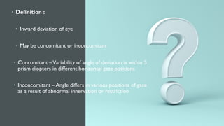
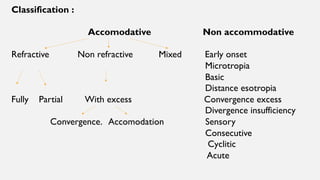
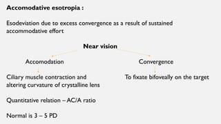
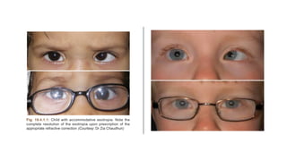
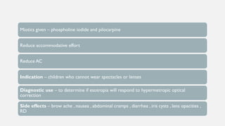
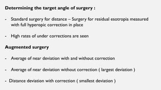
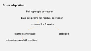
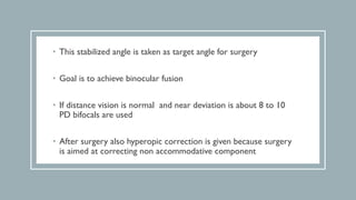
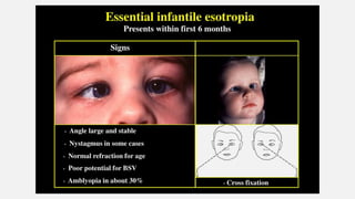
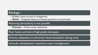
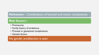
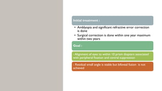
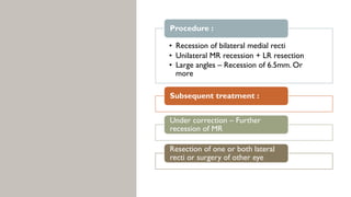
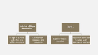
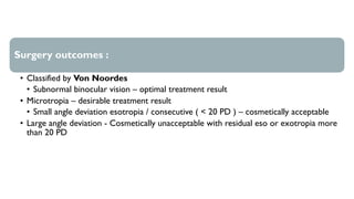
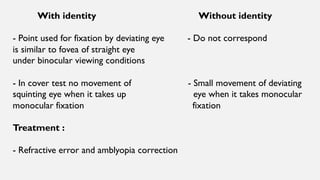
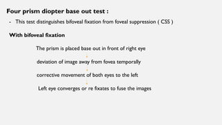
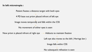
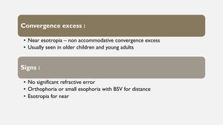
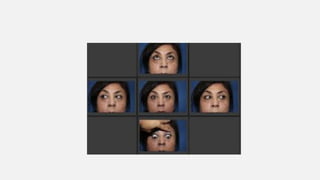
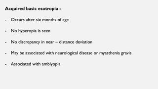
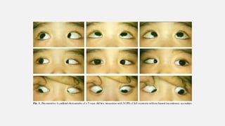
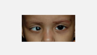
ESOTROPIA
DR.PRAKRITIYAGNAM.K
• Definition :
• Inward deviation of eye
• May be concomitant or inconcomitant
• Concomitant –Variability of angle of deviation is within 5
prism diopters in different horizontal gaze positions
• Inconcomitant – Angle differs in various positions of gaze
as a result of abnormal innervation or restriction
• Concomitant esotropia :
• Usually constant and present in early childhood or infancy
• Therefore often associated with amblyopia , poor binocular
fixation and poor stereopsis
• Fusional divergence helps to keep esotropia in check but innate
divergence amplitude in humans is weak ( 6 – 8 PD ) resulting in
poor control
Classification :
Accomodative Non accommodative
Refractive Non refractive Mixed Early onset
Microtropia
Basic
Distance esotropia
Fully Partial With excess Convergence excess
Divergence insufficiency
Convergence. Accomodation Sensory
Consecutive
Cyclitic
Acute
Accomodative esotropia :
Esodeviation due to excess convergence as a result of sustained
accommodative effort
Near vision
Accomodation Convergence
Ciliary muscle contraction and To fixate bifoveally on the target
altering curvature of crystalline lens
Quantitative relation – AC/A ratio
Normal is 3 – 5 PD
• Refractive accommodative esotropia :
• Excessive hypermetropia ( +2 to +7 D )
• AC / A ratio is normal
• Presents at ages 18 months to 3 years ( Range 6 months to 7 years )
• Fully accommodative – hypermetropia with esometropia when refractive error is
uncorrected
• After optical correction – deviation eliminated and BSV present in all directions
• Partially refractive – Deviation reduced but not eliminated by
full correction of hypermetropia
• Amblyopia with bilateral congenital superior oblique weakness
present
• Supression of squinting eye is present
• Sometimes ARC develop
Pathogenesis :
Uncorrected hyperopic error
Retinal blur
Excessive accommodative effort
Accomodative convergence with poor divergence amplitude
Esotropia
- In children who do not make an effort to clear retinal blur by
accommodative effort or if hypermetropia is too high.To overcome with
accommodation
Uncorrected hypermetropia
B/L ametropia but no esotropia
• Clinical features :
• Age of onset – Infancy to late childhood
• Size of deviation – 20 to 60 PD
• Amblyopia , stereopsis and binocularity may be compromised
• Complete evaluation to rule out any other neurological causes of
acquired esotropia
Diagnosis :
- Cycloplegic refraction and measurement of baselines at distance and near
Older children – Cyclopentolate 1%
Younger children – Atropine E/O
Management :
- Goals – Good alignment
Equal vision
Good stereopsis
Emmetropization
- Full hyperopic correction is given
- Aim is to reduce esotropia by 8 to 10 PD to develop peripheral fusion
- Amblyopia – full patching regime
Regular followup is
essential
Cycloplegic refraction ,
magnitude of deviation
and presence of fusion
is checked in each visit
Tapering of hyperopic
correction in steps of
0.5 D over a period of
6 – 8 months
Miotics given – phospholine iodide and pilocarpine
Reduce accommodative effort
Reduce AC
Indication – children who cannot wear spectacles or lenses
Diagnostic use – to determine if esotropia will respond to hypermetropic optical
correction
Side effects – brow ache , nausea , abdominal cramps , diarrhea , iris cysts , lens opacities ,
RD
Surgery :
Not recommended in
fully accommodative
refractive esotropia
Can be done in partially
accommodative
esotropia for residual
deviation after hyperopic
correction
Surgery done is bilateral
MR recession
Determining the target angle of surgery :
- Standard surgery for distance – Surgery for residual esotropia measured
with full hyperopic correction in place
- High rates of under corrections are seen
Augmented surgery
- Average of near deviation with and without correction
- Average of near deviation without correction ( largest deviation )
- Distance deviation with correction ( smallest deviation )
Prism adaptation :
Full hyperopic correction
Base out prisms for residual correction
assessed for 2 weeks
esotropia increased stabilized
prisms increased till stabilised
• This stabilized angle is taken as target angle for surgery
• Goal is to achieve binocular fusion
• If distance vision is normal and near deviation is about 8 to 10
PD bifocals are used
• After surgery also hyperopic correction is given because surgery
is aimed at correcting non accommodative component
• Non refractive accommodation :
• Esotropia with greater deviation for near
compared to distance
• With full hyperopic correction distance angle
can be controlled but significant residual near
angle is present
• AC/A ration is high
• For 1 D increase in accommodation there is
large increase in convergence
• Independent of refractive error
• Frequently hypermetropia coexists
sometimes myopia
Diagnosis :
- Cycloplegic refraction
- Measurement of AC / A
ratio by heterophoric or
gradient method
Management :
Bifocal glasses
- Bifocal add for residual near esotropia ( < 10 PD )
- Add relaxes accommodation thus reduces convergence
- Aim – to promote fusion and to reduce near angles
- Maximum can be added is +3 D
- Ideal is executive type bisecting the pupil
- Can be gradually reduced
Miotics :
Phospholine iodide 0.125 %
Ciliary spasm
Reduces peripheral accommodative demand and convergence
Surgery :
- Bilateral MR recession with posterior fixation suture ( or )
- Bilateral Faden procedure
Convergence excess Hypoaccomodative convergence
- Increased convergence - Weak accommodation
with normal accommodation
Increased effort
- AC/A ratio is normal
Over convergence
- Normal NPA - Remote NPA
- Straight eye with BSV for distance
- Esotropia for near with suppression
- Straight eyes through bifocals
Treatment :
• Refractive error correction
< 6 years – full cycloplegic refraction
> 8 years – without cycloplegia maximum plus is
prescribed
( manifest hypermetropia treated )
• Convergence excess – bifocals to relieve
accommodation and then accommodative
convergence
Bifocals – executive type
strength of lower segment is gradually
reduced and eliminated
• Surgery :
• Near deviation > far deviation – Bilateral MR
recession
• Near and far deviation same with equal vision in BE –
Unilateral MR recession with LR resection
• In residual amblyopia – surgery in amblyopic eye done
• In partially accommodative esotropia – surgery is best
delayed to avoid consecutive esotropia
• So aim is to correct residual squint with glasses
• Undercorrection – MR posterior fixation sutures –
Faden operation
• Early onset esotropia :
• - Infantile or congenital esotropia
• Idiopathic
• Develops within six months of life
• There will be no significant refractive error
• No limitation of ocular movements
• Upto four months infrequent episodes of
convergence are normal but ocular alignment
thereafter is abnormal
• Signs :
• Large angle of deviation ( 30 prism diopters )
and stable
• Fixation is alternating in primary position
• Cross fixation is seen in side gaze – Left eye
in right gaze and vice versa
• Refractive error is normal for the age ( +1 /
+2 D )
• Inferior oblique overaction may be present or
develops
• DVD develops in 80 % by the age of three
years
- Nystagmus is present
and is horizontal and
latent
Other associations :
• Mild amblyopia
• Apparent limitation of
abduction due to cross
fixation
• Absent or reduced
binocular vision
• Absence of nervous system
disorders
Etiology :
• Multiple causes can lead to misalignment
• Worth – Congenital absence of fusional potential at cortical levels
Restoring binocularity is not possible
• Chavasse – Primary motor dysfunction
Poor fusion and lack of high grade stereopsis
Sensory adaptation to abnormal visual stimulation during early
binocular development caused by motor misalignment
Helveston – Combination of fusional and motor components
Risk factors :
• Prematurity
• Family history of strabismus
• Prenatal or gestational complications
• Genetic factors
No gender predilection is seen
Abduction limitation is elicited by Dolls head manoever
Gentle spinning of child
Vestibular movement to opposite direction of spin
Refixation saccade in same direction
Full abduction is elicited
- Due to limited abduction and tight medial recti children cross fixate
- These manifest after one year of age combinedly or
individually
Classic triad for motor abnormality :
Inferior oblique
overaction
DissociatedVertical
Deviation
Latent nystagmus
IO overaction :
• Seen in 70 % of patients
• Overelevation of eye is supra adduction
DVD :
• Seen in 75 % of patients
• Elevation of non fixing eye when covered or with
visual inattention
• 3. Latent nystagmus :
• Seen in 50 % of patients
• Predominantly horizontal jerk
nystagmus elevated by occlusion of
either eye
• Slow phase is towards side of occluded
eye
• Also show persistent smooth pursuit
asymmetry throughout life
• Temporally directed smooth pursuit
eye movements are slow and lag
behind fixation target compared to
nasally directed movements
Evaluation :
• Visual acuity
• Ocular preference detection
• Measuring deviation by cover
tests or Krimpsky tests
• Ocular mobility examination
• Sensory evaluation
• Fundus evaluation
DD :
• Bilateral congenital sixth nerve palsy
• Sensory esotropia due to organic eye disease
• Nystagmus blockage syndrome
• Mechanical limitation of eye movements – Duanes syndrome ,
Mobius syndrome or strabismus fixus
Pseudoesotropia
Congenital fibrosis syndrome
Infantile Myasthenia Gravis
Esotropia secondary to neurological diseases like hydrocephalus ,
intracranial tumors
Initial treatment :
• Amblyopia and significant refractive error correction
is done
• Surgical correction is done within one year maximum
within two years
Goal :
- Alignment of eyes to within 10 prism diopters associated
with peripheral fixation and central suppression
- Residual small angle is stable but bifoveal fusion is not
achieved
• Recession of bilateral medial recti
• Unilateral MR recession + LR resection
• Large angles – Recession of 6.5mm. Or
more
Procedure :
Subsequent treatment :
Under correction – Further
recession of MR
Resection of one or both lateral
recti or surgery of other eye
Inferior oblique
overaction :
At age of 2 years
and other eye
within 6 months
Disinsertion ,
recession and
myectomy
DVD :
Superior rectus
recession
Botulinum toxin can
be used as an
alternate treatment
Surgery outcomes :
• Classified by Von Noordes
• Subnormal binocular vision – optimal treatment result
• Microtropia – desirable treatment result
• Small angle deviation esotropia / consecutive ( < 20 PD ) – cosmetically acceptable
• Large angle deviation - Cosmetically unacceptable with residual eso or exotropia more
than 20 PD
Regular followup done
postoperatively to
evaluate amblyopia ,
consecutive exotropia
or residual esotropia
If consecutive or
residual deviations are
large resurgery is
planned
If residual esotropia is
small full hyperopic
spectacle correction
can be given
Infantile esotropia can
develop good vision
but binocularity is poor
Microtropia :
• Small angle ( < 10 prism diopter )
squint
• Symptoms are rare unless with
associating decompensated
heterophoria
Signs :
• Prominent association with
anisometropia or hypermetropic
astigmatism of more ametropic eye is
seen
• Normal motor fusion is seen
• ARC is present with abnormal
binocular single vision
• Monocular fixation is eccentric to
fovea in deviating eye and central
suppression scotoma is present
• Stereopsis is reduced
• Types :
• With identity and without identity
• Diagnosis :
• - 4 prism diopter base out test
With identity Without identity
- Point used for fixation by deviating eye - Do not correspond
is similar to fovea of straight eye
under binocular viewing conditions
- In cover test no movement of - Small movement of deviating
squinting eye when it takes up eye when it takes monocular
monocular fixation fixation
Treatment :
- Refractive error and amblyopia correction
Four prism diopter base out test :
- This test distinguishes bifoveal fixation from foveal suppression ( CSS )
With bifoveal fixation
The prism is placed base out in front of right eye
deviation of image away from fovea temporally
corrective movement of both eyes to the left
Left eye converges or re fixates to fuse the images
In left microtropia :
Patient fixates a distance target with both eyes
4 PD base out prism placed infront of left eye
Image moves temporally and falls within the CSS
No movement of either eyes is seen
Now prism is placed infront of right eye Adducts to maintain fixation
Left eye also moves to the left ( Herings law )
Image falls within CSS
No subsequent refixation is seen
Convergence excess :
• Near esotropia – non accommodative convergence excess
• Usually seen in older children and young adults
Signs :
• No significant refractive error
• Orthophoria or small esophoria with BSV for distance
• Esotropia for near
- NORMAL OR LOW AC/A
RATIO
- NORMAL NPA
TREATMENT :
- BILATERAL MR RECESSION
Distance esotropia :
• In healthy young adults with
myopia
Signs :
• Intermittent or constant
esotropia for distance
• Minimal or no deviation for
near
• Normal bilateral abduction
• Fusional deviational
amplitude may be reduced
Absence of
neurological
disease
Treatment :
- Prisms until
spontaneous
resolution
- Surgery in
persistent
cases
Acquired basic esotropia :
- Occurs after six months of age
- No hyperopia is seen
- No discrepancy in near – distance deviation
- May be associated with neurological disease or myasthenia gravis
- Associated with amblyopia
Management :
- Amblyopia therapy
- Extraocular surgery for deviation
Acute ( late onset ) esotropia :
• At around 5 – 6 years
Etiology :
• ICSOL , orbital lesions , psychological
stress or emotional problems
• Underlying sixth nerve palsy must be
excluded
Signs :
• Sudden onset of diplopia and esotropia which is comitant
• Normal ocular motility without significant refractive error
Treatment – Prisms or occlusion of one eye to
obliviate diplopia
• If etiology can be delineated it is managed
• If deviation is stable for six months or more B/L MR recession or
resection – recession procedure of affected eye is done
Secondary ( sensory ) esotropia :
• Unilateral reduction in vision that
interferes or abolishes fusion
Causes :
. Corneal scarring
• Cataract
• Optic atrophy
• Optic N. hypoplasia
• Macular scarring
• Retinoblastoma
Esodeviation is more common in
children while exo is common inadults
Oblique muscle overaction is common
Severe degree of recalcitrant amblyopia
not responding to treatment is common
Management :
Treatment of cause
Amblyopia therapy
- Strabismus correction
Functional – if amblyopia treatment is successful
Cosmetic – chances of recurrence with time is
common
Surgery on amblyopic eye is preferable
- Oblique muscle dysfunction must be dealt with
Consecutive esotropia
:
Surgical overcorrection of
exodeviation
Due to loss or slipping of
lateral rectus muscle
- Complication in surgery
to oblique muscles –
more common in SO
weakening
Management :
- If deviation is large in immediate postoperative period with restrictive
ocular motility – lost or slipped muscle
MRI of orbit with EOM is done
Immediate exploration of and retrieving done
Muscle transposition surgery is done
Small overcorrections resolve over time
due to establishment of normal fusional
divergence phenomenon
Base out prisms , plus lenses and miotics
can aid in fusional divergence
If persistent diplopia is present alternate
occlusion of eyes is done
Constant significant deviation over 3 – 6
months requires resurgery
Cyclic esotropia :
Rare condition
Alternating manifest
esotropia with
suppression and BSV
lasting for 24 - 48 hours
Develops esotropia in
trophic phase alone
Remaining times there
will be straight gaze
Possible factor –
repetitive circadian
rhythm
Diagnosis :
Based on typical
history
Examination is done
in both phases
Repeated
examinations are
necessary
Usually progressive
and finally becomes
constant over several
months to years
Management :
If significant
hypertropia is present
– refractive
correction
Surgery is effective
even in cyclic phase –
performed for
maximum angle of
deviation
High myopic esotropia :
High myopia
Instability of muscle pulleys that stabilize SR and LR
Nasal displacement of SR and
Inferior displacement of LR
Esotropia with hypotropia
Heavy eye syndrome
Key to diagnosis – MRI
- Shows distorted muscle paths
- Displacement of SR muscle nasally – mechanical adduction and limitation
of abduction
- Displacement of LR inferiorly – mechanical depression with limitation of
elevation
- Orbital connective tissue degeneration and associated abnormalities are seen
• Management :
• Surgery – Large recession – resection procedures
• Disinsertion of medial rectus muscle
• Modified Jensen procedure with transposition of superior rectus
• inferior rectus
• Loop myopexy of Lateral rectus and Superior rectus muscles
with or without frontal muscle split performed along with medial rectus recession
• Other variants
• Traditional scleral fixation of sutures
• Use of silicone bands around two muscles
• Injection of botulinum toxin into medial rectus muscle along with myopexy
Divergence insufficiency :
Constant or
intermittent
Greater for distance
than near by atleast
10 PD
Normal versions and
ductions
Bifoveal fixation is
lost for distance but
present for near
AC /A ratio is low
CAUSES :
SUPRANUCLEAR
CONDITION
VERGENCE DISORDER
DEGENERATIVE
CONNECTIVE
TISSUE DISORDER
OF EXTRAOCULAR
MUSCLES
Nystagmus blockage syndrome :
• Condition with congenital nystagmus and straight eyes who may use
accommodative convergence to damp their nystagmus leading to esotropia
• Therefore in inattention– orthotropia + manifest nystagmus
• Near fixation – esotropia + dampened nystagmus
• The clinical nystagmus is inversely proportional to esotropia
• Characteristic sign – Appearance of nystagmus on attempted abduction of
either eye with a head turn in the direction of abducted eye
• Pupillary constriction during esotropic phase is seen
• Surgery :
• Bilateral MR recession with or without posterior fixating sutures
Or
• - Unilateral recession – resection procedures
THANK YOU !!!

Esotropia

  • 1.
  • 3.
    • Definition : •Inward deviation of eye • May be concomitant or inconcomitant • Concomitant –Variability of angle of deviation is within 5 prism diopters in different horizontal gaze positions • Inconcomitant – Angle differs in various positions of gaze as a result of abnormal innervation or restriction
  • 4.
    • Concomitant esotropia: • Usually constant and present in early childhood or infancy • Therefore often associated with amblyopia , poor binocular fixation and poor stereopsis • Fusional divergence helps to keep esotropia in check but innate divergence amplitude in humans is weak ( 6 – 8 PD ) resulting in poor control
  • 5.
    Classification : Accomodative Nonaccommodative Refractive Non refractive Mixed Early onset Microtropia Basic Distance esotropia Fully Partial With excess Convergence excess Divergence insufficiency Convergence. Accomodation Sensory Consecutive Cyclitic Acute
  • 6.
    Accomodative esotropia : Esodeviationdue to excess convergence as a result of sustained accommodative effort Near vision Accomodation Convergence Ciliary muscle contraction and To fixate bifoveally on the target altering curvature of crystalline lens Quantitative relation – AC/A ratio Normal is 3 – 5 PD
  • 7.
    • Refractive accommodativeesotropia : • Excessive hypermetropia ( +2 to +7 D ) • AC / A ratio is normal • Presents at ages 18 months to 3 years ( Range 6 months to 7 years ) • Fully accommodative – hypermetropia with esometropia when refractive error is uncorrected • After optical correction – deviation eliminated and BSV present in all directions
  • 9.
    • Partially refractive– Deviation reduced but not eliminated by full correction of hypermetropia • Amblyopia with bilateral congenital superior oblique weakness present • Supression of squinting eye is present • Sometimes ARC develop
  • 11.
    Pathogenesis : Uncorrected hyperopicerror Retinal blur Excessive accommodative effort Accomodative convergence with poor divergence amplitude Esotropia
  • 12.
    - In childrenwho do not make an effort to clear retinal blur by accommodative effort or if hypermetropia is too high.To overcome with accommodation Uncorrected hypermetropia B/L ametropia but no esotropia
  • 13.
    • Clinical features: • Age of onset – Infancy to late childhood • Size of deviation – 20 to 60 PD • Amblyopia , stereopsis and binocularity may be compromised • Complete evaluation to rule out any other neurological causes of acquired esotropia
  • 14.
    Diagnosis : - Cycloplegicrefraction and measurement of baselines at distance and near Older children – Cyclopentolate 1% Younger children – Atropine E/O
  • 15.
    Management : - Goals– Good alignment Equal vision Good stereopsis Emmetropization - Full hyperopic correction is given - Aim is to reduce esotropia by 8 to 10 PD to develop peripheral fusion - Amblyopia – full patching regime
  • 16.
    Regular followup is essential Cycloplegicrefraction , magnitude of deviation and presence of fusion is checked in each visit Tapering of hyperopic correction in steps of 0.5 D over a period of 6 – 8 months
  • 17.
    Miotics given –phospholine iodide and pilocarpine Reduce accommodative effort Reduce AC Indication – children who cannot wear spectacles or lenses Diagnostic use – to determine if esotropia will respond to hypermetropic optical correction Side effects – brow ache , nausea , abdominal cramps , diarrhea , iris cysts , lens opacities , RD
  • 18.
    Surgery : Not recommendedin fully accommodative refractive esotropia Can be done in partially accommodative esotropia for residual deviation after hyperopic correction Surgery done is bilateral MR recession
  • 19.
    Determining the targetangle of surgery : - Standard surgery for distance – Surgery for residual esotropia measured with full hyperopic correction in place - High rates of under corrections are seen Augmented surgery - Average of near deviation with and without correction - Average of near deviation without correction ( largest deviation ) - Distance deviation with correction ( smallest deviation )
  • 20.
    Prism adaptation : Fullhyperopic correction Base out prisms for residual correction assessed for 2 weeks esotropia increased stabilized prisms increased till stabilised
  • 21.
    • This stabilizedangle is taken as target angle for surgery • Goal is to achieve binocular fusion • If distance vision is normal and near deviation is about 8 to 10 PD bifocals are used • After surgery also hyperopic correction is given because surgery is aimed at correcting non accommodative component
  • 22.
    • Non refractiveaccommodation : • Esotropia with greater deviation for near compared to distance • With full hyperopic correction distance angle can be controlled but significant residual near angle is present • AC/A ration is high • For 1 D increase in accommodation there is large increase in convergence • Independent of refractive error • Frequently hypermetropia coexists sometimes myopia
  • 25.
    Diagnosis : - Cycloplegicrefraction - Measurement of AC / A ratio by heterophoric or gradient method
  • 26.
    Management : Bifocal glasses -Bifocal add for residual near esotropia ( < 10 PD ) - Add relaxes accommodation thus reduces convergence - Aim – to promote fusion and to reduce near angles - Maximum can be added is +3 D - Ideal is executive type bisecting the pupil - Can be gradually reduced
  • 27.
    Miotics : Phospholine iodide0.125 % Ciliary spasm Reduces peripheral accommodative demand and convergence Surgery : - Bilateral MR recession with posterior fixation suture ( or ) - Bilateral Faden procedure
  • 28.
    Convergence excess Hypoaccomodativeconvergence - Increased convergence - Weak accommodation with normal accommodation Increased effort - AC/A ratio is normal Over convergence - Normal NPA - Remote NPA - Straight eye with BSV for distance - Esotropia for near with suppression - Straight eyes through bifocals
  • 30.
    Treatment : • Refractiveerror correction < 6 years – full cycloplegic refraction > 8 years – without cycloplegia maximum plus is prescribed ( manifest hypermetropia treated ) • Convergence excess – bifocals to relieve accommodation and then accommodative convergence Bifocals – executive type strength of lower segment is gradually reduced and eliminated
  • 31.
    • Surgery : •Near deviation > far deviation – Bilateral MR recession • Near and far deviation same with equal vision in BE – Unilateral MR recession with LR resection • In residual amblyopia – surgery in amblyopic eye done • In partially accommodative esotropia – surgery is best delayed to avoid consecutive esotropia • So aim is to correct residual squint with glasses • Undercorrection – MR posterior fixation sutures – Faden operation
  • 32.
    • Early onsetesotropia : • - Infantile or congenital esotropia • Idiopathic • Develops within six months of life • There will be no significant refractive error • No limitation of ocular movements • Upto four months infrequent episodes of convergence are normal but ocular alignment thereafter is abnormal
  • 33.
    • Signs : •Large angle of deviation ( 30 prism diopters ) and stable • Fixation is alternating in primary position • Cross fixation is seen in side gaze – Left eye in right gaze and vice versa • Refractive error is normal for the age ( +1 / +2 D ) • Inferior oblique overaction may be present or develops • DVD develops in 80 % by the age of three years
  • 35.
    - Nystagmus ispresent and is horizontal and latent Other associations : • Mild amblyopia • Apparent limitation of abduction due to cross fixation • Absent or reduced binocular vision • Absence of nervous system disorders
  • 37.
    Etiology : • Multiplecauses can lead to misalignment • Worth – Congenital absence of fusional potential at cortical levels Restoring binocularity is not possible • Chavasse – Primary motor dysfunction Poor fusion and lack of high grade stereopsis Sensory adaptation to abnormal visual stimulation during early binocular development caused by motor misalignment
  • 38.
    Helveston – Combinationof fusional and motor components Risk factors : • Prematurity • Family history of strabismus • Prenatal or gestational complications • Genetic factors No gender predilection is seen
  • 39.
    Abduction limitation iselicited by Dolls head manoever Gentle spinning of child Vestibular movement to opposite direction of spin Refixation saccade in same direction Full abduction is elicited - Due to limited abduction and tight medial recti children cross fixate
  • 40.
    - These manifestafter one year of age combinedly or individually Classic triad for motor abnormality : Inferior oblique overaction DissociatedVertical Deviation Latent nystagmus
  • 41.
    IO overaction : •Seen in 70 % of patients • Overelevation of eye is supra adduction DVD : • Seen in 75 % of patients • Elevation of non fixing eye when covered or with visual inattention
  • 43.
    • 3. Latentnystagmus : • Seen in 50 % of patients • Predominantly horizontal jerk nystagmus elevated by occlusion of either eye • Slow phase is towards side of occluded eye • Also show persistent smooth pursuit asymmetry throughout life • Temporally directed smooth pursuit eye movements are slow and lag behind fixation target compared to nasally directed movements
  • 44.
    Evaluation : • Visualacuity • Ocular preference detection • Measuring deviation by cover tests or Krimpsky tests • Ocular mobility examination • Sensory evaluation • Fundus evaluation
  • 45.
    DD : • Bilateralcongenital sixth nerve palsy • Sensory esotropia due to organic eye disease • Nystagmus blockage syndrome • Mechanical limitation of eye movements – Duanes syndrome , Mobius syndrome or strabismus fixus
  • 46.
    Pseudoesotropia Congenital fibrosis syndrome InfantileMyasthenia Gravis Esotropia secondary to neurological diseases like hydrocephalus , intracranial tumors
  • 47.
    Initial treatment : •Amblyopia and significant refractive error correction is done • Surgical correction is done within one year maximum within two years Goal : - Alignment of eyes to within 10 prism diopters associated with peripheral fixation and central suppression - Residual small angle is stable but bifoveal fusion is not achieved
  • 48.
    • Recession ofbilateral medial recti • Unilateral MR recession + LR resection • Large angles – Recession of 6.5mm. Or more Procedure : Subsequent treatment : Under correction – Further recession of MR Resection of one or both lateral recti or surgery of other eye
  • 49.
    Inferior oblique overaction : Atage of 2 years and other eye within 6 months Disinsertion , recession and myectomy DVD : Superior rectus recession Botulinum toxin can be used as an alternate treatment
  • 50.
    Surgery outcomes : •Classified by Von Noordes • Subnormal binocular vision – optimal treatment result • Microtropia – desirable treatment result • Small angle deviation esotropia / consecutive ( < 20 PD ) – cosmetically acceptable • Large angle deviation - Cosmetically unacceptable with residual eso or exotropia more than 20 PD
  • 51.
    Regular followup done postoperativelyto evaluate amblyopia , consecutive exotropia or residual esotropia If consecutive or residual deviations are large resurgery is planned If residual esotropia is small full hyperopic spectacle correction can be given Infantile esotropia can develop good vision but binocularity is poor
  • 52.
    Microtropia : • Smallangle ( < 10 prism diopter ) squint • Symptoms are rare unless with associating decompensated heterophoria Signs : • Prominent association with anisometropia or hypermetropic astigmatism of more ametropic eye is seen • Normal motor fusion is seen • ARC is present with abnormal binocular single vision
  • 54.
    • Monocular fixationis eccentric to fovea in deviating eye and central suppression scotoma is present • Stereopsis is reduced • Types : • With identity and without identity • Diagnosis : • - 4 prism diopter base out test
  • 55.
    With identity Withoutidentity - Point used for fixation by deviating eye - Do not correspond is similar to fovea of straight eye under binocular viewing conditions - In cover test no movement of - Small movement of deviating squinting eye when it takes up eye when it takes monocular monocular fixation fixation Treatment : - Refractive error and amblyopia correction
  • 56.
    Four prism diopterbase out test : - This test distinguishes bifoveal fixation from foveal suppression ( CSS ) With bifoveal fixation The prism is placed base out in front of right eye deviation of image away from fovea temporally corrective movement of both eyes to the left Left eye converges or re fixates to fuse the images
  • 57.
    In left microtropia: Patient fixates a distance target with both eyes 4 PD base out prism placed infront of left eye Image moves temporally and falls within the CSS No movement of either eyes is seen Now prism is placed infront of right eye Adducts to maintain fixation Left eye also moves to the left ( Herings law ) Image falls within CSS No subsequent refixation is seen
  • 59.
    Convergence excess : •Near esotropia – non accommodative convergence excess • Usually seen in older children and young adults Signs : • No significant refractive error • Orthophoria or small esophoria with BSV for distance • Esotropia for near
  • 60.
    - NORMAL ORLOW AC/A RATIO - NORMAL NPA TREATMENT : - BILATERAL MR RECESSION
  • 62.
    Distance esotropia : •In healthy young adults with myopia Signs : • Intermittent or constant esotropia for distance • Minimal or no deviation for near • Normal bilateral abduction • Fusional deviational amplitude may be reduced
  • 63.
    Absence of neurological disease Treatment : -Prisms until spontaneous resolution - Surgery in persistent cases
  • 65.
    Acquired basic esotropia: - Occurs after six months of age - No hyperopia is seen - No discrepancy in near – distance deviation - May be associated with neurological disease or myasthenia gravis - Associated with amblyopia
  • 67.
    Management : - Amblyopiatherapy - Extraocular surgery for deviation
  • 68.
    Acute ( lateonset ) esotropia : • At around 5 – 6 years Etiology : • ICSOL , orbital lesions , psychological stress or emotional problems • Underlying sixth nerve palsy must be excluded
  • 69.
    Signs : • Suddenonset of diplopia and esotropia which is comitant • Normal ocular motility without significant refractive error Treatment – Prisms or occlusion of one eye to obliviate diplopia • If etiology can be delineated it is managed • If deviation is stable for six months or more B/L MR recession or resection – recession procedure of affected eye is done
  • 71.
    Secondary ( sensory) esotropia : • Unilateral reduction in vision that interferes or abolishes fusion Causes : . Corneal scarring • Cataract • Optic atrophy • Optic N. hypoplasia • Macular scarring • Retinoblastoma
  • 73.
    Esodeviation is morecommon in children while exo is common inadults Oblique muscle overaction is common Severe degree of recalcitrant amblyopia not responding to treatment is common Management : Treatment of cause Amblyopia therapy
  • 74.
    - Strabismus correction Functional– if amblyopia treatment is successful Cosmetic – chances of recurrence with time is common Surgery on amblyopic eye is preferable - Oblique muscle dysfunction must be dealt with
  • 75.
    Consecutive esotropia : Surgical overcorrectionof exodeviation Due to loss or slipping of lateral rectus muscle - Complication in surgery to oblique muscles – more common in SO weakening
  • 77.
    Management : - Ifdeviation is large in immediate postoperative period with restrictive ocular motility – lost or slipped muscle MRI of orbit with EOM is done Immediate exploration of and retrieving done Muscle transposition surgery is done
  • 78.
    Small overcorrections resolveover time due to establishment of normal fusional divergence phenomenon Base out prisms , plus lenses and miotics can aid in fusional divergence If persistent diplopia is present alternate occlusion of eyes is done Constant significant deviation over 3 – 6 months requires resurgery
  • 79.
    Cyclic esotropia : Rarecondition Alternating manifest esotropia with suppression and BSV lasting for 24 - 48 hours Develops esotropia in trophic phase alone Remaining times there will be straight gaze Possible factor – repetitive circadian rhythm
  • 80.
    Diagnosis : Based ontypical history Examination is done in both phases Repeated examinations are necessary Usually progressive and finally becomes constant over several months to years
  • 82.
    Management : If significant hypertropiais present – refractive correction Surgery is effective even in cyclic phase – performed for maximum angle of deviation
  • 83.
    High myopic esotropia: High myopia Instability of muscle pulleys that stabilize SR and LR Nasal displacement of SR and Inferior displacement of LR Esotropia with hypotropia Heavy eye syndrome
  • 85.
    Key to diagnosis– MRI - Shows distorted muscle paths - Displacement of SR muscle nasally – mechanical adduction and limitation of abduction - Displacement of LR inferiorly – mechanical depression with limitation of elevation - Orbital connective tissue degeneration and associated abnormalities are seen
  • 87.
    • Management : •Surgery – Large recession – resection procedures • Disinsertion of medial rectus muscle • Modified Jensen procedure with transposition of superior rectus • inferior rectus • Loop myopexy of Lateral rectus and Superior rectus muscles with or without frontal muscle split performed along with medial rectus recession
  • 88.
    • Other variants •Traditional scleral fixation of sutures • Use of silicone bands around two muscles • Injection of botulinum toxin into medial rectus muscle along with myopexy
  • 89.
    Divergence insufficiency : Constantor intermittent Greater for distance than near by atleast 10 PD Normal versions and ductions Bifoveal fixation is lost for distance but present for near AC /A ratio is low
  • 90.
  • 91.
    Nystagmus blockage syndrome: • Condition with congenital nystagmus and straight eyes who may use accommodative convergence to damp their nystagmus leading to esotropia • Therefore in inattention– orthotropia + manifest nystagmus • Near fixation – esotropia + dampened nystagmus • The clinical nystagmus is inversely proportional to esotropia
  • 92.
    • Characteristic sign– Appearance of nystagmus on attempted abduction of either eye with a head turn in the direction of abducted eye • Pupillary constriction during esotropic phase is seen • Surgery : • Bilateral MR recession with or without posterior fixating sutures Or • - Unilateral recession – resection procedures
  • 93.